Primary Newborn Care: Management of important problems
•
2 gefällt mir•1,445 views

1. Jaundice is caused by high levels of bilirubin in the blood and can become dangerous at very high levels when bilirubin enters the brain. 2. Physiological jaundice is mild jaundice that is common in newborns, while abnormal jaundice requires treatment and may be caused by infections, blood incompatibilities, or other issues. 3. Phototherapy uses light to change bilirubin into a form that can be excreted from the liver, lowering bilirubin levels to prevent brain damage in infants with dangerous jaundice.

Empfohlen
Weitere ähnliche Inhalte
Was ist angesagt?
Was ist angesagt? (20)
Andere mochten auch
Andere mochten auch (14)
Ähnlich wie Primary Newborn Care: Management of important problems
Ähnlich wie Primary Newborn Care: Management of important problems (20)
Mehr von Saide OER Africa
Mehr von Saide OER Africa (20)
Kürzlich hochgeladen
Kürzlich hochgeladen (20)
Primary Newborn Care: Management of important problems
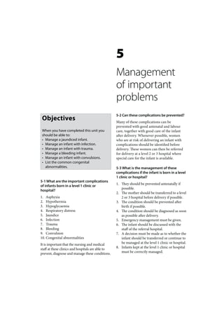
- 1. 5 Management of important problems 5-2 Can these complications be prevented? Objectives Many of these complications can be prevented with good antenatal and labour When you have completed this unit you care, together with good care of the infant should be able to: after delivery. Whenever possible, women • Manage a jaundiced infant. who are at risk of delivering an infant with • Manage an infant with infection. complications should be identified before • Manage an infant with trauma. delivery. These women can then be referred • Manage a bleeding infant. for delivery at a level 2 or 3 hospital where • Manage an infant with convulsions. special care for the infant is available. • List the common congenital abnormalities. 5-3 What is the management of these complications if the infant is born in a level 1 clinic or hospital? 5-1 What are the important complications 1. They should be prevented antenatally if of infants born in a level 1 clinic or possible. hospital? 2. The mother should be transferred to a level 1. Asphyxia 2 or 3 hospital before delivery if possible. 2. Hypothermia 3. The condition should be prevented after 3. Hypoglycaemia birth if possible. 4. Respiratory distress 4. The condition should be diagnosed as soon 5. Jaundice as possible after delivery. 6. Infection 5. Emergency management must be given. 7. Trauma 6. The infant should be discussed with the 8. Bleeding staff of the referral hospital. 9. Convulsion 7. A decision must be made as to whether the 10. Congenital abnormalities infant should be transferred or continue to It is important that the nursing and medical be managed at the level 1 clinic or hospital. staff at these clinics and hospitals are able to 8. Infants kept at the level 1 clinic or hospital prevent, diagnose and manage these conditions. must be correctly managed.
- 2. MANAGEMENT OF IMPOR TANT PROBLEMS 77 MANAGEMENT OF AN bilirubin increases in the serum and the newborn infant may become jaundiced as the INFANT WITH JAUNDICE excess bilirubin is deposited in the skin. After a few days the rate of conjugation in the liver increases and much more bilirubin is excreted. 5-4 What is jaundice? As a result, the amount of bilirubin in the Jaundice is a yellow colour of the skin caused serum slowly returns to the normal adult by deposits of bilirubin in the skin. Jaundice is a range and any jaundice disappears. clinical sign and not a laboratory measurement. Some of the bilirubin that is conjugated and excreted by the liver in the first weeks of life is 5-5 What is bilirubin? often broken down (uncongugated) by another Red cells in the blood contain a red pigment enzyme in the intestine. This bilirubin is then called haemoglobin, which carries oxygen. reabsorbed back into the blood stream, adding Red cells live for a few months only. Therefore, to the hyperbilirubinaemia. The reabsorption the body is continually forming new red cells of bilirubin from the intestine is greater in in the bone marrow and destroying old red starved and breast fed infants. cells in the liver and spleen. The haemoglobin in old red cells is broken down into a yellow 5-8 How is bilirubin measured? pigment called bilirubin. As newborn It is both difficult and inaccurate to assess infants normally have a high haemoglobin the concentration of bilirubin in the serum concentration, they produce a lot of bilirubin. by clinical examination of the degree of jaundice, especially in an infant with a dark 5-6 What is hyperbilirubinaemia? skin. Therefore, it is important to measure Hyperbilirubinaemia is defined as a the bilirubin concentration of the serum if concentration of total serum bilirubin that is an infant is very jaundiced. Usually a sample higher than the normal range. Normally the of blood is collected into a capillary tube and bilirubin concentration in the serum is low spun down to separate the serum from the at birth, as it has been rapidly removed by red cells. The total serum bilirubin (TSB) is the placenta during pregnancy. The bilirubin then measured with a bilirubinometer and concentration climbs steadily for the first few expressed in μmol/l. days after delivery, before returning to an adult level by 2 weeks. The total serum bilirubin (TSB) cannot be estimated accurately by assessing the degree of 5-7 How is bilirubin excreted? jaundice in the skin. After birth, bilirubin is carried by the blood stream to the liver where a special enzyme changes the bilirubin into a water 5-9 What is physiological jaundice? soluble form. This chemical process is called This is the mild jaundice that is seen in up to conjugation. Only when the bilirubin is water 50% of all healthy, term infants during the first soluble (i.e. conjugated) can the liver cells 2 weeks of life. Many of these infants are breast excrete it into the small bile ducts. From here fed. These infants are well and do not need any the conjugated bilirubin is carried in the bile treatment. Physiological jaundice in newborn to the small intestine, where it is broken down infants is the result of: further by bacteria and is excreted in the stool. 1. The normally high haemoglobin During the first weeks of life the enzyme concentration which results in a lot of system, that conjugates bilirubin in the liver, bilirubin being produced. functions slowly. Therefore, the amount of
- 3. 78 PRIMAR Y NEWBORN CARE 2. The slow rate of conjugation of bilirubin 3. When the infant has hepatitis due to: by the liver, which results in only small, • Congenital syphilis. amounts being excreted. • Septicaemia. 3. The reabsorption of bilirubin from the 4. When too much bilirubin is reabsorbed intestines during the first few weeks after from the intestines: birth. • Starved infants. • Some breast fed infants. All these factors usually disappear by 2 weeks and the jaundice disappears. It is not uncommon for otherwise healthy breast fed infants to remain jaundiced for more than 2 weeks. Many healthy infants have mild jaundice. All these conditions may cause an abnormally high TSB and a very jaundiced infant. 5-10 When is jaundice abnormal? 1. If the TSB increases above the upper limit 5-12 What is haemolytic disease of the of the normal range or the infant looks newborn? severely jaundiced. The phototherapy line Haemolytic disease of the newborn is a shown in Figure 1 is the upper limit of condition where antibodies from the mother normal for TSB. Jaundice of the palms and cross the placenta into the fetal blood stream. soles suggests severe jaundice. Here these antibodies destroy the fetal red 2. If the infant appears jaundiced in the first cells (i.e. haemolysis), causing anaemia and 24 hours. an increased production of bilirubin in 3. If the infant is still jaundiced after 1 month. the fetus and newborn infant. The 2 most 4. If the infant is clinically sick or anaemic. important causes of haemolytic disease of the It is very important to decide whether the newborn are: jaundice is physiological or abnormal. 1. ABO haemolytic disease. 2. Rhesus haemolytic disease. 5-11 What causes jaundice which is In haemolytic diseases of the newborn the abnormal and not physiological? blood group of the infant is different to that of 1. When too much bilirubin is produced the mother as it is inherited from the father. from haemoglobin because: • The concentration of haemoglobin is very high (i.e. polycythaemia). 5-13 What is ABO haemolytic disease? • Bilirubin is absorbed from a Red cells have blood group proteins on their cephalhaematoma or area of bruising. surface. A, B, O and D (Rhesus) are the • There is a very rapid break down of red most important blood group proteins. ABO blood cells (i.e. haemolysis). haemolytic disease occurs when the mother 2. When the excretion of bilirubin is too slow: is blood group O and her fetus is blood group • Preterm infants who have an immature A or B. For reasons unknown, some group liver. O mothers start producing antibodies to the • About 10% of clinically healthy, term A or B proteins. These antibodies cross the infants have slower conjugation than placenta and cause haemolysis in the fetus normal. by damaging the fetal red cells. With ABO • Congenital hypothyroidism. Due to haemolytic disease, the haemolysis is not the absence of a thyroid gland and low severe enough to cause anaemia in the fetus concentrations of thyroid hormone, but may cause severe jaundice and anaemia the enzymes in the liver function very in the newborn infant. slowly.
- 4. MANAGEMENT OF IMPOR TANT PROBLEMS 79 The haemolysis results in anaemia (low All women must have their blood group identified haemoglobin concentration and low packed during pregnancy and all Rhesus negative mothers cell volume) and jaundice in the newborn infant. An infant with ABO haemolytic must receive anti-d immunoglobulin after delivery. disease usually appears normal at delivery but becomes jaundiced within the first 24 5-16 When is jaundice dangerous? hours. The TSB may increase rapidly and reach dangerous levels. Due to the haemolysis, Jaundice can become dangerous when the the infant may also become anaemic. ABO concentration of bilirubin in the blood haemolytic disease cannot be prevented. becomes very high. Bilirubin may now enter the brain of the newborn infant and cause bilirubin encephalopathy (also Jaundice on day 1 suggests haemolytic disease. called kernicterus). The risk of bilirubin encephalopathy depends on the severity of the hyperbilirubinaemia. In well, term infants 5-14 What is Rhesus haemolytic disease? the TSB becomes dangerous and may cause Rhesus haemolytic disease is a form of bilirubin encephalopathy above 350 μmol/l haemolytic disease of the newborn, which may while in preterm infants the TSB becomes occur when the mother is Rhesus negative dangerous above 250 μmol/l. (she has no D protein on her red cells) and her fetus is Rhesus positive (it has D protein on its red cells). Rhesus haemolytic disease is A high serum concentration of bilirubin can caused by maternal antibodies to the D (i.e. damage the brain. Rhesus or Rh) protein on the red cells of the fetus. Only if fetal red cells accidentally cross Bilirubin encephalopathy presents with the placenta, and enter the mother’ s blood lethargy, a high pitched cry and convulsions. stream, will she produce antibodies to the D protein of her fetus. This process is known as 5-17 How can bilirubin encephalopathy be sensitisation and may occur in a Rhesus (Rh) prevented? negative woman during a delivery, miscarriage or placental abruption. Rhesus haemolytic By not allowing the TSB to reach dangerous disease is more severe than ABO haemolytic levels. A number of methods can be used to disease. Therefore, the fetus may develop reduce the TSB: severe anaemia and die before birth. If born 1. Give early milk feeds (to reduce bilirubin alive, the newborn infant rapidly becomes reabsoption from the intestine). jaundiced and anaemic. 2. Prevent preterm delivery. 3. Give anti-D immunoglobulin to all 5-15 How can you prevent Rhesus Rhesus negative mothers after delivery, a haemolytic disease? miscarriage or abruptio placentae. 4. Give phototherapy when the TSB All pregnant women must have their blood approaches dangerous levels. group tested at the start of antenatal care. 5. Do an exchange transfusion when All Rhesus negative women must be given phototherapy cannot keep the TSB below 100 μg (4 ml) of anti-D immunoglobulin by dangerous levels. intramuscular injection within 72 hours of delivery, miscarriage or placental abruption. This prevents sensitisation. Early milk feeds help lower the total serum bilirubin.
- 5. 80 PRIMAR Y NEWBORN CARE 5-18 What is phototherapy? 5-20 When should you give phototherapy? Phototherapy uses white or blue light to Whenever the TSB is above the normal range change bilirubin in the skin into a water and approaches dangerous levels, or if the soluble form of bilirubin. This water soluble infant appears very jaundiced. In practice a bilirubin is then carried in the blood to the simple chart is used to decide when to give liver, from where it can be excreted without phototherapy. If the TSB concentration having to be conjugated. Phototherapy is, reaches the phototherapy line, treatment therefore, able to lower the TSB. should be started. The phototherapy line is the same as the upper limit of normal for the 5-19 What equipment is used to give TSB and increases from birth to day 4 then phototherapy? levels off. Phototherapy is usually started earlier in preterm or sick infants. It is not Phototherapy is usually given with a necessary to give phototherapy to healthy, phototherapy unit, which consists of a row of term infants who are jaundiced with a TSB fluorescent tubes. Daylight tubes, white tubes below the phototherapy line. Therefore, or blue tubes are used. They should be changed phototherapy should not be given to well after being used for 1000 hours because their infants with physiological jaundice. It is very effectiveness decreases with time, even if they difficult to use phototherapy safely if the TSB still appear bright. A perspex (clear plastic) cannot be measured. sheet must be placed below the tubes to reduce heat and filter out any ultraviolet light. See Figure 1 for a phototherapy chart showing A perspex sheet also protects the infant if a the phototherapy line for term infants. fluorescent tube breaks or comes loose. Prophylactic phototherapy is given when the Although exposure to sunlight also lowers the TSB is still below the phototherapy line but TSB, an infant placed in the sun may rapidly either the TSB is expected to increase rapidly become too hot. Therefore, this form of or the infant is at an increased risk of bilirubin phototherapy must be used with great caution. encephalopathy. Therefore, prophylactic phototherapy is started immediately after birth if haemolytic disease of the newborn is suspected or diagnosed. Prophylactic TSB μmol/1 350 300 250 200 150 100 50 0 0 1 2 3 4 5 6 7 Age in days Figure I: Phototherapy chart showing the phototherapy line for term infants.
- 6. MANAGEMENT OF IMPOR TANT PROBLEMS 81 phototherapy is often given to preterm infants 2. The infant may pass large, loose, green when their TSB gets near the phototherapy line. stools. 3. The infant’s eyes pads may cover its nose or 5-21 How do you give phototherapy? prevent conjunctivitis being noticed. 4. Phototherapy may interfere with maternal 1. Switch on the phototherapy unit and make bonding if the infant is separated from the sure the tubes are all working. Check mother. the age of the tubes and ensure that the 5. The clinical jaundice may disappear even perspex sheet is in position. though the TSB remains high. 2. Place the infant naked in an incubator or 6. Skin rashes are common. bassinet so that the infant is about 40 cm from the phototherapy tubes. The infant must not wear a cap or nappy. Instead, a The total serum bilirubin should be measured in nappy can be placed under the infant. all infants receiving phototherapy. 3. Cover the infant’s eyes with pads as the bright light often worries the infant. If the TSB cannot be measured at the level 1 Remove the eye pads during feeding so clinic or hospital, a heparinized capillary tube that the eyes can be checked for infection of blood should be sealed at both ends with and to allow the infant and mother to see plasticine and placed in a box to keep light each other. away. It must be sent to the nearest laboratory, 4. Turn the infant over every hour. Frequent which should phone the result to the clinic or turning will make the phototherapy more hospital, as soon as it is available. While waiting effective as more skin is exposed to the light. for the result, phototherapy should be started. 5. Feed the infant milk, at least every 3 to 4 hours. Breast feed if possible. 5-24 When should an infant with jaundice 6. Monitor the infant’s skin temperature be tranferred? hourly, weigh daily and measure TSB daily or more frequently if it approaches 1. Whenever there are any signs of abnormal dangerous levels. jaundice, such as jaundice in the first 24 7. Allow the mother unrestricted visiting. hours, severe jaundice or jaundice in an If possible, the infant should be given ill infant, ie. when the jaundice is not phototherapy next to the mother in the physiological. postnatal ward. 2. If the TSB is more than 100 μmol/l above the phototherapy line. It is difficult to give phototherapy safely if the 3. If the TSB is above 350 μmol/l. TSB cannot be measured. These infants should be discussed with the 5-22 For how long should you give staff of the referral hospital before transfer. phototherapy? Continue phototherapy until the TSB has been INFECTION IN THE under the phototherapy line for 24 hours. Sometimes the TSB rises above the line again NEWBORN INFANT after the phototherapy has been stopped. If so, the phototherapy should be restarted. 5-25 What infections are important in newborn infants? 5-23 What are the problems with phototherapy? 1. Conjunctivitis 2. Umbilical cord infection 1. The infant may become too hot or too cold. 3. Skin infection 4. Oral thrush
- 7. 82 PRIMAR Y NEWBORN CARE 5. Septicaemia prevents conjunctivitis resulting from 6. Congenital syphilis infection with Gonococcus during delivery. 7. HIV infection 7. If formula feeds are being used, the aseptic preparation of formula feeds, and the 5-26 Why is infection common in newborn boiling of cups, bottles and teats is essential infants? to prevent contaminated feeds. Use a cup rather than a bottle to give formula feeds. Because they have an immature immune 8. Stethoscopes and other instruments system and are exposed to infectious should be sprayed with an antiseptic spray organisms during pregnancy, delivery and before an infant is examined. in the nursery. The risk of infection in the 9. Immunization of all pregnant women newborn infant is much higher than in older with tetanus toxoid prevents neonatal children or adults. Infection is important as tetanus complicating cord infection. it is one of the commonest causes of death in 10. Avoid overcrowding in nurseries by infants during the first few months of life. keeping normal infants with their mothers whenever possible. Infection is a common cause of death in young infants. Breast milk protects against infections. It is not necessary to restrict visits of parents 5-27 How can you prevent infection in and family in the nursery provided that strict newborn infants? hand washing and hand spraying is enforced. There are many simple ways in which infections There is no need for visitors to wear masks can be prevented in the newborn infant: or gowns. If possible, family and staff with 1. Do not separate mothers and their infants coughs, colds and ‘flu should not handle unless absolutely necessary. Use skin-to- infants. Herpes infection (fever blisters) can skin care. cause a very serious infection. Infants should 2. Breast feeding. Breast milk contains cells not be kissed. and antibodies, which help to protect the infant from infections. 5-28 What are the signs of conjunctivitis? 3. Vernix has antibacterial properties Conjunctivitis presents with: and, therefore, should not be washed off routinely after delivery. It is rapidly 1. A discharge from the eyes (mucous or pus). absorbed by the skin. 2. Redness of the conjunctivae. 4. Hand spraying or hand washing before 3. Oedema of the eyelids. touching an infant is the most important The degree of conjunctivitis can be divided method of preventing the spread of clinically into mild, moderate and severe: infection in the nursery. Before handling an infant spray your hands with an 1. Mild conjunctivitis consists of a slight antiseptic spray (e.g. D-germ). There is no discharge. The eyelashes tend to stick evidence that gowns or masks reduce cross together. infection. In the home, the family must 2. Moderate conjunctivitis presents with wash their hands well after using the toilet. redness of the conjunctivae with an 5. Routine care of the umbilical stump with obvious discharge. Pus is present in the eye alcohol (surgical spirits) helps prevent when the lids are separated. infection. 3. Severe conjunctivitis has a marked 6. Routine prophylactic eye care after discharge with oedema of the eyelids. Pus delivery with chloromycetin ointment spurts from the eye and runs down the cheeks when the eyelids are opened. In
- 8. MANAGEMENT OF IMPOR TANT PROBLEMS 83 the most severe cases, it is not possible to alone are inadequate for treating severe separate the eyelids due to the swelling. The conjunctivitis as the infection may have pus may soften the cornea and cause it to already spread to involve the whole eye. perforate (burst), severly damaging the eye. Start this treatment before referring the infant urgently to a level 2 or 3 hospital Conjunctivitis is usually mild. It is difficult for further management. to clinically identify the organism causing conjunctivitis although severe conjunctivitis is usually caused by Gonococcus from the Gonococcus causes severe conjunctivitis which mother’s cervix and vagina. Therefore, the may result in blindness. management depends on the severity rather than the cause. 5-30 What are the signs of an infected 5-29 What is the management of umbilical cord? conjunctivitis? A healthy umbilical cord stump is white 1. Mild conjunctivitis can usually be treated and soft at delivery. With good cord care it by cleaning the eye with saline or warm becomes dark brown and dehydrated within water at feeding times or when the lashes a few days, and at no stage does it smell become sticky. A local antibiotic is usually offensive or produce pus. The aim of good not needed. cord care is to dry the cord and keep it clean. 2. Moderate conjunctivitis should be treated Infection of the umbilical cord (omphalitis) by cleaning the eye and then putting in presents with: chloromycetin ointment 3 hourly or more frequently if needed. 1. An offensive (smelly) cord with a discharge 3. Severe conjunctivitis is a medical of pus. emergency as it can lead to blindness if 2. Failure of the cord to become dehydrated not promptly and efficiently treated. The (i.e. the cord remains wet and soft). infection is usually due to the Gonococcus 3. Redness of the skin around the base of the and treatment consists of irrigating the eye cord (a flare). and giving parenteral penicillin: The commonest site of infection is at the base • The pus must be washed out of where the cord meets the skin. When the the eye with saline, warm water or infection is localised to the cord only, there is penicillin drops. This must be started no oedema of the skin around the base of the immediately and repeated frequently cord and the infant is generally well. Umbilical enough to keep the eye clear of pus. cord infection may spread to the anterior The simplest way of irrigating the eye is abdominal wall from where it may cause to use a vacolitre of normal saline and a peritonitis or septicaemia. Signs that the an administration set. Penicillin drops infection of the umbilical cord has extended to can also be used. They can be made up the abdominal wall are: in the nursery by adding 1 ml of benzyl penicillin to 50 ml sterile water or 1. Redness and oedema of the skin around normal saline. The mixture must not be the base of the cord and spreading onto the kept for more than 24 hours. abdomen (cellulitis). • Benzyl penicillin intravenously 2. Abdominal distension often with decreased 6 hourly or procaine penicillin bowel sounds and vomiting (peritonitis). intramuscularly 12 hourly must be 3. The infant is generally unwell with the given for 3 days. The dose of both features of septicaemia. benzyl and procaine penicillin is 50 000 Cellulitis, peritonitis and septicaemia are units/kg per day. Penicillin eye drops serious infections and the infant may die if
- 9. 84 PRIMAR Y NEWBORN CARE not treated immediately with intramuscular 5-33 How do you manage tetanus? or intravenous antibiotics. Infection of the Tetanus can be prevented by: umbilical cord may also cause tetanus in the newborn infant. 1. Good cord care. 2. Immunising all pregnant women with 5-31 How do you treat umbilical cord tetanus toxoid if tetanus is common in infection? the region. All children should be fully immunised. With good preventative cord care, infection of the umbilical cord should not occur. Prevention The emergency treatment of tetanus consists of: consists of routine applications of alcohol 1. Keeping the airway clear and giving oxygen. (surgical spirits) to the cord every 6 hours until 2. Not stimulating the infant. it is dehydrated. Antibiotic powder is not used. 3. Stopping spasms with 1 mg diazepam Do not put aspirin or other home remedies on (Valium) rectally. This may have to be the cord. Never cover the cord with the nappy repeated until the spasms stop. You may or a bandage as this keeps it moist. have to mask and bag ventilate the infant. If the infection is localised to the umbilical 4. Transferring the infant urgently to the cord, and there are no signs of cellulitis, nearest level 2 and 3 hospital. peritonitis, septicaemia or tetanus, then treatment consists simply of cleaning the 5-34 What are the signs and causes of skin cord with surgical spirits every 3 hours to infection? clear the infection and hasten dehydration. The 2 commonest forms of skin infection in Neither local nor systemic antibiotics are the newborn infant are: needed. Special attention must be paid to the folds around the base of the cord, which often 1. Impetigo caused by Staphylococcus, remain moist. Within 24 hours the infection which presents as pus filled blisters should have cleared. Keep a careful watch usually seen around the umbilicus or in for signs that the infection may have spread the nappy area. beyond the umbilicus. 2. A monilial rash caused by a fungus (Candida or Monilia). This almost always 5-32 What is tetanus? occurs in the nappy area and presents as a red, slightly raised, ‘velvety’ rash which Tetanus in the newborn infant (tetanus is most marked in the skin creases. In neonatorum) is caused by bacteria, which contrast, a nappy rash due to irritation of infect dead tissues such as the umbilical cord. the skin by stool and urine usually affects Tetanus bacteria usually occurs in soil and the exposed areas of the skin and not the faeces, which may be placed on the cord or creases. other wounds as a traditional practice. They produces a powerful toxin that affects the A sweat rash, due to excessive sweating, may nervous system. look like an infection. It presents as small, clear blisters on the forehead or a fine red rash on the Tetanus presents with: neck and trunk. Treat, by washing the infant to 1. Increased muscle tone (spasm), especially remove the sweat, and prevent overheating. of the jaw muscles and abdomen. 2. Generalised muscle spasms and 5-35 How do you treat skin infections? convulsions, often precipitated by Pay strict attention to hand washing and stimulation such as handling or loud noises. spraying, and do not routinely wash off vernix. 3. Respiratory failure and death in untreated Then skin infection should not be a problem infants, due to spasm of the respiratory in a nursery. muscles.
- 10. MANAGEMENT OF IMPOR TANT PROBLEMS 85 1. Impetigo is treated by washing the infant prevent bacterial contamination of bottles but in chlorhexidine (e.g. Bioscrub) or a red may not kill Candida. Dummies should be soap (e.g. Lifebouy) twice a day for 5 days. boiled or thrown away. If the infant should become unwell and show any signs of septicaemia, then urgent 5-37 What are the signs of septicaemia? treatment with parenteral antibiotics is indicated. The clinical signs of septicaemia are often 2. A monilial rash should be treated with non-specific, making the early diagnosis of topical mycostatin (Nystatin) cream and septicaemia difficult. The common clinical the area should not be covered. Allow the signs are: infant to sleep on a nappy and keep the 1. Lethargy and appearing generally unwell. infected area of skin exposed to the air. A 2. Poor feeding. The infant may also fail to little sunshine will also help but do not let gain or even lose weight. the infant get too hot or sunburned. 3. Abdominal distension and vomiting. 4. Pallor (appear pale), jaundice and purpura 5-36 What are the clinical signs and (small red or blue spots due to bleeding management oral thrush? into the skin). 5. Recurrent apnoea. Oral thrush is caused by a fungus (Candida 6. Hypothermia. or Monilia). It presents as a patchy, white 7. Oedema or sclerema (a woody feel to the coating on the tongue and mucus membrane skin). of the mouth. Unlike a deposit of milk curds, sometime seen after a feed, thrush can not be The infant may also have signs of pneumonia, easily wiped away. Mild thrush is very common, meningitis or necrotising enterocolitis. especially in breast fed infants and usually requires no treatment. In contrast, with severe 5-38 How should you treat septicaemia? thrush the tongue and mucus membrane are red and covered with a thick white layer of Management of septicaemia consists of: fungus. The infant feeds poorly due to a painful 1. General supportive care of a sick infant (i.e. mouth. The infant appears miserable and may keep the infant warm, monitor the vital lose weight or even become dehydrated. signs, give oxygen and intravenous fluids if Severe thrush should be treated with 1 ml necessary). mycostatin drops (Nystatin) into the mouth 2. Antibiotics. Usually ceftriaxone or after each feed. Mycostatin ointment can also cefotaxime 100 mg/kg/day IM alone, or be used and should be wiped onto the oral benzyl penicillin 50 000 units/kg/day IM mucus membrane with a swab or clean finger. or IV plus gentamicin 7.5 mg/kg/day IM or Treatment should be continued for a week. IV. Intramuscular ceftriaxone, gentamicin Gentian violet can be used on the thrush if and benzathine penicillin are given daily, mycostatin is not available. In a breast fed while intravenous benzyl penicillin and infant the source usually is monilial infection cefotaxime are given 8-hourly. of the mother’s nipples. Mycostatin ointment 3. Discuss the infant urgently with the should be smeared on the nipple and areolae referral hospital and arrange transfer. after each feed. If the mother has a monilial vaginal discharge, this should be treated 5-39 What are the signs of congenital with mycostatin vaginal cream to reduce the syphilis? amount of Candida on the mother’s skin. In An infant born with congenital syphilis may bottle fed infants, the bottles and teats must have one or more of the following signs: be boiled after the feed. Disinfectant solutions such as Milton and Jik are very useful to 1. Low birth weight 2. Blisters and peeling of the hands and feet
- 11. 86 PRIMAR Y NEWBORN CARE 3. A distended abdomen due to an enlarged infected with HIV usually appear healthy. liver and spleen Weeks to months or even years later they will 4. Pallor due to anaemia develop signs of HIV infection. 5. Purpura (petechiae) due to too few The risk of HIV infection in the infant is greatly platelets reduced with antiretroviral prophylaxis. All 6. Jaundice due to hepatitis HIV-exposed infants must be identified at birth 7. Respiratory distress due to pneumonia and correctly managed. 8. A heavy, pale placenta weighing more than a fifth of the weight of the infant Some infants that have recently acquired TRAUMA IN THE NEWBORN congenital syphilis may have no clinical signs yet. If untreated, most of these asymptomatic INFANT infants will develop clinical signs of syphilis within a few months. Infants with congenital 5-42 What are the important types of syphilis will have a positive VDRL or RPR test. trauma in the newborn infant? 5-40 How do you treat congenital syphilis? 1. Cephalhaematoma 2. Brachial plexus injury (Erb’s palsy) The method of treatment depends on whether 3. Bruising the infant has or has not clinical signs of congenital syphilis: 5-43 What is a cephalhaematoma? 1. If the infant has clinical signs of syphilis A cephalhaematoma is a collection of blood give 50 000 units/kg of procaine penicillin under the periosteum of the parietal bone of daily by intramuscular injection for 10 the skull. It is common, appears within hours days. These infants are often very sick and of delivery as a soft swelling on the side of need good general supportive care in a the head, and may be on one or both sides. level 2 hospital. A cephalhaematoma is caused by damage 2. If the mother has untreated syphilis or has to capillaries under the periosteum and, not received a full course of treatment (3 therefore, never extends beyond the edges weekly doses of benzathine penicillin), and of the bone. Cephalhaematomas are usually the infant has no clinical signs of syphilis, small and need no treatment. The absorption then the infant can be treated with a single of blood may cause jaundice, however, which intramuscular dose of 50 000 units/kg of may require treatment by phototherapy. benzathine penicillin. It can take up to 3 months before the 3. If the mother has received a full course cephalhaematoma disappears. Never aspirate of penicillin and the infant has no signs a cephalhaematoma as it may result in further of syphilis, then the infant requires no bleeding or infection. treatment. 5-41 Can an infant get AIDS? Never aspirate or drain a cephalhaematoma. Yes. If a woman with HIV infection falls pregnant, or gets infected with HIV during In contrast, a subaponeurotic haemorrhage pregnancy or while still breast feeding, the is a collection of blood under the scalp. infant may also become infected with HIV. If Fortunately a subaponeurotic haemorrhage is the mother is HIV positive, the risk of infection not common as it rapidly results in shock due in the infant is about 25%. HIV transmission to blood loss. from mother to infant usually takes place during labour and delivery. At birth infants
- 12. MANAGEMENT OF IMPOR TANT PROBLEMS 87 5-44 What is a brachial plexus injury? THE MANAGEMENT A brachial plexus injury (or Erb’s palsy) is OF BLEEDING IN THE caused by excessive pulling on the head and neck during delivery. The infant is usually large NEWBORN INFANT and born at term with difficulty delivering the shoulders. Brachial palsy may also complicate a poorly managed breech delivery. By over 5-46 What is the commonest cause of stretching the neck, the brachial plexus of bleeding? nerves in the infant’s neck is damaged. Bleeding from the cut umbilical cord due to Immediately after birth it is noticed that the a slipped cord clamp or cord tie. Therefore, it infant does not move one arm due to weakness is very important to make sure that the cord at the shoulder and elbow. The arm remains is clamped or tied correctly, or the infant may fully extended and held beside the body. The bleed severely. infant is unable to flex that arm at the elbow or lift the arm off the bed. Movement of the hand 5-47 What is haemorrhagic disease of the and fingers is normal, however. The infant newborn? also has a markedly asymmetrical Moro reflex. Haemorrhagic disease of the newborn is Unless there is an associated fracture, there is bleeding due to a lack of vitamin K, which no tenderness, pain or swelling of the arm. is needed by the infant to produce clotting Usually the weakness is much better by a week factors in the liver. During pregnancy the fetus and full movement and power return by a does not get much vitamin K from the mother month. If the weakness is not much improved and there is not much vitamin K in breast by a week, refer the infant to a level 2 or 3 milk. hospital for assessment. Keeping the arm Infants with haemorrhagic disease of the above the head will not help recovery. newborn usually bleed from their umbilical A facial palsy presenting with weakness of one cord, vomit blood or have blood in their side of the face after delivery is not common stools during the first week of life. If severe, and usually recovers within a few days. the infants can bleed to death. Any infant presenting with bleeding must be given 1 mg 5-45 What causes bruising? of Konakion (vitamin K) and urgently referred to a level 2 or 3 unit. If very pale and shocked, Bruising is common after difficult deliveries, the infant must first be resuscitated and given especially breech delivery in a preterm infant. intravenous fluid. The bruise is due to bleeding into the skin. A tight umbilical cord around the neck commonly 5-48 How do you prevent haemorrhagic causes severe congestion and bruising of the disease of the newborn? face. The bruise fades after a week or two and needs no treatment. The absorbed blood may By giving all infants 1 mg (0.1 ml) of Konakion cause jaundice, requiring phototherapy. by intramuscular injection after delivery. This is best given into the lateral thigh (NOT into Rarely, fractures of the clavicle (collar bone), the buttock). humerus or even femur occur after a very difficult delivery. These fractures usually heal well without splinting. Paracetamol (Panado All babies must be given intramuscular konakion syrup 2.5 ml) should be given for pain relief in after delivery. all fractures.
- 13. 88 PRIMAR Y NEWBORN CARE Oral Konakion should not be used, as it addition, jitteriness can be stopped by holding cannot be relied on to prevent haemorrhagic that limb. disease unless it is given repeatedly. 5-51 What are the important causes of 5-49 What is purpura? convulsions? Purpura (or petechiae) are small bleeds under The important causes of convulsions in the the skin presenting as pink or blue spots. newborn infant are: Purpura usually only occurs over part of the 1. Fetal hypoxia (hypoxia during labour) body and is caused by pulling and squeezing 2. Hypoglycaemia of the arms or legs during a difficult delivery. 3. Meningitis It disappears after a few days. If purpura occurs over the whole body there is probably Convulsions in the first few days of life are some abnormality with the infant’s platelets. usually due to hypoxia during labour. These infants must be urgently referred to a level 2 or 3 hospital for investigation and 5-52 How do you treat a convulsion? treatment. Do not confuse purpura with the blue patches (Mongolian spots) commonly 1. Clear the mouth and throat by suction seen over the back. and remove any vomited milk. Then give oxygen by face mask. Mask and bag ventilation will be needed if the infant is not breathing or has central cyanosis. MANAGEMENT OF 2. Empty the stomach by a nasogastric tube CONVULSIONS (FITS) to prevent vomiting. 3. Stop the convulsion with phenobarbitone 20 mg/kg given intravenously, if possible, 5-50 How can you recognise a convulsion or by intramuscular injection. If the fit in a newborn infant? does not stop in 15 minutes, diazepam (Valium) 0.5 mg/kg can be given rectally A convulsion (fit) may present as: with a syringe and nasogastric tube. 1. Twitching of part of the body (e.g. a hand), 4. Measure the blood glucose concentration one side of the body, or the whole body (a and treat hypoglycaemia, if present, before generalised fit). transferring the infant. 2. Extension (spasm) of part of the body (e.g. 5. All infants that have had a fit must an arm) or the whole body. transferred urgently to a level 2 or 3 3. Abnormal movements (e.g. mouthing hospital for investigation and treatment. movements, turning the eyes to one side or cycling movements of the legs). 4. Apnoea alone. CONGENITAL It is often very difficult to recognise a ABNORMALITIES convulsion in a newborn infant as infants usually do not have a grand mal fit (generalised extension followed by jerking movements) as 5-53 What are the common congenital seen in older children and adults. abnormalities? Jitteriness and the movements normal infants 1. Extra fingers: These are usually attached make while asleep must not be confused with by a thin thread and can be tied off with convulsions. Unlike convulsions, jitteriness suture material. One of the parents often can be stimulated by handling the infant. In has also had extra fingers at birth. 2. Hypospadias in boys: The underside of the foreskin is missing, the penis is bent
- 14. MANAGEMENT OF IMPOR TANT PROBLEMS 89 down and the opening of the urethra is 2. Why does the infant not have jaundice not at the end of the penis. These infants caused by ABO or Rhesus haemolytic should be referred, as should infants with disease? undescended testes at term. Infants with Because both the mother and infant have the abnormal genitalia and uncertain gender same ABO and Rhesus blood groups. must be referred urgently. 3. Birth marks: Blue marks over the lower back (Mongolian spots) are common and 3. Does this infant have hyper- disappear in a few years. Bright red, raised bilirubinaemia? Give reasons for your strawberry spots appear in the first few answer. weeks. They enlarge for a few months and No, this infant does not have then disappear by 5 years. hyperbilirubinaemia because the TSB falls 4. Clubbed feet: They cannot be twisted back within the normal range for day 3. into a normal position unlike feet that are simple squashed by too little amniotic 4. What is the correct management of this fluid. The infant must be referred for infant? treatment. 5. Cleft lip or palate: These infants may need The infant should be managed as for a healthy, to be fed by tube or cup for the first few normal infant except that the TSB should be days. They must be referred for treatment. repeated daily until it starts to fall. 6. Bowel abnormalities: Infants who dribble or choke because they cannot swallow 5. Should this infant receive phototherapy? must be transferred urgently as must No. There is no reason for phototherapy. infants who vomit bile, develop a distended abdomen or have no anus. 7. Infants with an abnormal face: Infants 6. Should the mother stop breast feeding? with Down syndrome and fetal alcohol Explain your answer. syndrome can be recognised at birth by No, she should continue to breast feed. experienced staff. They have an abnormal Although breast feeding may result in a appearance to their face and may have slightly higher TSB, it is not necessary to stop other abnormalities. They must be referred breast feeding. for a specialist opinion. CASE STUDY 1 CASE STUDY 2 The mother delivers a 2000 g infant at home. A well, breast fed, term infant develops On day 2 the infant develops bilateral purulent jaundice on day 3. The TSB (total serum conjunctivitis. When he is brought to the local bilirubin) is 120 μmol/l, which falls into the clinic his eyelids are swollen. Otherwise the normal range for day 3. Both the mother and infant is well with no other abnormal signs. infant are blood group 0 positive. However, it is noticed that the mother was VDRL positive during her pregnancy and was 1. What is the probable cause of this not treated. infant’s jaundice? This infant probably has physiological jaundice caused by slow bilirubin conjugation by the liver and increased bilirubin reabsorption by the intestines.
- 15. 90 PRIMAR Y NEWBORN CARE 1. What is the probable cause of the 7. What is the treatment if the infant conjunctivitis? appears well but the mother has untreated syphilis? Gonococcus. This is the commonest cause of severe conjunctivitis. The infant was probably If the infant has no clinical signs of syphilis infected during delivery. the treatment is a single intramuscular dose of 50 000 units/kg benzathine penicillin. 2. How could the conjunctivitis have been If the infant had clinical signs of syphilis prevented? the treatment would be procaine penicillin 50 000 units/kg IM daily for 10 days. By placing chloromycetin ointment into the infant’s eyes after delivery. CASE STUDY 3 3. Why should you call this severe conjunctivitus? An infant weighing 5000 g is born in a level Because the eyelids are swollen and the eyes 2 hospital. The shoulders are delivered with are filled with pus. great difficulty. After birth it is noticed that the infant does not move her right arm much and 4. What is the danger of severe has an asymmetrical Moro reflex. conjunctivitis? 1. What do you think is wrong with her arm? The cornea may become soft and perforate, causing blindness. She probably has a brachial plexus injury (Erb’s palsy) caused by excessive downward 5. What is the correct treatment of severe traction (pulling) on the neck during the conjunctivitis? difficult delivery of the shoulders. The eyes must be washed out with saline 2. How would you confirm this diagnosis? or water. They should then be washed out or irrigated repeatedly until the pus stops The infant will have weakness of the shoulder forming. In addition, procaine penicillin 100 and elbow and will be unable to lift her arm 000 units must be given by intramuscular off the bed or flex the elbow against gravity. injection daily for 3 days. Only when the eyes Movement and power in the hand will be are clean and the first dose of penicillin has normal. Unless there is a fracture, there should been given should the infant be referred to be no tenderness. hospital for further treatment. 3. Will the weakness recover? 6. Why is it important to know that the Usually the weakness is much improved by a mother had a positive VDRL test during week. pregnancy? Because it indicates that she probably has 4. What is the correct treatment? syphilis. If the mother has not been fully treated, the infant must be treated as he may If the weakness is not much better after a have asymptomatic syphilis infection. week, the infant must be referred to a level 2 or 3 hospital for further management.
- 16. MANAGEMENT OF IMPOR TANT PROBLEMS 91 CASE STUDY 4 3. What is Konakion? Vitamin K1. This must be given to all infants A preterm infant weighing 1500 g is born at at birth by intramuscular injection into the home. The infant is transferred to hospital but thigh. the staff forget to give Konakion. On day 5 the infant passes a lot of fresh blood in the stool, 4. What is the correct management of this has a small dark brown vomit and appears pale. infant? Give 1 mg Konakion immediately. Start an 1. Why does this infant have blood in the intravenous infusion if the infant appears pale vomitus and stool? and shocked. Discuss the infant with the staff The infant probably has haemorrhagic disease at the referral hospital and transfer the infant of the newborn. as soon as possible. 2. Is this condition preventable? Yes. Haemorrhagic disease of the newborn should not be seen if Konakion is given routinely to all infants.