SlideShare ist ein Scribd-Unternehmen logo
1 von 47
Anatomy of the PeriodontiumAnatomy of the Periodontium
PeriodontiumPeriodontium
The tissues that surround and support theThe tissues that surround and support the
teeth are known as the periodontium.teeth are known as the periodontium.
It includes:It includes:
1. Gingiva1. Gingiva
2. Periodontal ligament2. Periodontal ligament
3. Alveolarbone3. Alveolarbone
4. Cementum4. Cementum
Periodontium: peri = around . odontous =Periodontium: peri = around . odontous =
toothtooth
Periodontics: the art and science ofPeriodontics: the art and science of
studying the periodontium.studying the periodontium.
Periodontist: specialist in periodonticsPeriodontist: specialist in periodontics
1) Gingiva1) Gingiva
 Gingiva:Gingiva:
The fibrous investing tissue coveredThe fibrous investing tissue covered
byby keratinized epithelium. The gingiva is onekeratinized epithelium. The gingiva is one
ofof the soft tissues that line the oral cavity.the soft tissues that line the oral cavity.
All the soft tissue in the mouth are KnownAll the soft tissue in the mouth are Known
as theas the
the oral mucosa, it isthe oral mucosa, it is
divided into three different types:divided into three different types:
Types of gingiva:Types of gingiva:
1.1. Marginal gingiva (free orMarginal gingiva (free or
unattached)unattached)
2.2. Attached gingiva (firmlyAttached gingiva (firmly
attached to underlying toothattached to underlying tooth
and bone)and bone)
3.3. Inter-dental gingiva( locatedInter-dental gingiva( located
between adjacent teeth.between adjacent teeth.
4.4. Gingival sulcusGingival sulcus
Clinical featuresClinical features
Microscopic featuresMicroscopic features
ClinicalClinical
• 1)Mucogingival junction.1)Mucogingival junction.
• 2)2)Interdental gingiva.Interdental gingiva.
• 3)Free gingival groove3)Free gingival groove
• 4)4)Attached gingivaAttached gingiva..
• 5)Alveolar mucosa5)Alveolar mucosa
• 6)6) Marginal gingiva.Marginal gingiva.
• 1:Enamel.1:Enamel.
• 2:gingival margin.2:gingival margin.
• 3:gingival sulcus.3:gingival sulcus.
• 4:free gingival groove.4:free gingival groove.
• 5:lveolar bone.5:lveolar bone.
• 6:CEJ6:CEJ
• 7Cementum7Cementum
• 8:PDL8:PDL
• Between 2 and 4 is free gingivaBetween 2 and 4 is free gingiva
 Gingival sulcus:Gingival sulcus:
** It isashallow fissureIt isashallow fissure
betweenbetween marginal gingival andmarginal gingival and
theenamel or cementum.theenamel or cementum.
** Clinical normalClinical normal
gingivalgingival sulcusdepth = 2-3 mmsulcusdepth = 2-3 mm
measured with periodontalmeasured with periodontal
probe.probe.
 Interdental gingiva:Interdental gingiva:
• Its that part of the gingiva that occupy theIts that part of the gingiva that occupy the
inter-proximal space, created by adjacent teethinter-proximal space, created by adjacent teeth
in contact. Therefore, shape determined byin contact. Therefore, shape determined by
contact relationship with teeth, and width ofcontact relationship with teeth, and width of
proximal surfaces.proximal surfaces.
• If the contours are flat, interproximalIf the contours are flat, interproximal
contacts, the gingiva will be narrow andcontacts, the gingiva will be narrow and
short. If the proximal contours are moreshort. If the proximal contours are more
convex with a small coronally positionedconvex with a small coronally positioned
contact area, the interdental gingiva will becontact area, the interdental gingiva will be
broad and high.broad and high.
The interdental gingiva can beThe interdental gingiva can be
pyramidal and have col shape.pyramidal and have col shape.
 Buccolingual dimension the inter-Buccolingual dimension the inter-
dental gingival terminates coronally withdental gingival terminates coronally with
separate buccal and lingual peaks ofseparate buccal and lingual peaks of
tissueasthegingival col.tissueasthegingival col.
 Gingival Groove (4):Gingival Groove (4):
It is a shallow, v-shaped orIt is a shallow, v-shaped or
indentation that is closely associatedindentation that is closely associated
with apical extent of freegingiva.with apical extent of freegingiva.
 The understanding of clinical features ofThe understanding of clinical features of
periodontium is enhanced by a knowledgeperiodontium is enhanced by a knowledge
of histological component of tissue.of histological component of tissue.
Microscopic (Histological)Microscopic (Histological)
FeaturesFeatures
• The gingiva consist of central core ofThe gingiva consist of central core of
connective tissue covered by stratifiedconnective tissue covered by stratified
squamous epithelium.squamous epithelium.
 Gingival Epithelium:Gingival Epithelium:
1) Oral (outer) Epithelium.1) Oral (outer) Epithelium.
2) Sulcular Epithelium2) Sulcular Epithelium
3) Junctional Epithelium3) Junctional Epithelium
 Oral Epithelium:Oral Epithelium:
• Keratinized or parakeratinized consist ofKeratinized or parakeratinized consist of
four layers:four layers:
• 1) Stratum basale,Basal cell layer (The deepest1) Stratum basale,Basal cell layer (The deepest
cells)cells)
• 2) Spinosum cell layer2) Spinosum cell layer
• 3) Stratum granulosum cell layer3) Stratum granulosum cell layer
• 4) Stratum corneum(Keratinized, para, or4) Stratum corneum(Keratinized, para, or
nonkeratinized) cell layernonkeratinized) cell layer
Layers of Squamous epitheliumLayers of Squamous epithelium
 SulcularEpithelium:SulcularEpithelium:
It lines the gingival sulcus facingIt lines the gingival sulcus facing
the tooth similar to oral epitheliumthe tooth similar to oral epithelium
except the 2except the 2ndnd
layer (it lacks granularlayer (it lacks granular
cell layer)cell layer)
It acts as semipermeable membrane fromIt acts as semipermeable membrane from
the bacteria and tissue fluid from thethe bacteria and tissue fluid from the
gingivaseepsinto thesulcus.gingivaseepsinto thesulcus.
 Junctional Epithelium:Junctional Epithelium:
Forms the core of epithelium. It isForms the core of epithelium. It is
thickest in coronal part. Is single or multiplethickest in coronal part. Is single or multiple
layer of non-keratinized cells adhering to toothlayer of non-keratinized cells adhering to tooth
surface and face the gingiva by means of basalsurface and face the gingiva by means of basal
lamina.lamina.
• Basal Laminaconsist of:Basal Laminaconsist of:
LaminalucidaLaminalucida
LaminadensalLaminadensal
Details of the sulcular junctional epithelium areasDetails of the sulcular junctional epithelium areas
 Connective Tissue:Connective Tissue:
Is known as the lamina propria. DividedIs known as the lamina propria. Divided
into two layers: (1) the papillary layerinto two layers: (1) the papillary layer
adjacent to epithelium, (2) reticular layer,adjacent to epithelium, (2) reticular layer,
contiguouswith theperiosteum.contiguouswith theperiosteum.
– Lamina propria consist of:Lamina propria consist of:
• Collagen fibersCollagen fibers
• Intercellular ground substanceIntercellular ground substance
• CellsCells
• Blood vesselsBlood vessels
• NervesNerves
Gingival collagen fiber.Gingival collagen fiber.
• 1.circular fibers1.circular fibers
• 2.dentogingival fibers.2.dentogingival fibers.
• 3.alveologingival3.alveologingival
fibers.fibers.
• 4.periostogingival4.periostogingival
fibers.fibers.
Histological charecterstics of the gingiva(FaciolingualHistological charecterstics of the gingiva(Faciolingual
section of the periodontium)section of the periodontium)
 The collagen fibers help to hold the marginal gingivaThe collagen fibers help to hold the marginal gingiva
tightly against the tooth and provide a firm junction oftightly against the tooth and provide a firm junction of
the attached gingiva to the underlying tooth root andthe attached gingiva to the underlying tooth root and
alveolar bone.alveolar bone.
Thefibersaregrouped:Thefibersaregrouped:
1. Gingivodental1. Gingivodental
2. Circular2. Circular
3. Transeptal3. Transeptal
4. Alveolo gingival4. Alveolo gingival
5. Inter-radicular fibers5. Inter-radicular fibers
6. Intra-papillary fibers6. Intra-papillary fibers
The most prominent cells found in theThe most prominent cells found in the
gingival connective tissue:gingival connective tissue:
1. Plasmacells1. Plasmacells
2. Fibroblasts2. Fibroblasts
3. Mast cells3. Mast cells
4. Lymphocytes4. Lymphocytes
VascularSupply:VascularSupply:
It’s derived from the branches of theIt’s derived from the branches of the
superiorand inferioralveolararteries:superiorand inferioralveolararteries:
1. Greater palatineartery1. Greater palatineartery
2. Buccal artery2. Buccal artery
3. Sublingual artery3. Sublingual artery
4. Mental artery4. Mental artery
 The lymphatic drainage usuallyThe lymphatic drainage usually
follows the blood supply, the majorfollows the blood supply, the major
portion of the lymph drainage from theportion of the lymph drainage from the
gingiva going to the submandibulargingiva going to the submandibular
lymph nodes.lymph nodes.
 Gingival Fluid:Gingival Fluid:
The gingival (crevicular) fluid isThe gingival (crevicular) fluid is
continually secreted from the gingivalcontinually secreted from the gingival
connective tissues into the sulcus throughconnective tissues into the sulcus through
thesulcular epithelial wall.thesulcular epithelial wall.
This fluid helps to mechanically cleanThis fluid helps to mechanically clean
the sulcus and in addition, possess anti-the sulcus and in addition, possess anti-
microbial properties and antibodies thatmicrobial properties and antibodies that
enhance the resistance of the gingiva toenhance the resistance of the gingiva to
gingivitis.gingivitis.
Clincal RelationshipClincal Relationship
Clinical Descriptive CriteriaClinical Descriptive Criteria
 Gingival color:Gingival color:
Itspink dependson:Itspink dependson:
-- amount of melaninamount of melanin
- thicknessof epithelium- thicknessof epithelium
- degreeof keratinization- degreeof keratinization
- vascularity- vascularity
 Gingival contour:Gingival contour:
The inter-dental gingiva is generallyThe inter-dental gingiva is generally
pointed. However, the contours of thepointed. However, the contours of the
gingiva vary depending upon the shape ofgingiva vary depending upon the shape of thethe
teeth, the buccolingual position of theteeth, the buccolingual position of the
teeth in the arch, and the size of the inter-teeth in the arch, and the size of the inter-
proximal embrasurespace.proximal embrasurespace.
 Gingival Consistency:Gingival Consistency:
Usually resilient and firm because of theUsually resilient and firm because of the
dense collagenous nature of the gingivaldense collagenous nature of the gingival
connectivetissue.connectivetissue.
 Gingival Surface Texture:Gingival Surface Texture:
Being stippled like an orange peel orBeing stippled like an orange peel or
smooth and shiny. Degree of stipplingsmooth and shiny. Degree of stippling
varies considerably among patients and invaries considerably among patients and in
different partsof thesamemouth.different partsof thesamemouth.
Nerve Supply:Nerve Supply:
 Derived from maxillary and mandibularDerived from maxillary and mandibular
branchesof thetrigeminal nerve.branchesof thetrigeminal nerve.
Periodontal LigamentPeriodontal Ligament
Periodontal ligamentPeriodontal ligament
• Connective tissue around and attach teethConnective tissue around and attach teeth
to the alveolar bone.to the alveolar bone.
• Consist of bundles of fibers, according toConsist of bundles of fibers, according to
their directions:their directions:
1)Alveolar crest group1)Alveolar crest group
2)Horizontal group2)Horizontal group
3)Oblique group.3)Oblique group.
4)Apical fibers.4)Apical fibers.
• The ends of the princible fibers areThe ends of the princible fibers are
embeded in cementum on the tooth sideembeded in cementum on the tooth side
and in the alveolar bone proper on theand in the alveolar bone proper on the
opposite side. The embeded portions ofopposite side. The embeded portions of
the princible fibers are the Sharpey’sthe princible fibers are the Sharpey’s
fibersfibers
FunctionsFunctions
• Physical (mechanical)Physical (mechanical)
• FormativeFormative
• NutritionalNutritional
• SensorySensory
The physical functions of pdlThe physical functions of pdl
• Transmission of occlusal forces to the bone.Transmission of occlusal forces to the bone.
• Attachment of the teeth to the bone.Attachment of the teeth to the bone.
• Maintainance of the gingival tissues in theirMaintainance of the gingival tissues in their
relationship to the teeth.relationship to the teeth.
• Resistence to the impact of occlusalResistence to the impact of occlusal
forces(shock obsorption).forces(shock obsorption).
• Provision of a soft tissue casing(to protectProvision of a soft tissue casing(to protect
vessels and nerves from injury by mechanicalvessels and nerves from injury by mechanical
forces).forces).
Any question?Any question?
CementumCementum
Cementum:Cementum:
 Calcified tissue covers the root of the teethCalcified tissue covers the root of the teeth
and provide attachment to the periodontaland provide attachment to the periodontal
ligament.ligament.
 Consist of collagen fibers in a groundConsist of collagen fibers in a ground
substance consist of 45-50% inorganicsubstance consist of 45-50% inorganic
materials. 50-55% organic materialsmaterials. 50-55% organic materials
 Width vary from 60-68 microns in coronalWidth vary from 60-68 microns in coronal
third 150-200 micronsin apical third.third 150-200 micronsin apical third.
 Width increase with age. 95 microns atWidth increase with age. 95 microns at
ageage 20. 215 micronsat age6020. 215 micronsat age60
 Two types of cementum a cellular (coronalTwo types of cementum a cellular (coronal
portion of the root) Cellular apical portionportion of the root) Cellular apical portion
of root and in furcation areas of multi-of root and in furcation areas of multi-
rootedrooted teeth.teeth.
Cemento Enamel JunctionCemento Enamel Junction::
The area where cementum and enamelThe area where cementum and enamel
meet (cervical area).meet (cervical area).
Threedifferent relationship:Threedifferent relationship:
60-50%60-50%  cementum overlapsenamelcementum overlapsenamel
30%30%  edgeto edgeedgeto edge
5%-10%5%-10%  cementum fail to meetcementum fail to meet
enamel resulting in exposedenamel resulting in exposed
dentinedentine
• 11:dentine.2:acellular:dentine.2:acellular
cementum.3:pdl withcementum.3:pdl with
fibroblast.4:cementoblastfibroblast.4:cementoblast
• Cellular cementum.Cellular cementum.
Cementocytes within theCementocytes within the
lacuna of cementumlacuna of cementum
Anatomy part 1

Weitere ähnliche Inhalte

Was ist angesagt?

Periodontium
PeriodontiumPeriodontium
Periodontium
UE
 
Ultrastructure of gingiva
Ultrastructure of gingiva Ultrastructure of gingiva
Ultrastructure of gingiva
naseemashraf2
 
Periodontium
PeriodontiumPeriodontium
Periodontium
amberifti
 
Clinical features of gingiva
Clinical features of gingivaClinical features of gingiva
Clinical features of gingiva
Nadia Dhiman
 

Was ist angesagt? (20)

Junctions in the oral mucosa
Junctions in the oral mucosaJunctions in the oral mucosa
Junctions in the oral mucosa
 
Anatomy and physiology of periodontuim
Anatomy and physiology of periodontuimAnatomy and physiology of periodontuim
Anatomy and physiology of periodontuim
 
Normal periodontium
Normal periodontiumNormal periodontium
Normal periodontium
 
486 junctional epithelium
486 junctional epithelium486 junctional epithelium
486 junctional epithelium
 
oral mucous membranes-6 /certified fixed orthodontic courses by Indian dental...
oral mucous membranes-6 /certified fixed orthodontic courses by Indian dental...oral mucous membranes-6 /certified fixed orthodontic courses by Indian dental...
oral mucous membranes-6 /certified fixed orthodontic courses by Indian dental...
 
The Normal Periodontium
The Normal PeriodontiumThe Normal Periodontium
The Normal Periodontium
 
gingiva
 gingiva gingiva
gingiva
 
Anatomy of the Periodontium
Anatomy of the PeriodontiumAnatomy of the Periodontium
Anatomy of the Periodontium
 
Gingiva
GingivaGingiva
Gingiva
 
gingiva
gingivagingiva
gingiva
 
Clinical Periodontology - Introduction & Anatomy
Clinical Periodontology - Introduction & AnatomyClinical Periodontology - Introduction & Anatomy
Clinical Periodontology - Introduction & Anatomy
 
Periodontium
PeriodontiumPeriodontium
Periodontium
 
Periodontium 1
Periodontium 1Periodontium 1
Periodontium 1
 
Macroscopic features of gingiva
Macroscopic features of gingivaMacroscopic features of gingiva
Macroscopic features of gingiva
 
Ultrastructure of gingiva
Ultrastructure of gingiva Ultrastructure of gingiva
Ultrastructure of gingiva
 
Gingiva / rotary endodontic courses by indian dental academy
Gingiva / rotary endodontic courses by indian dental academyGingiva / rotary endodontic courses by indian dental academy
Gingiva / rotary endodontic courses by indian dental academy
 
Periodontium
PeriodontiumPeriodontium
Periodontium
 
Clinical features of gingiva
Clinical features of gingivaClinical features of gingiva
Clinical features of gingiva
 
Periodontium
PeriodontiumPeriodontium
Periodontium
 
Cement bone
Cement boneCement bone
Cement bone
 

Andere mochten auch (10)

Seminario Rio de Janeiro 2012 Andrew moger
Seminario Rio de Janeiro 2012 Andrew mogerSeminario Rio de Janeiro 2012 Andrew moger
Seminario Rio de Janeiro 2012 Andrew moger
 
Inzhener programmist
Inzhener programmistInzhener programmist
Inzhener programmist
 
Above the line odalys chavez
Above the line odalys chavezAbove the line odalys chavez
Above the line odalys chavez
 
1tym
1tym1tym
1tym
 
Gen amsterdam volkskrant
Gen amsterdam volkskrantGen amsterdam volkskrant
Gen amsterdam volkskrant
 
Methiopropamine research chemical
Methiopropamine research chemicalMethiopropamine research chemical
Methiopropamine research chemical
 
Similac coupons-may2013
Similac coupons-may2013Similac coupons-may2013
Similac coupons-may2013
 
Presentation2 (4)
Presentation2 (4)Presentation2 (4)
Presentation2 (4)
 
Los Tres Triangulos de Validación para una Startup
Los Tres Triangulos de Validación para una StartupLos Tres Triangulos de Validación para una Startup
Los Tres Triangulos de Validación para una Startup
 
TRABALHO DE GESTÃO DE CAMPANHAS DE MARKETING DIGITAL.
TRABALHO DE GESTÃO DE CAMPANHAS DE MARKETING DIGITAL.TRABALHO DE GESTÃO DE CAMPANHAS DE MARKETING DIGITAL.
TRABALHO DE GESTÃO DE CAMPANHAS DE MARKETING DIGITAL.
 

Ähnlich wie Anatomy part 1

Structural anatomy and development of periodontium
Structural anatomy and development of periodontiumStructural anatomy and development of periodontium
Structural anatomy and development of periodontium
PeriSrivani
 

Ähnlich wie Anatomy part 1 (20)

Gingiva
Gingiva Gingiva
Gingiva
 
Structural anatomy and development of periodontium
Structural anatomy and development of periodontiumStructural anatomy and development of periodontium
Structural anatomy and development of periodontium
 
Gingiva
Gingiva Gingiva
Gingiva
 
Gingiva ppt
Gingiva pptGingiva ppt
Gingiva ppt
 
Gingiva in health and disease
Gingiva in health and diseaseGingiva in health and disease
Gingiva in health and disease
 
examination_of_periodontium.ppt
examination_of_periodontium.pptexamination_of_periodontium.ppt
examination_of_periodontium.ppt
 
Oral cavity anatomy and histology
Oral cavity anatomy and histologyOral cavity anatomy and histology
Oral cavity anatomy and histology
 
Gingiva copy
Gingiva copyGingiva copy
Gingiva copy
 
Gingiva
GingivaGingiva
Gingiva
 
Gingiva
GingivaGingiva
Gingiva
 
Gingiva (2)
Gingiva (2)Gingiva (2)
Gingiva (2)
 
Prostho perio/ orthodontic practice/ orthodontic continuing education
Prostho   perio/ orthodontic practice/ orthodontic continuing educationProstho   perio/ orthodontic practice/ orthodontic continuing education
Prostho perio/ orthodontic practice/ orthodontic continuing education
 
pegah pourmand
pegah pourmand pegah pourmand
pegah pourmand
 
Width of attached gingiva and its significance
Width of attached gingiva and its significance Width of attached gingiva and its significance
Width of attached gingiva and its significance
 
Periodontium seminar
Periodontium seminar Periodontium seminar
Periodontium seminar
 
Gingiva / dental implant courses by Indian dental academy 
Gingiva / dental implant courses by Indian dental academy Gingiva / dental implant courses by Indian dental academy 
Gingiva / dental implant courses by Indian dental academy 
 
GINGIVA SEMINAR .pptx
GINGIVA SEMINAR .pptxGINGIVA SEMINAR .pptx
GINGIVA SEMINAR .pptx
 
Pulp lecture-2017
Pulp lecture-2017Pulp lecture-2017
Pulp lecture-2017
 
GINGIVA.pptx
GINGIVA.pptxGINGIVA.pptx
GINGIVA.pptx
 
Gingiva
GingivaGingiva
Gingiva
 

Kürzlich hochgeladen

Kürzlich hochgeladen (20)

How to Troubleshoot Apps for the Modern Connected Worker
How to Troubleshoot Apps for the Modern Connected WorkerHow to Troubleshoot Apps for the Modern Connected Worker
How to Troubleshoot Apps for the Modern Connected Worker
 
Powerful Google developer tools for immediate impact! (2023-24 C)
Powerful Google developer tools for immediate impact! (2023-24 C)Powerful Google developer tools for immediate impact! (2023-24 C)
Powerful Google developer tools for immediate impact! (2023-24 C)
 
ProductAnonymous-April2024-WinProductDiscovery-MelissaKlemke
ProductAnonymous-April2024-WinProductDiscovery-MelissaKlemkeProductAnonymous-April2024-WinProductDiscovery-MelissaKlemke
ProductAnonymous-April2024-WinProductDiscovery-MelissaKlemke
 
Scaling API-first – The story of a global engineering organization
Scaling API-first – The story of a global engineering organizationScaling API-first – The story of a global engineering organization
Scaling API-first – The story of a global engineering organization
 
The 7 Things I Know About Cyber Security After 25 Years | April 2024
The 7 Things I Know About Cyber Security After 25 Years | April 2024The 7 Things I Know About Cyber Security After 25 Years | April 2024
The 7 Things I Know About Cyber Security After 25 Years | April 2024
 
Exploring the Future Potential of AI-Enabled Smartphone Processors
Exploring the Future Potential of AI-Enabled Smartphone ProcessorsExploring the Future Potential of AI-Enabled Smartphone Processors
Exploring the Future Potential of AI-Enabled Smartphone Processors
 
How to Troubleshoot Apps for the Modern Connected Worker
How to Troubleshoot Apps for the Modern Connected WorkerHow to Troubleshoot Apps for the Modern Connected Worker
How to Troubleshoot Apps for the Modern Connected Worker
 
A Domino Admins Adventures (Engage 2024)
A Domino Admins Adventures (Engage 2024)A Domino Admins Adventures (Engage 2024)
A Domino Admins Adventures (Engage 2024)
 
Mastering MySQL Database Architecture: Deep Dive into MySQL Shell and MySQL R...
Mastering MySQL Database Architecture: Deep Dive into MySQL Shell and MySQL R...Mastering MySQL Database Architecture: Deep Dive into MySQL Shell and MySQL R...
Mastering MySQL Database Architecture: Deep Dive into MySQL Shell and MySQL R...
 
Apidays Singapore 2024 - Building Digital Trust in a Digital Economy by Veron...
Apidays Singapore 2024 - Building Digital Trust in a Digital Economy by Veron...Apidays Singapore 2024 - Building Digital Trust in a Digital Economy by Veron...
Apidays Singapore 2024 - Building Digital Trust in a Digital Economy by Veron...
 
Strategize a Smooth Tenant-to-tenant Migration and Copilot Takeoff
Strategize a Smooth Tenant-to-tenant Migration and Copilot TakeoffStrategize a Smooth Tenant-to-tenant Migration and Copilot Takeoff
Strategize a Smooth Tenant-to-tenant Migration and Copilot Takeoff
 
Workshop - Best of Both Worlds_ Combine KG and Vector search for enhanced R...
Workshop - Best of Both Worlds_ Combine  KG and Vector search for  enhanced R...Workshop - Best of Both Worlds_ Combine  KG and Vector search for  enhanced R...
Workshop - Best of Both Worlds_ Combine KG and Vector search for enhanced R...
 
HTML Injection Attacks: Impact and Mitigation Strategies
HTML Injection Attacks: Impact and Mitigation StrategiesHTML Injection Attacks: Impact and Mitigation Strategies
HTML Injection Attacks: Impact and Mitigation Strategies
 
Bajaj Allianz Life Insurance Company - Insurer Innovation Award 2024
Bajaj Allianz Life Insurance Company - Insurer Innovation Award 2024Bajaj Allianz Life Insurance Company - Insurer Innovation Award 2024
Bajaj Allianz Life Insurance Company - Insurer Innovation Award 2024
 
TrustArc Webinar - Unlock the Power of AI-Driven Data Discovery
TrustArc Webinar - Unlock the Power of AI-Driven Data DiscoveryTrustArc Webinar - Unlock the Power of AI-Driven Data Discovery
TrustArc Webinar - Unlock the Power of AI-Driven Data Discovery
 
04-2024-HHUG-Sales-and-Marketing-Alignment.pptx
04-2024-HHUG-Sales-and-Marketing-Alignment.pptx04-2024-HHUG-Sales-and-Marketing-Alignment.pptx
04-2024-HHUG-Sales-and-Marketing-Alignment.pptx
 
What Are The Drone Anti-jamming Systems Technology?
What Are The Drone Anti-jamming Systems Technology?What Are The Drone Anti-jamming Systems Technology?
What Are The Drone Anti-jamming Systems Technology?
 
AWS Community Day CPH - Three problems of Terraform
AWS Community Day CPH - Three problems of TerraformAWS Community Day CPH - Three problems of Terraform
AWS Community Day CPH - Three problems of Terraform
 
Boost Fertility New Invention Ups Success Rates.pdf
Boost Fertility New Invention Ups Success Rates.pdfBoost Fertility New Invention Ups Success Rates.pdf
Boost Fertility New Invention Ups Success Rates.pdf
 
Tech Trends Report 2024 Future Today Institute.pdf
Tech Trends Report 2024 Future Today Institute.pdfTech Trends Report 2024 Future Today Institute.pdf
Tech Trends Report 2024 Future Today Institute.pdf
 

Anatomy part 1

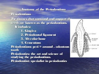
  • 1. Anatomy of the PeriodontiumAnatomy of the Periodontium PeriodontiumPeriodontium The tissues that surround and support theThe tissues that surround and support the teeth are known as the periodontium.teeth are known as the periodontium. It includes:It includes: 1. Gingiva1. Gingiva 2. Periodontal ligament2. Periodontal ligament 3. Alveolarbone3. Alveolarbone 4. Cementum4. Cementum Periodontium: peri = around . odontous =Periodontium: peri = around . odontous = toothtooth Periodontics: the art and science ofPeriodontics: the art and science of studying the periodontium.studying the periodontium. Periodontist: specialist in periodonticsPeriodontist: specialist in periodontics
  • 3.  Gingiva:Gingiva: The fibrous investing tissue coveredThe fibrous investing tissue covered byby keratinized epithelium. The gingiva is onekeratinized epithelium. The gingiva is one ofof the soft tissues that line the oral cavity.the soft tissues that line the oral cavity. All the soft tissue in the mouth are KnownAll the soft tissue in the mouth are Known as theas the the oral mucosa, it isthe oral mucosa, it is divided into three different types:divided into three different types:
  • 4. Types of gingiva:Types of gingiva: 1.1. Marginal gingiva (free orMarginal gingiva (free or unattached)unattached) 2.2. Attached gingiva (firmlyAttached gingiva (firmly attached to underlying toothattached to underlying tooth and bone)and bone) 3.3. Inter-dental gingiva( locatedInter-dental gingiva( located between adjacent teeth.between adjacent teeth. 4.4. Gingival sulcusGingival sulcus
  • 5. Clinical featuresClinical features Microscopic featuresMicroscopic features
  • 6. ClinicalClinical • 1)Mucogingival junction.1)Mucogingival junction. • 2)2)Interdental gingiva.Interdental gingiva. • 3)Free gingival groove3)Free gingival groove • 4)4)Attached gingivaAttached gingiva.. • 5)Alveolar mucosa5)Alveolar mucosa • 6)6) Marginal gingiva.Marginal gingiva.
  • 7. • 1:Enamel.1:Enamel. • 2:gingival margin.2:gingival margin. • 3:gingival sulcus.3:gingival sulcus. • 4:free gingival groove.4:free gingival groove. • 5:lveolar bone.5:lveolar bone. • 6:CEJ6:CEJ • 7Cementum7Cementum • 8:PDL8:PDL • Between 2 and 4 is free gingivaBetween 2 and 4 is free gingiva
  • 8.  Gingival sulcus:Gingival sulcus: ** It isashallow fissureIt isashallow fissure betweenbetween marginal gingival andmarginal gingival and theenamel or cementum.theenamel or cementum. ** Clinical normalClinical normal gingivalgingival sulcusdepth = 2-3 mmsulcusdepth = 2-3 mm measured with periodontalmeasured with periodontal probe.probe.
  • 9.  Interdental gingiva:Interdental gingiva: • Its that part of the gingiva that occupy theIts that part of the gingiva that occupy the inter-proximal space, created by adjacent teethinter-proximal space, created by adjacent teeth in contact. Therefore, shape determined byin contact. Therefore, shape determined by contact relationship with teeth, and width ofcontact relationship with teeth, and width of proximal surfaces.proximal surfaces. • If the contours are flat, interproximalIf the contours are flat, interproximal contacts, the gingiva will be narrow andcontacts, the gingiva will be narrow and short. If the proximal contours are moreshort. If the proximal contours are more convex with a small coronally positionedconvex with a small coronally positioned contact area, the interdental gingiva will becontact area, the interdental gingiva will be broad and high.broad and high.
  • 10.
  • 11. The interdental gingiva can beThe interdental gingiva can be pyramidal and have col shape.pyramidal and have col shape.  Buccolingual dimension the inter-Buccolingual dimension the inter- dental gingival terminates coronally withdental gingival terminates coronally with separate buccal and lingual peaks ofseparate buccal and lingual peaks of tissueasthegingival col.tissueasthegingival col.
  • 12.  Gingival Groove (4):Gingival Groove (4): It is a shallow, v-shaped orIt is a shallow, v-shaped or indentation that is closely associatedindentation that is closely associated with apical extent of freegingiva.with apical extent of freegingiva.
  • 13.  The understanding of clinical features ofThe understanding of clinical features of periodontium is enhanced by a knowledgeperiodontium is enhanced by a knowledge of histological component of tissue.of histological component of tissue.
  • 14. Microscopic (Histological)Microscopic (Histological) FeaturesFeatures • The gingiva consist of central core ofThe gingiva consist of central core of connective tissue covered by stratifiedconnective tissue covered by stratified squamous epithelium.squamous epithelium.
  • 15.  Gingival Epithelium:Gingival Epithelium: 1) Oral (outer) Epithelium.1) Oral (outer) Epithelium. 2) Sulcular Epithelium2) Sulcular Epithelium 3) Junctional Epithelium3) Junctional Epithelium
  • 16.  Oral Epithelium:Oral Epithelium: • Keratinized or parakeratinized consist ofKeratinized or parakeratinized consist of four layers:four layers: • 1) Stratum basale,Basal cell layer (The deepest1) Stratum basale,Basal cell layer (The deepest cells)cells) • 2) Spinosum cell layer2) Spinosum cell layer • 3) Stratum granulosum cell layer3) Stratum granulosum cell layer • 4) Stratum corneum(Keratinized, para, or4) Stratum corneum(Keratinized, para, or nonkeratinized) cell layernonkeratinized) cell layer
  • 17. Layers of Squamous epitheliumLayers of Squamous epithelium
  • 18.  SulcularEpithelium:SulcularEpithelium: It lines the gingival sulcus facingIt lines the gingival sulcus facing the tooth similar to oral epitheliumthe tooth similar to oral epithelium except the 2except the 2ndnd layer (it lacks granularlayer (it lacks granular cell layer)cell layer) It acts as semipermeable membrane fromIt acts as semipermeable membrane from the bacteria and tissue fluid from thethe bacteria and tissue fluid from the gingivaseepsinto thesulcus.gingivaseepsinto thesulcus.
  • 19.  Junctional Epithelium:Junctional Epithelium: Forms the core of epithelium. It isForms the core of epithelium. It is thickest in coronal part. Is single or multiplethickest in coronal part. Is single or multiple layer of non-keratinized cells adhering to toothlayer of non-keratinized cells adhering to tooth surface and face the gingiva by means of basalsurface and face the gingiva by means of basal lamina.lamina. • Basal Laminaconsist of:Basal Laminaconsist of: LaminalucidaLaminalucida LaminadensalLaminadensal
  • 20. Details of the sulcular junctional epithelium areasDetails of the sulcular junctional epithelium areas
  • 21.  Connective Tissue:Connective Tissue: Is known as the lamina propria. DividedIs known as the lamina propria. Divided into two layers: (1) the papillary layerinto two layers: (1) the papillary layer adjacent to epithelium, (2) reticular layer,adjacent to epithelium, (2) reticular layer, contiguouswith theperiosteum.contiguouswith theperiosteum. – Lamina propria consist of:Lamina propria consist of: • Collagen fibersCollagen fibers • Intercellular ground substanceIntercellular ground substance • CellsCells • Blood vesselsBlood vessels • NervesNerves
  • 22. Gingival collagen fiber.Gingival collagen fiber. • 1.circular fibers1.circular fibers • 2.dentogingival fibers.2.dentogingival fibers. • 3.alveologingival3.alveologingival fibers.fibers. • 4.periostogingival4.periostogingival fibers.fibers.
  • 23. Histological charecterstics of the gingiva(FaciolingualHistological charecterstics of the gingiva(Faciolingual section of the periodontium)section of the periodontium)
  • 24.  The collagen fibers help to hold the marginal gingivaThe collagen fibers help to hold the marginal gingiva tightly against the tooth and provide a firm junction oftightly against the tooth and provide a firm junction of the attached gingiva to the underlying tooth root andthe attached gingiva to the underlying tooth root and alveolar bone.alveolar bone. Thefibersaregrouped:Thefibersaregrouped: 1. Gingivodental1. Gingivodental 2. Circular2. Circular 3. Transeptal3. Transeptal 4. Alveolo gingival4. Alveolo gingival 5. Inter-radicular fibers5. Inter-radicular fibers 6. Intra-papillary fibers6. Intra-papillary fibers
  • 25. The most prominent cells found in theThe most prominent cells found in the gingival connective tissue:gingival connective tissue: 1. Plasmacells1. Plasmacells 2. Fibroblasts2. Fibroblasts 3. Mast cells3. Mast cells 4. Lymphocytes4. Lymphocytes
  • 26. VascularSupply:VascularSupply: It’s derived from the branches of theIt’s derived from the branches of the superiorand inferioralveolararteries:superiorand inferioralveolararteries: 1. Greater palatineartery1. Greater palatineartery 2. Buccal artery2. Buccal artery 3. Sublingual artery3. Sublingual artery 4. Mental artery4. Mental artery
  • 27.  The lymphatic drainage usuallyThe lymphatic drainage usually follows the blood supply, the majorfollows the blood supply, the major portion of the lymph drainage from theportion of the lymph drainage from the gingiva going to the submandibulargingiva going to the submandibular lymph nodes.lymph nodes.
  • 28.  Gingival Fluid:Gingival Fluid: The gingival (crevicular) fluid isThe gingival (crevicular) fluid is continually secreted from the gingivalcontinually secreted from the gingival connective tissues into the sulcus throughconnective tissues into the sulcus through thesulcular epithelial wall.thesulcular epithelial wall. This fluid helps to mechanically cleanThis fluid helps to mechanically clean the sulcus and in addition, possess anti-the sulcus and in addition, possess anti- microbial properties and antibodies thatmicrobial properties and antibodies that enhance the resistance of the gingiva toenhance the resistance of the gingiva to gingivitis.gingivitis.
  • 30. Clinical Descriptive CriteriaClinical Descriptive Criteria  Gingival color:Gingival color: Itspink dependson:Itspink dependson: -- amount of melaninamount of melanin - thicknessof epithelium- thicknessof epithelium - degreeof keratinization- degreeof keratinization - vascularity- vascularity
  • 31.  Gingival contour:Gingival contour: The inter-dental gingiva is generallyThe inter-dental gingiva is generally pointed. However, the contours of thepointed. However, the contours of the gingiva vary depending upon the shape ofgingiva vary depending upon the shape of thethe teeth, the buccolingual position of theteeth, the buccolingual position of the teeth in the arch, and the size of the inter-teeth in the arch, and the size of the inter- proximal embrasurespace.proximal embrasurespace.
  • 32.  Gingival Consistency:Gingival Consistency: Usually resilient and firm because of theUsually resilient and firm because of the dense collagenous nature of the gingivaldense collagenous nature of the gingival connectivetissue.connectivetissue.  Gingival Surface Texture:Gingival Surface Texture: Being stippled like an orange peel orBeing stippled like an orange peel or smooth and shiny. Degree of stipplingsmooth and shiny. Degree of stippling varies considerably among patients and invaries considerably among patients and in different partsof thesamemouth.different partsof thesamemouth.
  • 33. Nerve Supply:Nerve Supply:  Derived from maxillary and mandibularDerived from maxillary and mandibular branchesof thetrigeminal nerve.branchesof thetrigeminal nerve.
  • 35. Periodontal ligamentPeriodontal ligament • Connective tissue around and attach teethConnective tissue around and attach teeth to the alveolar bone.to the alveolar bone. • Consist of bundles of fibers, according toConsist of bundles of fibers, according to their directions:their directions: 1)Alveolar crest group1)Alveolar crest group 2)Horizontal group2)Horizontal group 3)Oblique group.3)Oblique group. 4)Apical fibers.4)Apical fibers.
  • 36.
  • 37. • The ends of the princible fibers areThe ends of the princible fibers are embeded in cementum on the tooth sideembeded in cementum on the tooth side and in the alveolar bone proper on theand in the alveolar bone proper on the opposite side. The embeded portions ofopposite side. The embeded portions of the princible fibers are the Sharpey’sthe princible fibers are the Sharpey’s fibersfibers
  • 38.
  • 39. FunctionsFunctions • Physical (mechanical)Physical (mechanical) • FormativeFormative • NutritionalNutritional • SensorySensory
  • 40. The physical functions of pdlThe physical functions of pdl • Transmission of occlusal forces to the bone.Transmission of occlusal forces to the bone. • Attachment of the teeth to the bone.Attachment of the teeth to the bone. • Maintainance of the gingival tissues in theirMaintainance of the gingival tissues in their relationship to the teeth.relationship to the teeth. • Resistence to the impact of occlusalResistence to the impact of occlusal forces(shock obsorption).forces(shock obsorption). • Provision of a soft tissue casing(to protectProvision of a soft tissue casing(to protect vessels and nerves from injury by mechanicalvessels and nerves from injury by mechanical forces).forces).
  • 43. Cementum:Cementum:  Calcified tissue covers the root of the teethCalcified tissue covers the root of the teeth and provide attachment to the periodontaland provide attachment to the periodontal ligament.ligament.  Consist of collagen fibers in a groundConsist of collagen fibers in a ground substance consist of 45-50% inorganicsubstance consist of 45-50% inorganic materials. 50-55% organic materialsmaterials. 50-55% organic materials
  • 44.  Width vary from 60-68 microns in coronalWidth vary from 60-68 microns in coronal third 150-200 micronsin apical third.third 150-200 micronsin apical third.  Width increase with age. 95 microns atWidth increase with age. 95 microns at ageage 20. 215 micronsat age6020. 215 micronsat age60  Two types of cementum a cellular (coronalTwo types of cementum a cellular (coronal portion of the root) Cellular apical portionportion of the root) Cellular apical portion of root and in furcation areas of multi-of root and in furcation areas of multi- rootedrooted teeth.teeth.
  • 45. Cemento Enamel JunctionCemento Enamel Junction:: The area where cementum and enamelThe area where cementum and enamel meet (cervical area).meet (cervical area). Threedifferent relationship:Threedifferent relationship: 60-50%60-50%  cementum overlapsenamelcementum overlapsenamel 30%30%  edgeto edgeedgeto edge 5%-10%5%-10%  cementum fail to meetcementum fail to meet enamel resulting in exposedenamel resulting in exposed dentinedentine
  • 46. • 11:dentine.2:acellular:dentine.2:acellular cementum.3:pdl withcementum.3:pdl with fibroblast.4:cementoblastfibroblast.4:cementoblast • Cellular cementum.Cellular cementum. Cementocytes within theCementocytes within the lacuna of cementumlacuna of cementum