SlideShare a Scribd company logo
1 of 12
Download to read offline
OUTLINE
                                                 A. The Pulpotomy Technique
                                                   1- Diagnosis
                                                   2- Indications / Contraindications
                Pulp Therapy for Primary Teeth     3- Step-by-step pulpotomy technique
                                                      Step- by-
                                                   4- Mechanism of action of formocresol
                                                   5- Alternatives to formocresol
                                                 B. The Pulpectomy Technique
                                                   1- Rationale for pulpectomy
             Reporter:   許修銘                       2- Indications / Contraindications
                                                   3- Root canal filling material
                                                   4- Types of pulpectomy techniques
                2004/03/30                         5- Success rates for primary tooth pulpectomies


                                  Introduction                                               Introduction

Preservation of primary teeth in the arch        Use of pulp therapy to conserve carious
  – Management of developing dentition             primary teeth
  – Nurturing a positive attitude in               – Preserve pulp involved primary molar
    children towards dental health                   when missing permanent successor
                                                   – Prevent possible aberrant habits
                                                   – Maintain masticatory function
                                                   – Preserve aesthetics
                                                   – Future dental attitudes


                                                 A pulpotomy is the procedure of
             A. The Pulpotomy Technique           removing coronal part of pulp
                                                  tissue, inflamed or infected as
                                                  a result of deep caries, &
                                                  maintenance of vital radicular
                                                  pulp tissue
A1- Diagnosis
                                                                               A1-

1/5 dilution of                                     Primary tooth with deep caries
  the original Buckley’s formocresol
                Buckley’
                                                             OD (with GIC)
                                                                  or
                                                              Pulpotomy
                          Cresol   40mL
              NISHIKA
                          Formalin 40mL
Root canal disinfectant   Ethanol 20mL


                                    A1- Diagnosis
                                    A1-                                        A1- Diagnosis
                                                                               A1-

                                                    The reason for this is that
                                                     caries in primary teeth
                                                     compromises pulp very early on,
                                                     with pulp inflammation setting in
                                                     even before pulp is exposed


                                    A1- Diagnosis
                                    A1-                                        A1- Diagnosis
                                                                               A1-

Hobson (1970)                                       Duggal et al (1999)
   In over 50% of the primary molars                  –The need for pulp therapy for
                                                       most primary molars where
   Loss of marginal ridge                              proximal caries has involved the
     irreversible pulp inflammation                    marginal ridge
                                                      –The importance of early
                                                       diagnosis of proximal caries with
                                                       the use of bitewing radiographs
A1- Diagnosis
                                                  A1-                                                                  A1- Diagnosis
                                                                                                                       A1-


                                                                                    Proximal caries that involved less than half the
                                                                                   intercuspal distance from buccal to lingual cusp


                                                  A1- Diagnosis
                                                  A1-                                                                  A1- Diagnosis
                                                                                                                       A1-


 By the time the caries exposes the pulp, the inflammation is irreversible
                                                              irreversible
                  Direct pulp capping is contraindicated


                                               A2- Indications
                                               A2-                                                     A2- Contraindications
                                                                                                       A2-

Large caries with substantial loss (≧1/3 )
                                    (≧                                       An unrestorable tooth
  of marginal ridge in restorable tooth                                      Bi- or trifurcation involvement
                                                                             Bi-
Tooth free of radicular pulpitis                                             Less than 2/3 of root remaining
At least 2/3 of root remaining                                               Presence of abscess or fistula
Absence of abscess or fistula                                                Permanent successor close to eruption
No inter-radicular bone loss
    inter-                                                                   Medical contraindications
No evidence of internal resorption                                             – Heart disease
Instances where extraction is C/I                                              – Immuno-compromised children
                                                                                 Immuno-
A3- Step-by-step
                          A3- Step- by-                                A3- Step-by-step
                                                                       A3- Step- by-

Step 1: Administer local analgesia with      Step 2: Isolate tooth with rubber dam
       the use of a topical analgesic


    Nerve block        Buccal infiltration


                          A3- Step-by-step
                          A3- Step- by-                                A3- Step-by-step
                                                                       A3- Step- by-

Step 3: Remove caries &                      Step 4: Remove roof of pulp chamber
        determine site of pulp exposure


                          A3- Step-by-step
                          A3- Step- by-                                A3- Step-by-step
                                                                       A3- Step- by-

Step 5: Remove coronal pulp with             Step 6: Apply FC on a pledget of
      large excavator or large round bur             cotton wool for 4 minutes
A3- Step-by-step
                              A3- Step- by-                                A3- Step-by-step
                                                                           A3- Step- by-

Step 7: Remove FC pledget after 4 mins           Step 8: Fill pulp chamber with cement
  & check that haemorrhage has stopped


                              A3- Step-by-step
                              A3- Step- by-                                A3- Step-by-step
                                                                           A3- Step- by-

Step 9: Restore tooth with SSC                   Step 10: Take a post-OP radiograph
                                                                 post-


                              A3- Step-by-step
                              A3- Step- by-                                A3- Step-by-step
                                                                           A3- Step- by-

Follow-up
Follow-
  –Regularly reviewed both clinically &
   radiographically 6-monthly
                     6-
  –Appearance of rarefaction of bone                              Pre-OP
                                                                  Pre-                   Post-OP
                                                                                         Post-
   in furcation area or
   a worsening of bone condition
   in furcation
   usually signifies failure of the procedure
                                                                    3M                     12 M
A4- Mechanism of action of FC
          A4-                                          A4- Mechanism of action of FC
                                                       A4-


FC acts through                               Reported
                     formaldehyde,
 aldehyde group of formaldehyde,             success rate
 forming bonds with side-groups
                      side-                     of FC
 of amino acids of both bacterial             pulpotomy
 proteins & remaining pulp tissue
Both bactericidal & devitalizing
 agent


                    A5- Alternatives to FC
                    A5-                                       A5- Alternatives to FC
                                                              A5-

Concern about possible toxicity of           Ferric sulphate [Fe2(SO4)3, 15.5%]
 FC, both locally & systemically               – Excellent haemostatic agent
Alternatives                                     (ferric ion-protein complex)
                                                         ion-
  – Ferric sulphate [Fe2(SO4)3]                – As effective as FC
  – Glutaraldehyde                             – No “fixative” effect
                                                      fixative”
  – Calcium hydroxide
  – Other experimental methods


                    A5- Alternatives to FC
                    A5-                                       A5- Alternatives to FC
                                                              A5-

Glutaraldehyde                               Calcium hydroxide
  – Introduced by s’Gravenmade (1975)          – Poor (around 60%) success rate
  – Better fixative agent                      – Extensive internal resorption
  – Toxic properties                             below amputation
    • Allergic reactions
    • Eye irritation
A5- Alternatives to FC
                 A5-

Other experimental methods
  – Electrosurgery                                     B. The Pulpetomy Technique
  – CO2 lasers
  – Enriched collagen solution


                                                     B1- Rationale for pulpectomy
                                                     B1-

It is true that some primary teeth          Gain access to the root canals
   do have a complex root                   Remove as much dead &
                                            Remove
   morphology (with many fine                 infected material as possible
   accessory root cancals),
                   cancals),                Fill the root canals with a suitable
   but this does not contraindicate           material
   pulpectomy
                                            Maintain primary tooth in a non-
                                                                          non-
                                              infected state


                          B2- Indications
                          B2-                                B2- Contraindications
                                                             B2-

Irreversible inflammation                   Unrestorable crown
  extending to radicular pulp               Advanced pathological root
Primary teeth with necrotic pulps            resorption
Evidence of furcation pathology             Medical contraindications
                                              – Heart disease
Presence of an abscess
                                              – Immuno-compromised
                                                Immuno-
                                                children
B3- Root canal filling material
            B3-                                                  B3- Root canal filling material
                                                                 B3-

Being totally resorbed at the
 same rate as the roots
  – Pure zinc oxide & eugenal mixed
    as a slurry
  – Maisto’s paste
    Maisto’
  – Iodoform paste                                                                               3 M later
  – Vitapex


            B3- Root canal filling material
            B3-                                                       B4- Types of pulpectomy
                                                                      B4-

Ca(OH)2-Iodoform Mixture                                    One-stage / single-visit
   - Vitapex, Endoflas
                                                                  pulpectomy
   - Machida (1983): Ca(OH)2-iodoform mixture to
     be a nearly ideal primary tooth filling material
   1) easy to apply
   2) resorbs at a slightly faster rate                       Two-stage / two-visit
      than that of the roots                                      pulpedctomy
   3) has no toxic effects on the
      permanent successor
   4) radiopaque


           B4- Single-visit of pulpectomy
           B4-                                                   B4- Single-visit of pulpectomy
                                                                 B4-

Indications                                             Step 1: Give local analgesia &
  – Presence of inflamed but vital                               isolate tooth with rubber dam
    radicular pulp
  – An asymptomatic primary tooth
    with necrotic pulp tissue without
    any associated acute symptoms,
    such as cellulitis
  – Presence of a chronic buccal
    lesion without any active
    discharge or acute symptoms
B4- Single-visit of pulpectomy
         B4-                                            B4- Single-visit of pulpectomy
                                                        B4-

Step 2: Remove caries &                     Step 3: Remove roof of pulp chamber,
         identify exposure site                      & identify opening of root canals


         B4- Single-visit of pulpectomy
         B4-                                            B4- Single-visit of pulpectomy
                                                        B4-

Step 4: Take a diagnostic radiograph        Step 5: Clean out root canals with H files
         with files in the root canals               & remove remnants of pulp tissue
                                                     & irrigate canals with saline


                                                               Within 1-2 mm
                                                                       1-
                                                                    File lightly
                                                      Reaming is not advisable
                                                  File to no more than size 30


         B4- Single-visit of pulpectomy
         B4-                                            B4- Single-visit of pulpectomy
                                                        B4-

Step 6: Dry root canals with paper points   Step 7: Select a spiral root canal filler of
  & place a pledget of FC in pulp chamber               appropriate size
    for 4 minutes
B4- Single-visit of pulpectomy
            B4-                                             B4- Single-visit of pulpectomy
                                                            B4-

Step 8: Mix ZnO & eugenol as a slurry,             Step 9: Fill pulp chamber with cement
            & spin it into root canals using
            spiral root canal filler


            B4- Single-visit of pulpectomy
            B4-                                             B4- Single-visit of pulpectomy
                                                            B4-

Step 10: Restore the tooth with SSC                Step 11: Take a post-op radiograph to
                                                                   post-
                                                              check root filling


             B4- Single-visit of pulpectomy
             B4- ingle-                                      B4- Single-visit of pulpectomy
                                                             B4- ingle-

Follow-up
Follow-
   –Regularly reviewed both clinically &
    radiographically 6-monthly
                     6-
                                                                    Pre-OP
                                                                    Pre-              3 M later


   Pre-OP
   Pre-                Post-OP
                       Post-           6 M later
                                                                    Post-OP
                                                                    Post-            12 M later
B4- Single-visit of pulpectomy
           B4- ingle-


                                                                                                 92/08/21
                                                                                                 (F/U 9M)

                 Pre-OP
                 Pre-                       Pre-OP
                                            Pre-
                                                         91/11/12
                                                     (Root canal filling)
                                                                                                 92/12/29
                                                                                                 (F/U 13M)


               6 M later                   Post-OP
                                           Post-


           B4- Single-visit of pulpectomy
           B4- ingle-                                                       B4- Two-visit of pulpectomy
                                                                            B4-

Spiral root filler                                     Indications
                                                           – Presence of an acute abscess
                                                             with or without associated
                                                             cellulitis
                                                           – Presence of active & persistent
                                                             discharge from the root canals


             B4- Two-visit of pulpectomy
             B4-                                                            B4- Two-visit of pulpectomy
                                                                            B4-

Visit 1: Emergency management of                       Visit 2: Final root canal filling
         the acute abscess                                 – 7~10 days later
  – Gaining drainage through carious cavity                – Rubber dam
    or puncturing fistula                                       Access root canals
  – LA Filed to drain                                           Pulpectomy procedure
        FC pledget IRM
  – Antibiotics: 2-dose regimen of amoxycillin
                 2-
B4- Two-visit of pulpectomy
   B4-                           B5- Success rates
                                 B5-


謝謝聆聽
敬請指正

More Related Content

Viewers also liked

Diagnosis Of Pulpal Pathology In Pedodontics
Diagnosis Of Pulpal Pathology In PedodonticsDiagnosis Of Pulpal Pathology In Pedodontics
Diagnosis Of Pulpal Pathology In PedodonticsDr. Shirin
 
Pulp therapy for primary and young teeth
Pulp therapy for primary and young teethPulp therapy for primary and young teeth
Pulp therapy for primary and young teethSaeed Bajafar
 
Morphology of primary teeth
Morphology of primary teethMorphology of primary teeth
Morphology of primary teethBanMaraby
 
Difference between primary and permanent teeth
Difference between primary and permanent teethDifference between primary and permanent teeth
Difference between primary and permanent teethprincesoni3954
 
Pulp Therapy In Pediatric Dentistry Revised 2
Pulp Therapy In Pediatric Dentistry Revised 2Pulp Therapy In Pediatric Dentistry Revised 2
Pulp Therapy In Pediatric Dentistry Revised 2jinishnath
 
Nonvital pulp therapy in pediatric dentistry
Nonvital pulp therapy in pediatric dentistryNonvital pulp therapy in pediatric dentistry
Nonvital pulp therapy in pediatric dentistryPriyank Pareek
 
Difference between pulpotomy and pulpectomy
Difference between pulpotomy and pulpectomyDifference between pulpotomy and pulpectomy
Difference between pulpotomy and pulpectomyOwais92
 
Direct and Indirect pulp capping
Direct and Indirect pulp cappingDirect and Indirect pulp capping
Direct and Indirect pulp cappingAnju Thomas
 
Tooth Morphology Basics
Tooth Morphology BasicsTooth Morphology Basics
Tooth Morphology Basicshchidmd
 
Pulp treatment modalities
Pulp treatment modalitiesPulp treatment modalities
Pulp treatment modalitiesmythreyeethakur
 
PULP AND PERIAPICAL LESIONS OF THE TOOTH ppt
PULP AND PERIAPICAL LESIONS OF THE TOOTH pptPULP AND PERIAPICAL LESIONS OF THE TOOTH ppt
PULP AND PERIAPICAL LESIONS OF THE TOOTH pptK BHATTACHARJEE
 
Morphology of primary teeth pedodontics
Morphology of primary teeth  pedodonticsMorphology of primary teeth  pedodontics
Morphology of primary teeth pedodonticsdeepeshkariwala
 
difference between primary and secondary tooth
difference between primary and secondary toothdifference between primary and secondary tooth
difference between primary and secondary toothAntara Narang
 
pulp therapy in pediatric dentistry
pulp therapy in pediatric dentistrypulp therapy in pediatric dentistry
pulp therapy in pediatric dentistryalaa Mohamed
 

Viewers also liked (18)

Diagnosis Of Pulpal Pathology In Pedodontics
Diagnosis Of Pulpal Pathology In PedodonticsDiagnosis Of Pulpal Pathology In Pedodontics
Diagnosis Of Pulpal Pathology In Pedodontics
 
Pulp therapy for primary and young teeth
Pulp therapy for primary and young teethPulp therapy for primary and young teeth
Pulp therapy for primary and young teeth
 
Dentin
DentinDentin
Dentin
 
Morphology of primary teeth
Morphology of primary teethMorphology of primary teeth
Morphology of primary teeth
 
Difference between primary and permanent teeth
Difference between primary and permanent teethDifference between primary and permanent teeth
Difference between primary and permanent teeth
 
Pulp Therapy In Pediatric Dentistry Revised 2
Pulp Therapy In Pediatric Dentistry Revised 2Pulp Therapy In Pediatric Dentistry Revised 2
Pulp Therapy In Pediatric Dentistry Revised 2
 
Nonvital pulp therapy in pediatric dentistry
Nonvital pulp therapy in pediatric dentistryNonvital pulp therapy in pediatric dentistry
Nonvital pulp therapy in pediatric dentistry
 
pulpectomy-pedo
pulpectomy-pedopulpectomy-pedo
pulpectomy-pedo
 
Diseases of pulp
Diseases of pulpDiseases of pulp
Diseases of pulp
 
Difference between pulpotomy and pulpectomy
Difference between pulpotomy and pulpectomyDifference between pulpotomy and pulpectomy
Difference between pulpotomy and pulpectomy
 
Direct and Indirect pulp capping
Direct and Indirect pulp cappingDirect and Indirect pulp capping
Direct and Indirect pulp capping
 
Tooth Morphology Basics
Tooth Morphology BasicsTooth Morphology Basics
Tooth Morphology Basics
 
Pulp treatment modalities
Pulp treatment modalitiesPulp treatment modalities
Pulp treatment modalities
 
PULP AND PERIAPICAL LESIONS OF THE TOOTH ppt
PULP AND PERIAPICAL LESIONS OF THE TOOTH pptPULP AND PERIAPICAL LESIONS OF THE TOOTH ppt
PULP AND PERIAPICAL LESIONS OF THE TOOTH ppt
 
Morphology of tooth
Morphology of tooth Morphology of tooth
Morphology of tooth
 
Morphology of primary teeth pedodontics
Morphology of primary teeth  pedodonticsMorphology of primary teeth  pedodontics
Morphology of primary teeth pedodontics
 
difference between primary and secondary tooth
difference between primary and secondary toothdifference between primary and secondary tooth
difference between primary and secondary tooth
 
pulp therapy in pediatric dentistry
pulp therapy in pediatric dentistrypulp therapy in pediatric dentistry
pulp therapy in pediatric dentistry
 

Similar to Pulp Therapy Techniques for Primary Teet

Acs0201 Oral Cavity Lesions
Acs0201 Oral Cavity LesionsAcs0201 Oral Cavity Lesions
Acs0201 Oral Cavity Lesionsmedbookonline
 
Radiographic assessment of dental caries
Radiographic assessment of dental cariesRadiographic assessment of dental caries
Radiographic assessment of dental cariesDrMohamedEkram
 
Pathology of the periapex
Pathology of the periapexPathology of the periapex
Pathology of the periapexSaeed Bajafar
 
Lect.5 pulp treatment final
Lect.5 pulp treatment finalLect.5 pulp treatment final
Lect.5 pulp treatment finalAmeer Al-Ameedee
 

Similar to Pulp Therapy Techniques for Primary Teet (6)

Acs0201 Oral Cavity Lesions
Acs0201 Oral Cavity LesionsAcs0201 Oral Cavity Lesions
Acs0201 Oral Cavity Lesions
 
Lesson
LessonLesson
Lesson
 
Lesson
LessonLesson
Lesson
 
Radiographic assessment of dental caries
Radiographic assessment of dental cariesRadiographic assessment of dental caries
Radiographic assessment of dental caries
 
Pathology of the periapex
Pathology of the periapexPathology of the periapex
Pathology of the periapex
 
Lect.5 pulp treatment final
Lect.5 pulp treatment finalLect.5 pulp treatment final
Lect.5 pulp treatment final
 

Pulp Therapy Techniques for Primary Teet

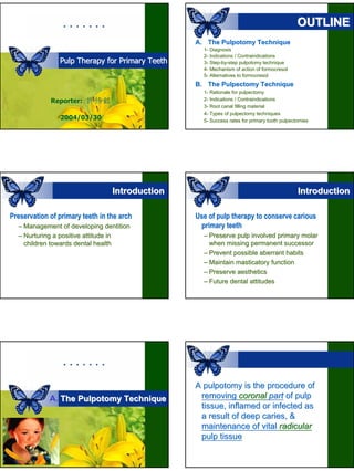
  • 1. OUTLINE A. The Pulpotomy Technique 1- Diagnosis 2- Indications / Contraindications Pulp Therapy for Primary Teeth 3- Step-by-step pulpotomy technique Step- by- 4- Mechanism of action of formocresol 5- Alternatives to formocresol B. The Pulpectomy Technique 1- Rationale for pulpectomy Reporter: 許修銘 2- Indications / Contraindications 3- Root canal filling material 4- Types of pulpectomy techniques 2004/03/30 5- Success rates for primary tooth pulpectomies Introduction Introduction Preservation of primary teeth in the arch Use of pulp therapy to conserve carious – Management of developing dentition primary teeth – Nurturing a positive attitude in – Preserve pulp involved primary molar children towards dental health when missing permanent successor – Prevent possible aberrant habits – Maintain masticatory function – Preserve aesthetics – Future dental attitudes A pulpotomy is the procedure of A. The Pulpotomy Technique removing coronal part of pulp tissue, inflamed or infected as a result of deep caries, & maintenance of vital radicular pulp tissue
  • 2. A1- Diagnosis A1- 1/5 dilution of Primary tooth with deep caries the original Buckley’s formocresol Buckley’ OD (with GIC) or Pulpotomy Cresol 40mL NISHIKA Formalin 40mL Root canal disinfectant Ethanol 20mL A1- Diagnosis A1- A1- Diagnosis A1- The reason for this is that caries in primary teeth compromises pulp very early on, with pulp inflammation setting in even before pulp is exposed A1- Diagnosis A1- A1- Diagnosis A1- Hobson (1970) Duggal et al (1999) In over 50% of the primary molars –The need for pulp therapy for most primary molars where Loss of marginal ridge proximal caries has involved the irreversible pulp inflammation marginal ridge –The importance of early diagnosis of proximal caries with the use of bitewing radiographs
  • 3. A1- Diagnosis A1- A1- Diagnosis A1- Proximal caries that involved less than half the intercuspal distance from buccal to lingual cusp A1- Diagnosis A1- A1- Diagnosis A1- By the time the caries exposes the pulp, the inflammation is irreversible irreversible Direct pulp capping is contraindicated A2- Indications A2- A2- Contraindications A2- Large caries with substantial loss (≧1/3 ) (≧ An unrestorable tooth of marginal ridge in restorable tooth Bi- or trifurcation involvement Bi- Tooth free of radicular pulpitis Less than 2/3 of root remaining At least 2/3 of root remaining Presence of abscess or fistula Absence of abscess or fistula Permanent successor close to eruption No inter-radicular bone loss inter- Medical contraindications No evidence of internal resorption – Heart disease Instances where extraction is C/I – Immuno-compromised children Immuno-
  • 4. A3- Step-by-step A3- Step- by- A3- Step-by-step A3- Step- by- Step 1: Administer local analgesia with Step 2: Isolate tooth with rubber dam the use of a topical analgesic Nerve block Buccal infiltration A3- Step-by-step A3- Step- by- A3- Step-by-step A3- Step- by- Step 3: Remove caries & Step 4: Remove roof of pulp chamber determine site of pulp exposure A3- Step-by-step A3- Step- by- A3- Step-by-step A3- Step- by- Step 5: Remove coronal pulp with Step 6: Apply FC on a pledget of large excavator or large round bur cotton wool for 4 minutes
  • 5. A3- Step-by-step A3- Step- by- A3- Step-by-step A3- Step- by- Step 7: Remove FC pledget after 4 mins Step 8: Fill pulp chamber with cement & check that haemorrhage has stopped A3- Step-by-step A3- Step- by- A3- Step-by-step A3- Step- by- Step 9: Restore tooth with SSC Step 10: Take a post-OP radiograph post- A3- Step-by-step A3- Step- by- A3- Step-by-step A3- Step- by- Follow-up Follow- –Regularly reviewed both clinically & radiographically 6-monthly 6- –Appearance of rarefaction of bone Pre-OP Pre- Post-OP Post- in furcation area or a worsening of bone condition in furcation usually signifies failure of the procedure 3M 12 M
  • 6. A4- Mechanism of action of FC A4- A4- Mechanism of action of FC A4- FC acts through Reported formaldehyde, aldehyde group of formaldehyde, success rate forming bonds with side-groups side- of FC of amino acids of both bacterial pulpotomy proteins & remaining pulp tissue Both bactericidal & devitalizing agent A5- Alternatives to FC A5- A5- Alternatives to FC A5- Concern about possible toxicity of Ferric sulphate [Fe2(SO4)3, 15.5%] FC, both locally & systemically – Excellent haemostatic agent Alternatives (ferric ion-protein complex) ion- – Ferric sulphate [Fe2(SO4)3] – As effective as FC – Glutaraldehyde – No “fixative” effect fixative” – Calcium hydroxide – Other experimental methods A5- Alternatives to FC A5- A5- Alternatives to FC A5- Glutaraldehyde Calcium hydroxide – Introduced by s’Gravenmade (1975) – Poor (around 60%) success rate – Better fixative agent – Extensive internal resorption – Toxic properties below amputation • Allergic reactions • Eye irritation
  • 7. A5- Alternatives to FC A5- Other experimental methods – Electrosurgery B. The Pulpetomy Technique – CO2 lasers – Enriched collagen solution B1- Rationale for pulpectomy B1- It is true that some primary teeth Gain access to the root canals do have a complex root Remove as much dead & Remove morphology (with many fine infected material as possible accessory root cancals), cancals), Fill the root canals with a suitable but this does not contraindicate material pulpectomy Maintain primary tooth in a non- non- infected state B2- Indications B2- B2- Contraindications B2- Irreversible inflammation Unrestorable crown extending to radicular pulp Advanced pathological root Primary teeth with necrotic pulps resorption Evidence of furcation pathology Medical contraindications – Heart disease Presence of an abscess – Immuno-compromised Immuno- children
  • 8. B3- Root canal filling material B3- B3- Root canal filling material B3- Being totally resorbed at the same rate as the roots – Pure zinc oxide & eugenal mixed as a slurry – Maisto’s paste Maisto’ – Iodoform paste 3 M later – Vitapex B3- Root canal filling material B3- B4- Types of pulpectomy B4- Ca(OH)2-Iodoform Mixture One-stage / single-visit - Vitapex, Endoflas pulpectomy - Machida (1983): Ca(OH)2-iodoform mixture to be a nearly ideal primary tooth filling material 1) easy to apply 2) resorbs at a slightly faster rate Two-stage / two-visit than that of the roots pulpedctomy 3) has no toxic effects on the permanent successor 4) radiopaque B4- Single-visit of pulpectomy B4- B4- Single-visit of pulpectomy B4- Indications Step 1: Give local analgesia & – Presence of inflamed but vital isolate tooth with rubber dam radicular pulp – An asymptomatic primary tooth with necrotic pulp tissue without any associated acute symptoms, such as cellulitis – Presence of a chronic buccal lesion without any active discharge or acute symptoms
  • 9. B4- Single-visit of pulpectomy B4- B4- Single-visit of pulpectomy B4- Step 2: Remove caries & Step 3: Remove roof of pulp chamber, identify exposure site & identify opening of root canals B4- Single-visit of pulpectomy B4- B4- Single-visit of pulpectomy B4- Step 4: Take a diagnostic radiograph Step 5: Clean out root canals with H files with files in the root canals & remove remnants of pulp tissue & irrigate canals with saline Within 1-2 mm 1- File lightly Reaming is not advisable File to no more than size 30 B4- Single-visit of pulpectomy B4- B4- Single-visit of pulpectomy B4- Step 6: Dry root canals with paper points Step 7: Select a spiral root canal filler of & place a pledget of FC in pulp chamber appropriate size for 4 minutes
  • 10. B4- Single-visit of pulpectomy B4- B4- Single-visit of pulpectomy B4- Step 8: Mix ZnO & eugenol as a slurry, Step 9: Fill pulp chamber with cement & spin it into root canals using spiral root canal filler B4- Single-visit of pulpectomy B4- B4- Single-visit of pulpectomy B4- Step 10: Restore the tooth with SSC Step 11: Take a post-op radiograph to post- check root filling B4- Single-visit of pulpectomy B4- ingle- B4- Single-visit of pulpectomy B4- ingle- Follow-up Follow- –Regularly reviewed both clinically & radiographically 6-monthly 6- Pre-OP Pre- 3 M later Pre-OP Pre- Post-OP Post- 6 M later Post-OP Post- 12 M later
  • 11. B4- Single-visit of pulpectomy B4- ingle- 92/08/21 (F/U 9M) Pre-OP Pre- Pre-OP Pre- 91/11/12 (Root canal filling) 92/12/29 (F/U 13M) 6 M later Post-OP Post- B4- Single-visit of pulpectomy B4- ingle- B4- Two-visit of pulpectomy B4- Spiral root filler Indications – Presence of an acute abscess with or without associated cellulitis – Presence of active & persistent discharge from the root canals B4- Two-visit of pulpectomy B4- B4- Two-visit of pulpectomy B4- Visit 1: Emergency management of Visit 2: Final root canal filling the acute abscess – 7~10 days later – Gaining drainage through carious cavity – Rubber dam or puncturing fistula Access root canals – LA Filed to drain Pulpectomy procedure FC pledget IRM – Antibiotics: 2-dose regimen of amoxycillin 2-
  • 12. B4- Two-visit of pulpectomy B4- B5- Success rates B5- 謝謝聆聽 敬請指正