MALDI-TOF MS for Protein ID from 2D Gels
•Als PPT, PDF herunterladen•
8 gefällt mir•4,504 views

good

Empfohlen
Weitere ähnliche Inhalte
Was ist angesagt?
Was ist angesagt? (20)
Andere mochten auch
Andere mochten auch (20)
Ähnlich wie MALDI-TOF MS for Protein ID from 2D Gels
Ähnlich wie MALDI-TOF MS for Protein ID from 2D Gels (20)
Mehr von Dr.M.Prasad Naidu
Mehr von Dr.M.Prasad Naidu (20)
Kürzlich hochgeladen
Kürzlich hochgeladen (20)
MALDI-TOF MS for Protein ID from 2D Gels
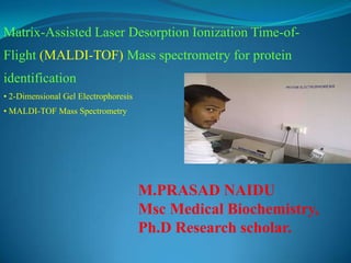
- 1. Matrix-Assisted Laser Desorption Ionization Time-of- Flight (MALDI-TOF) Mass spectrometry for protein identification • 2-Dimensional Gel Electrophoresis • MALDI-TOF Mass Spectrometry M.PRASAD NAIDU Msc Medical Biochemistry, Ph.D Research scholar.
- 2. The age of X-omics and biotechnology: • Genomics: Human genome project • Transcriptomics: cDNA microarray • Proteomics: Development and involvement of mass spectrometry MALDI-TOF MSTandem mass spectrometer (MS/MS)cDNA microarray Celera Genomics Inc.
- 4. 2-Dimension Electrophoresis (2-DE) for Protein Separation Speaker: C. C. Wu Date: 31/10/2001 The core technology of proteomics is 2- DE: At present, there is no other technique which is capable of resolving thousands of proteins in one separation procedure.
- 5. Isoelectric point (pI): Isoelectric point is the pH of a solution at which the net charge of protein is zero. In electrophoresis there is no motion of the particles in an electric field at the isoelectric point. Netcharge -3 -2 -1 0 1 2 3 2 3 4 5 6 7 8 9 10 11 pH Isoelectric point NH3 + COOH NH3 + COOH pH < pI Positive charge NH3 + COO- NH3 + COO- pH = pI NH2 COO- NH2 COO- pH > pI Negative charge
- 6. sample pH 9 - pH 3 + Isoelectric focusing (1st dimension) General principle and protocol of 2-Dimension Electrophoresis MW pH gradient SDS-PAGE Ampholytes polyacrylamide 2nd dimension
- 7. Traditional Equipment for Isoelectric focusing (IEF): Ampholytes polyacrylamide Cathode (- ) electrode solution Anode (+) electrode solution
- 8. Traditional 2-Dimensional Electrophoresis Anode (+) electrode solution Cathode (-) electrode solution Disadvantage: cathodic drift Ampholyte polyacrylamide pH 3 pH 3 pH 3 pH 9 pH 7 pH 5 Time
- 9. Immobilized pH Gradient (IPG) Polyacrylamide gel Acidic buffering group: Basic buffering group:CH2 = CH-C-NH-R O COO- NH3 + Acrylamide monomer
- 10. Gradient maker plastic support film Production of Immobilized pH Gradient (IPG) strip A C B F E Dacidic basic pH 3 pH 10
- 11. IPGphor (IEF System) Amersham Pharmacia Biotech Inc. Protein IEF Cell Bio-Rad Laboratories Equipment for Isoelectric focusing (IEF):
- 12. Lysis solution: 8M Urea 4% NP-40 or CHAPS 40mM Tris base Sample preparation Cell line Lysis solution Sonication vacuum Lysis solution Centrifugation Measurement of [protein] 2-DE sample
- 13. IPG strip rehydration and sample loading 2-DE sample Rehydration solution Rehydration solution: 8M Urea 2% NP-40 or CHAPS 2% IPG buffer (Ampholyte) 0.28% DTT Trace Bromophenol blue IPG strip holder Position the IPG strip
- 14. IPG strip rehydration and sample loading Strip holder Cathode (-) electrode Anode (+) electrode 30 voltage 12hr
- 15. First dimension: Isoelectric focusing 1. Place electrode pads (?) 2. 200 V step-n-hold 1.5hr 3. 500 V step-n-hold 1.5hr 4. 1000 V gradient 1500vhr 5. 8000 V gradient (?) 36000vhr Time Voltage Holder cover IPG strip Electrode Electrode pads
- 16. Second dimension: SDS-PAGE • SDS equilibration • SDS-PAGE SDS equilibration buffer 50 mM Tris-HCl 6 M Urea 30% Glycerol 2% SDS Trace Bromophenol SDS SDS-PAGE SDS-PAGE 0.5% agarose in running buffer SDS-PAGE Marker in paper IPG strip
- 17. Protocol of silver stain: 50% methanol 25% acetic acid 4hr ddH2O x 3 times 30min/time 0.004% DTT solution 30min 0.1% AgNO3 30min ddH2O 30 sec 3% Na2CO3 0.0185% formaldehyde 2.3M citric acid 5% acetic acid 25% methanol
- 18. 2-DE separation of soluble E. coli proteins