

Empfohlen
Weitere ähnliche Inhalte
Was ist angesagt?
Was ist angesagt? (19)
Andere mochten auch
Ähnlich wie Cat scratch disease
Ähnlich wie Cat scratch disease (20)
Kürzlich hochgeladen
Kürzlich hochgeladen (20)
Cat scratch disease
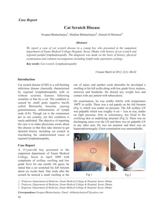
- 1. Abstract We report a case of cat scratch disease in a young boy who presented in the outpatient department of Enam Medical College Hospital, Savar, Dhaka with history of cat scratch and regional painful lymphadenopathy. The diagnosis was made on the basis of history, physical examination and common investigations including lymph node aspiration cytology. Key words: Cat scratch, Lymphadenopathy J Enam Med Col 2012; 2(1): 40-42 Introduction Cat scratch disease (CSD) is a self-limiting infectious disease classically characterised by regional lymphadenopathy with or without systemic features following scratches or bite by a cat. The condition is caused by small gram negative bacilli called Bartonella henselae causing granulomatous inflammation of lymph node (LN). Though cat is the commonest pet in our country, yet this condition is rarely published. The objective of reporting the case is to make physicians aware about this disease so that they take interest to get detailed history including cat scratch in elucidating the undetermined cause of regional lymphadenopathy. Case Report A 15-year-old boy presented to the outpatient department of Enam Medical College, Savar in April 2009 with complaints of axillary swelling and low grade fever for one month. On query, he stated that a cat scratched his left forearm about six weeks back. One week after the scratch he noticed a small swelling at the site of injury and another week thereafter he developed a swelling in his left axilla along with low grade fever, malaise, anorexia and headache. He denied any weight loss and contact with any patient with tuberculosis. On examination, he was mildly febrile with temperature 1000F in axilla. There was a red papule on the left forearm (Fig 1) which was tender on pressure. The left axillary LN was palpable which was roughly 4 cm × 3cm in size, tender on light pressure, firm in consistency, not fixed to the overlying skin or underlying structure (Fig 2). There was no discharging sinus over the LN and there was no palpable LN in any other area. He was not anaemic and there was no hepatosplenomegaly. Chest examination was unremarkable. Fig 2Fig 1 40 1. Professor, Department of Medicine, Enam Medical College & Hospital, Savar, Dhaka. 2. Professor, Department of Medicine, Enam Medical College & Hospital, Savar, Dhaka. 3. Registrar, Department of Medicine, Enam Medical College & Hospital, Savar, Dhaka. Correspondence Swapna Bhattacharjee, Email: shbhattacharjee07@yahoo.com Case Report Swapna Bhattacharjee1, Shekhar Bhattacharjee2, Ahmed Al Montasir3 Cat Scratch Disease
- 2. His initial workup showed ESR 80 mm in 1st hour, Hb 11.0 gm/dL, total WBC count 12,500/cu mm with neutrophil 75%, lymphocyte 20% and eosinophil 04%, red blood cell and platelet counts were normal. Chest radiography was unremarkable and tuberculin test was insignificant with induration of 4 mm after 72 hours. Fine needle aspiration cytology (FNAC) of left axillary LN showed granulomatous inflammation with collection of epithelioid cells, lymphocytes, histiocytes with small areas of focal necrosis, simulating tubercular lymphadenitis. After initial evaluation, he was prescribed ciprofloxacin 500mg twice daily for one week. On follow-up after two months he was found healthy and his fever and LN regressed completely. Discussion The clinical syndrome of CSD was first reported in 1950 by Debre et al, but the causative agent, small gram negative bacilli was discovered in 1983 by Wear et al and is currently known as Bartonella henselae.1,2 Cats are the major reservoir of B. henselae. Once transmitted to human via cat’s saliva or scratch, B. henselae invades CD 3/4+ haematopoietic progenitor cells. The response to infection depends on immune status of the host. In immunocompetent individual, the response is granulomatous inflammation and suppuration as compared to vasoproliferative response in immunocompromised patients.3 The disease usually occurs in children and young adults with a median age 15 years.4 CSD usually presents as a benign and self-limiting lymphadenitis after a cat scratch or bite, but may progress to a severe, systemic or recurrent form producing occuloglandular syndrome, encephalopathy, convulsion, osteomyelitis, retinitis, arthritis, hepatitis, splenitis, erythema nodosum, pulmonary nodules and pleurisy, specially in immunocompromised patients.5 One week after inoculation by a cat scratch or bite an erythematous papule that appears at the puncture site persists for 1 to 3 weeks.6 Regional lymphadenopathy develops 1–3 weeks after inoculation. Eighty five percent of patients have single lymph node involved and systemic lymphadenopathy is exceptional.7 Axillay and epitrochlear LN are involved in most cases (46%) followed by LN of head and neck (26%) and the groin (17%) areas.8 The lymphadenopathy usually resolves within several weeks and the lesion heals without scar formation. Cat scratch disease is usually diagnosed clinically and special tests are rarely necessary.9 Carithers developed ‘Rule of Five’ as a diagnostic tool in his original series.8 Points are given to each of the four criteria: 1 point for lymphadenopathy, 2 for cat exposure, 2 for presence of an inoculation site and 2 for a positive skin test with CSD antigen. Total of 5 points strongly suggest CSD while 7 points make the diagnosis confirmed. Lymph node histology which is a diagnostic criterion suggested by others shows a constant pathological hallmark of granulomatous inflammation with focal necrosis similar to other granulomatous diseases like tuberculosis, brucellosis etc.3,10 Now-a-days diagnosis can be confirmed by immunofluroscent assay (IFA), tissue culture and PCR of lymphnode aspirate.11 Typical CSD is a self-limiting illness that resolves spontaneously within 2–6 months. Although usually not indicated in most cases but those patients with painful large lymphnode may benefit from a short course of antibiotics like azithromycin, ciprofloxacin, rifampicin, trimethoprim etc.12 Conclusion Importance of cat scratch disease lies in the fact that although it is a self-limiting benign condition caused by traumatic cat contact but reluctance to take detailed history may lead to erroneous diagnosis of other granulomatous lymphadenitis particularly tuberculosis in our country. References 1. Debre R, Lamy M, Jammet M, Costil L, Mozzicona P. La maladie des griffes de chat. Bull Mem Soc Med Hop Paris 1950; 66: 76-79. 2. Wear DJ, Margileth AM, Hadfield TL, Fischer GW, Schlagei CJ, King FM. Cat-scratch disease: a bacterial infection. Science 1983; 221: 1403-1405. 3. Bass JW, Vincent JM, Person DA. The expanding spectrum of Bartonella infections: II. cat-scratch disease. Pediatr Infect Dis J 1997; 16(2): 163-179. 41 J Enam Med Col Vol 2 No 1 January 2012
- 3. 4. Margileth AM. Cat-scratch disease. Adv Pediatr Infect Dis 1993; 8: 1-21. 5. Batts S, Demers D. Spectrum and treatment of cat- scratch disease. Pediatr Infect Dis J 2004; 23: 1161- 1162. 6. Chian CA, Arrese JE, Pierard GE. Skin manifestations of Bartonella infections. Int J Dermatol 2002; 41(8): 461-466. 7. Lamps LW, Scott MA. Cat-scratch disease: historic, clinical and pathologic perspectives. Am J Clin Pathol 2004; 121(Suppl): S71-80. 8. Carthers HA. Cat-scratch disease: an overview based on a study of 1200 patients. Am J Dis Child 1985; 139(11): 1124-1133. 9. Schwartz SB, Chambers FH. In: Current medical diagnosis and treatment. 49th edn. New York: McGraw-Hill, 2010: 1321. 10. Margileth AM. Recent advances in diagnosis and treatment of cat scratch disease. Curr Infect Dis Rep. 2000; 2(2): 141-146. 11. MetzKor-Cotter E, Kletter Y, AvoidorB et al. Longterm serological analysis and clinical follow up of patients with cat-scratch disease. Clin Infect Dis 2003; 37: 1149-1154. 12. Rolain JM, Brouqui P, Koehler JE et al. Recommendations for treatment of human infections caused by Bartonella species. Antimicrobial Agents Chemother 2004; 48: 1921-1923. 42 J Enam Med Col Vol 2 No 1 January 2012