Glissonian approach for laparoscopic liver resections
•
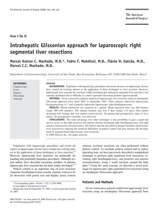
1 gefällt mir•1,482 views

Technique for intrahepatic Glissonian approach for laparoscopic right segmental liver resections is presented. http://www.drmarcel.com.br

Empfohlen
Empfohlen
Weitere ähnliche Inhalte
Was ist angesagt?
Was ist angesagt? (20)
Ähnlich wie Glissonian approach for laparoscopic liver resections
Ähnlich wie Glissonian approach for laparoscopic liver resections (12)
Kürzlich hochgeladen
Kürzlich hochgeladen (20)
Glissonian approach for laparoscopic liver resections
- 1. The American Journal of Surgery (2008) 196, e38 – e42 How I Do It Intrahepatic Glissonian approach for laparoscopic right segmental liver resections Marcel Autran C. Machado, M.D.*, Fabio F. Makdissi, M.D., Flávio H. Galvão, M.D., Marcel C.C. Machado, M.D. Department of Gastroenterology, University of São Paulo, Rua Evangelista Rodrigues 407, 05463-000 São Paulo, Brazil KEYWORDS: Abstract Llaparoscopy; BACKGROUND: Experience with laparoscopic procedures and recent advances in laparoscopic devices Liver; have created an evolving interest in the application of these techniques to liver resection. However, Surgery; laparoscopic liver resection has not been widely developed and anatomical segmental liver resection is not Glissonian; currently performed due to difficulty to control segmental Glissonean pedicles laparoscopically. Anatomy METHODS: Seven consecutive patients underwent laparoscopic liver resection using an intrahepatic Glissonian approach from April 2007 to September 2007. Three patients underwent laparoscopic bisegmentectomy 6 –7 and 4 patients underwent laparoscopic right hemihepatectomy. RESULTS: Blood transfusion was required in 1 patient. Mean operation time was 460 minutes (range 300 – 630 minutes). The median hospital stay was 5 days (range 3– 8 days). One patient developed bile leakage that was treated conservatively. No patient had postoperative signs of liver failure. No postoperative mortality was observed. CONCLUSIONS: The main advantage over other techniques is the possibility to gain a rapid and precise access to the right posterior and anterior sheaths facilitating right hemihepatectomy, and right anterior and posterior sectionectomies. We believe that the described technique facilitates laparoscopic liver resection by reducing the technical difficulties in pedicle control and may increase the develop- ment of segment-based laparoscopic liver resections. © 2008 Elsevier Inc. All rights reserved. Experience with laparoscopic procedures and recent ad- whereas sectional resections are often performed without vances in laparoscopic devices have created an evolving inter- pedicle control. To facilitate pedicle control and to reduce est in the application of these techniques to liver resection.1,2 operating time, we have previously described a standardized However, laparoscopic liver resections are technically de- technique to perform right segmental liver resections, in- manding and potentially hazardous procedures. Although sev- cluding right hemihepatectomy, and posterior and anterior eral authors have described increasing numbers of patients, sectionectomies, using 3 small incisions around the hilar laparoscopic liver resection has not been widely developed.2–5 plate.6 Using the same concept, we describe a novel tech- Pedicle control is an important step of liver resection. nique for laparoscopic right segmental liver resections using Anatomic hemihepatectomies usually requires extensive hi- an intrahepatic Glissonian approach. lar dissection with portal vein and hepatic artery control, * Corresponding author. Tel./fax: 55-11-3256-4098. Patients and Methods E-mail address: dr@drmarcel.com.br Manuscript received September 25, 2007; revised manuscript October Seven consecutive patients underwent laparoscopic liver 19, 2007 resection using an intrahepatic Glissonian approach from 0002-9610/$ - see front matter © 2008 Elsevier Inc. All rights reserved. doi:10.1016/j.amjsurg.2007.10.027
- 2. M.A.C. Machado et al. Laparoscopic right segmental liver resections e39 April 2007 to September 2007. There were 7 women with a ration of the abdominal cavity and ultrasound liver exami- mean age of 46.8 years (range 23–75 years). Four patients nation are performed. had liver metastasis and 3 patients had hepatocellular ade- After cholecystectomy, a small (3 mm) anterior incision noma with a mean size of 7.6 cm (range 6 –10 cm). The is made in front of the hilum (shown at A in Figure 2a). A surgical procedure, postoperative course, and outpatient fol- second incision (B in Figure 2a) is performed on the right low-up were evaluated and the following data collected edge of the gallbladder. A third incision is made perpendic- prospectively: duration of surgery, average time to inflow ular to the hepatic hilum in the segment 7, where it connects pedicles control, perioperative transfusions, postoperative to the caudate lobe (C in Figure 2a). By combining 2 of complications, and hospital stay. The interval timing to these 3 incisions it is possible to control the portal pedicle of control portal pedicles was defined by the time between right liver (A to C), anterior section (A to B), and posterior beginning of intrahepatic dissection of Glissonian sheaths section (B to C). and establishment of ischemic delineation. For right hemihepatectomy (segments 5, 6, 7, and 8) a large laparoscopic vascular clamp is introduced through Operative technique incisions A and C to occlude right Glissonian pedicle as displayed in the Figure 2b. The vascular clamp is then The patient is placed in a left semi-lateral decubitus replaced by an endoscopic vascular stapling device, the position (Figure 1a) with the surgeon standing between the right liver ischemic delineation is confirmed, and the stapler patient’s legs. This technique uses 5 trocars. A 12-mm is fired. For right posterior sectionectomy (segments 6 and trocar is placed 3 cm above the umbilicus. Pneumoperito- 7) the same maneuver is done using incisions B and C, and neum is established at a pressure of 12 mm Hg. The other 4 for right anterior sectionectomy (segments 5 and 8) we can trocars are located as shown in Figure 1b. The round liga- use a combination of incisions A and B (Figures 2c and 2d). ment is transected using laparoscopic coagulation shears All of these steps are performed without the Pringle (LCS; Ethicon Endo Surgery Industries, Cincinnati, OH), maneuver and without hand assistance. Liver transection and the falciform and coronary ligaments are then dissected and vascular control of the hepatic veins are accomplished to expose the suprahepatic inferior vena cava (IVC). Explo- with harmonic scalpel and endoscopic stapling device as appropriate. The specimen is extracted through a suprapubic incision. One round 19-F Blake abdominal drain (Ethicon, Inc, Cincinnati, OH) was left in place in all patients. In order to safely perform the described technique, the knowledge of liver anatomy is imperative. Careful analyses of preopera- tive imaging and intraoperative ultrasound are needed to determine portal anatomy. Results Three patients underwent laparoscopic bisegmentectomy 6 –7 (Figure 3) and 4 laparoscopic right hemihepatectomy (Figure 4). Blood transfusion was required in 1 patient (1 unit). Mean operative time consumed to achieve complete control of pedicles was 5.8 minutes (range 4 –12 minutes) and mean operation time was 460 minutes (range 300 – 630 minutes). The median hospital stay was 5 days (range 3– 8 days). One patient developed bile leakage that was treated conser- vatively. No patient had postoperative signs of liver failure. No postoperative mortality was observed. Three patients were operated on for benign primary tu- mor while the remaining 4 were operated on due to malig- nant secondary tumors. These 4 patients had negative sur- gical margin, and were greater than 1 cm in all patients. Two patients exhibited moderate post-chemotherapy steato- Figure 1 Diagrams of patient position and trocar placement for hepatitis and 1 had mild steatosis. Intraoperative laparo- laparoscopic right liver resections. (a) Patient is placed in a left scopic liver ultrasound confirmed the site and size of the semi-lateral decubitus position. (b) Three 12-mm trocars (red) and lesions diagnosed by computed tomography scan and/or two 5-mm trocars (blue) are needed. magnetic resonance imaging.
- 3. e40 The American Journal of Surgery, Vol 196, No 4, October 2008 Figure 2 Schematic view of intrahepatic Glissonian access for laparoscopic right liver resections. (a) Incisions for the intrahepatic approach of Glissonian pedicles. A anterior incision in front of the hilum. B incision on the right edge of gallbladder bed. C vertical incision on the segment 7 perpendicular to the hepatic hilum. (b) Right hemihepatectomy—a large laparoscopic vascular clamp is introduced through incisions A and C to occlude right Glissonian pedicle. (c) Right posterior sectionectomy— combining incisions B and C it is possible to occlude the Glissonian pedicle of segments 6 and 7. (d) Right anterior sectionectomy— combining incisions A and B and using the same maneuver it is possible to occlude the Glissonian pedicle of segments 5 and 8. Figure 3 Laparoscopic right posterior sectionectomy (resection of segments 6 and 7). (a) A large laparoscopic vascular clamp is introduced through incisions B and C. (b) Glissonian pedicle is occluded and ischemic delineation of right posterior section (S6 and S7) is obtained. (c) Schematic view—vascular clamp is replaced by a vascular stapler. (d) Schematic view—stapler is fired and ischemic delineation of right posterior segments is seen.
- 4. M.A.C. Machado et al. Laparoscopic right segmental liver resections e41 only hemihepatectomy but also sectional liver resections. Although not yet performed, laparoscopic right anterior sectionectomy (segments 5 and 8) may be facilitated using the present technique. This novel technique can expand the indication for lapa- roscopic segment-based liver resection, sparing liver paren- chyma. Preservation of liver parenchyma should always be attempted in order to prevent postoperative liver failure and to increase the opportunity for performing repeated resec- tions in cases of recurrent malignancy.10 Several authors have described right hepatectomies using dissection of the right hepatic artery, duct, and portal vein separately,5,11 which is tedious and time-consuming, and may jeopardize vascular and biliary control if anatomical variation is present. In order to overcome these facts, some authors report laparoscopic Glissonian approach. Topal et al12 used a technique similar to the intrahepatic posterior ap- proach7 using incision of liver parenchyma posterior and anterior to the hilum and insertion of an endoscopic vascular stapling device under cholangiography guidance. Cho et al13 described a laparoscopic hand-assisted procedure with dis- section of the hepatic parenchyma covering the bifurcation of the right and posterior Glissonian pedicles. The present technique, based on small incisions following specific ana- tomical landmarks, allows a straight forward control of Figure 4 Laparoscopic right hemihepatectomy (resection of Glissonian pedicle without hilar or parenchymal dissection segments 5, 6, 7, and 8). (a) Intraoperative view of ischemic and no need for ultrasound or cholangiography guidance. It delineation of the right liver. (b) Schematic view—stapler is fired is important to comment that intraoperative ultrasound re- and ischemic delineation of right liver is obtained. mains a necessary tool for safe hepatic surgery and should be routinely used regardless of the technical approach. The described technique precludes encircling of the Glissonian Comments pedicles, thus simplifying the procedure and minimizing bleeding from this blunt maneuver, which is much more The development of segment-based resection using in- difficult to perform laparoscopically. trahepatic Glissonian access made it possible to develop The main advantage over other techniques is the possi- techniques to identify and isolate the right and left segmen- bility of gaining rapid and precise access to the right pos- tal Glissonian pedicles.6 – 8 These techniques permit a tai- terior and anterior sheaths, facilitating right hemihepatec- lored liver resection by removing only the liver segments tomy, and right anterior and posterior sectionectomies. involved by the underlying disease. Respect of anatomic We believe that the described technique facilitates lapa- landmarks of liver segments during resection prevents im- roscopic liver resection by reducing the technical difficulties pairment of the vascularization of the remaining paren- in pedicle control and may increase the development of chyma and excessive bleeding. segment-based laparoscopic liver resections. Recent advances in laparoscopic techniques have re- sulted in a growing indication for laparoscopic liver resec- tion.1–5 Most laparoscopic liver procedures are right or left References hemihepatectomies, left lateral segmentectomies, and non- anatomical liver resections.3 Anatomical segmental liver 1. Koffron A, Geller D, Gamblin TC, et al. Laparoscopic liver surgery: resection is not currently performed due to technical diffi- shifting the management of liver tumors. Hepatology 2006;44:1694 – culties in controlling segmental Glissonian pedicles laparo- 700. 2. Dagher I, Proske JM, Carloni A, et al. Laparoscopic liver resection: scopically. results for 70 patients. Surg Endosc 2007;21:619 –24. The Pringle maneuver is the main step during laparo- 3. Gagner M, Rogula T, Selzer D. Laparoscopic liver resection: benefits scopic liver resection and it is used to avoid major bleeding and controversies. Surg Clin North Am 2004;84:451– 62. but can be associated with prolonged ischemic time. Laurent 4. Vibert E, Perniceni T, Levard H, et al. Laparoscopic liver resection. Br J Surg 2006;93:67–72. et al9 showed that laparoscopic liver resections are associ- 5. Koffron AJ, Auffenberg G, Kung R, et al. Evaluation of 300 minimally ated with higher ischemic times. The present technique invasive liver resections at a single institution: less is more. Ann Surg precludes the use of the Pringle maneuver and permits not 2007;246:385–92.
- 5. e42 The American Journal of Surgery, Vol 196, No 4, October 2008 6. Machado MA, Herman P, Machado MC. A standardized technique for 10. Nishio H, Hamady ZZ, Malik HZ, et al. Outcome following repeat right segmental liver resections. Arch Surg 2003;138:918 –20. liver resection for colorectal liver metastases. Eur J Surg Oncol 2007; 7. Launois B, Jamieson GG. The posterior intrahepatic approach for 33:729 –34. hepatectomy or removal of segments of the liver. Surg Gynecol Obstet 11. O’Rourke N, Fielding G. Laparoscopic right hepatectomy: surgical 1992;174:155– 8. technique. J Gastrointest Surg 2004;8:213– 6. 8. Machado MA, Herman P, Figueira ER, et al. Intrahepatic Glissonian 12. Topal B, Aerts R, Penninckx F. Laparoscopic intrahepatic Glissonian access for segmental liver resection in cirrhotic patients. Am J Surg approach for right hepatectomy is safe, simple, and reproducible. Surg 2006;192:388 –92. Endosc 2007;21:2111. 9. Laurent A, Cherqui D, Lesurtel M, et al. Laparoscopic liver resection 13. Cho A, Asano T, Yamamoto H, et al. Laparoscopy-assisted hepatic for subcapsular hepatocellular carcinoma complicating chronic liver lobectomy using hilar Glissonean pedicle transection. Surg Endosc disease. Arch Surg 2003;138:763–9. 2007;21:1466 – 8.