1) The vertebral column develops from segments of mesenchyme tissue called sclerotomes. These migrate and surround the spinal cord, forming the primordium of each vertebra.
2) Intervertebral discs develop between sclerotome segments from cells that do not proliferate. The nucleus pulposus forms in the center, surrounded by the annulus fibrosus.
3) Cartilage replaces the mesenchyme by the 6th week. Ossification begins in the 8th week, with primary centers forming in the centrum and neural arches. Secondary centers form after puberty.
Tata AIG General Insurance Company - Insurer Innovation Award 2024
Development Of Vertebral Column Group 1
1. Group 1 – SEM IV – 2010
The Development Of The Vertebral Column
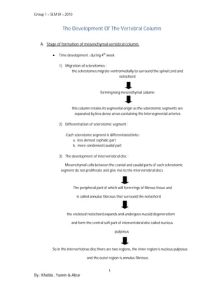
A. Stage of formation of mesenchymal vertebral column.
Time development : during 4th week
1) Migration of sclerotomes :
the sclerotomes migrate ventromedially to surround the spinal cord and
notochord
forming long mesenchymal column
this column retains its segmental origin as the sclerotomic segments are
separated by less dense areas containing the intersegmental arteries
2) Differentiation of sclerotomic segment :
-Each sclerotome segment is differentiated into :
a. less densed cephalic part
b. more condensed caudal part
3) The development of intervertebral disc :
Mesenchymal cells between the cranial and caudal parts of each sclerotomic
segment do not proliferate and give rise to the intervertebral discs
The peripheral part of which will form rings of fibrous tissue and
is called annulus fibrosus that surround the notochord
the enclosed notochord expands and undergoes nucoid degeneratiom
and form the central soft part of intervertebral disc called nucleus
pulposus
So in the intervertebrae disc there are two regions, the inner region is nucleus pulposus
and the outer region is annulus fibrosus.
1
By : Khalida , Yasmin & Abrar
2. Group 1 – SEM IV – 2010
4) The development of the body (the centrum) of the vertebra :
The remaining caudal condensed part of each scelorotomic
segment joins the cephalic less condensed part of scelorotomic segment caudal
Form mesenchymal centrum ( the primordium of the body of vertebra)
The notochord degenerates and disappears
5) The development of neural arch :
Scelorotomic tissue migrates backwards from both sides of the centrum of
vertebra to surround the neural tube
The neural spine forms at the point of meeting of neural arch,posteriorly.
Sclerotomic tissue also extends laterally from both sides of the centrum to form
2 processes (costal process ventrally, transverse process dorsally)
B. Stage of formation of cartilaginous vertebral column
Time of appearance of chondrofication centres : during 6th week
Process of chondrofication :
2 centres of chondrofication appear in centrum of vertebra
They fused together at the end of embryonic period (8th week)
Centres of chondrofication appear in neural arches
They fuse with each other and with the centrum
The spinous and transverse processes develop from extension of
chondrofication centres in the neural arch
2
By : Khalida , Yasmin & Abrar
3. Group 1 – SEM IV – 2010
C. Stage of ossification of vertebral column
Primary ossification centers Secondary ossification centers
End of 8th week Time of After puberty
development
3 primary ossification centers Number 5 secondary ossifation cernters
i. 1 in the centrum i. 1 for the tip of the spinous
ii. 1 in each half of neural arch process
ii. 1 for the tip of each transverse
process
iii. 1 for the superior rim of the
vertebral body
iv. 1 for inferior rim of the
vertebral body
Process of ossification: Fate of notochord:
At birth each vertebra consists 1. The most cranial part of the
of 3 bony parts connected by notochord is incorporated in the
cartilages. basilar part of the occipital bone
The bony halves of the vertebral ant the posterior part of body of
arch fuse together during the 1st sphenoid bone.
3 to 5 years. 2. The parts of the notochord in the
The arches articulate with the bodies of the vertebrae
centrum at cartilaginous degeneration and disappear.
neurocentral joints. 3. The parts of the notochord in
These joints disappear when the between the bodies of the
vertebral arch fuses with the vertebrae undergo mucoid
centrum during the 3rd to 6th degeneration to form the nucleus
year. pulposus.
Fate of costal processes:
1. In cervical region
they form the anterior and
lateral boundary of the foramen
transversum.
2. In thoracic region
they form the ribs.
3. In lumbar region
they fuse with the transverse
processes.
4. In upper sacral region
they unite tgether to form the
anterior portion of the ala of
sacrum.
3
By : Khalida , Yasmin & Abrar
4. Group 1 – SEM IV – 2010
Curvature of the vertebral column
1. Primary curvature
In the fetus the entire vertebral column is flexed
anteriorly like a "C"
anterior concavity
maintained at birth
seen in the thoracic and sacral regions.
2. Secondary curvatures
develop after birth
seen in the cervical and lumbar region
appears at the 3rd to 6th month when the child holds its head up and the lumbar
curvature in the 6th month to 2 year range when the child sits up
Anomalies of vertebral column
1. Spina bifida
Incomplete fusion of the halves of the vertebral arches resulting in a midline defect
usually in the lumbosacral region
Types : a) Spina bifida occulta( closed): 20%
b) Spina bifida manifesta (open): 80%
-Meningocele: CSF only
-Meningomyelocele( CSF+ Nerve fibers)
2. Hemivertebra
Failure of one of the chondrofication centres to appear and subsequent failure of
half of the vertebra to form producing scoliosis.
3. Scralization of the fifth lumbar vertebra
5th lumbar vertebra is fused with the sacrum and as a result the number of lumbar
vertebrae is 4 and the sacrum is formed of 6 vertebrae.
4. Lumbrization of the first piece of sacrum
Separation of the first piece of the sacrum to form a separate vertebra so the
number of lumbar vertebrae is 6 and the sacrum is only formed of 4 sacral
vertebrae.
5. Congenital Kyphosis
due to either failure of segmentation or defect in formation of vertebral body
elements.
N/B : For pictures, please refer to group 1 student presentation.
4
By : Khalida , Yasmin & Abrar