3. Trace the flow of blood into and out of the orbit and orbital structures. Internal carotid a. Ophthalmic a. Lacrimal a. Post. ethmoidal a. Ant. ethmoidal a. Supratrochlear a. Supra-orbital a. Lacrimal gland Facial v. Sup. Ophthalmic v. Cavernous sinus Pterygoid plexus Inf. Ophthalmic v. Retromandibular v.
4. Identify the extra-ocular muscles. Side view Frontal views Levator palpebrae Superior oblique Inferior rectus Superior rectus Lateral rectus (cut) Inferior oblique Medial rectus Optic nerve LR SO IR IO MR SR LP LP SR SO IO IR MR LR LR SO IR IO MR SR LP
5. Indicate the nerve supply to each. V III IV VI V 1 V 2 V 3 II Frontal n. Supratrochlear n. Supra-orbital n. Lacrimal n. Sensory nerves are branches of the ophthalmic division of the trigeminal - V 1 Motor nerves are branches of cranial nerves III, IV, and VI V III IV VI II VI Nasociliary n. Lacrimal n. Ciliary ganglion Short ciliary nn. Ethmoidal nn. Frontal n. (cut) Infratrochlear n. Long ciliary nn.
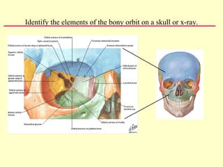
6.
7.
8. Predict the functional deficit resulting from damage to each nerve. There is a very cool web site that allows you to simulate the eye motions expected from lesions to different cranial nerves and different eye muscles. Check it out at http://cim.ucdavis.edu/Eyes/Version1/eyesim.htm This patient has a damaged right trochlear nerve (IV). The right superior oblique muscle is paralyzed. Note that movements to the right are not affected, but movements down and, especially, down and to the left result in asymmetrical eye movements. The right eye is not able to move down and in.

![Indicate the structures lying superior, inferior, medial and lateral to the eyeball and the position of the eyeball relative to the bony orbit. Eyeball is oriented anteroposteriorly but orbits are angled outward Horizontal CT ,[object Object],[object Object],[object Object],Coronal CT ,[object Object],[object Object],[object Object]](data:image/gif;base64,R0lGODlhAQABAIAAAAAAAP///yH5BAEAAAAALAAAAAABAAEAAAIBRAA7)