Management of prerenal arf part two
•Als DOCX, PDF herunterladen•
4 gefällt mir•837 views

TO MY WIFE WHO SUPPORT ME ALL THE WAY ,STOOD BESIDE ME ALL THE TIME , ENCOURAGE ME TO BE VALUABLE IN MY COUNTRY ......HOPPING ALLAH ACCEPT MY AIMS

Empfohlen
Empfohlen
Weitere ähnliche Inhalte
Was ist angesagt?
Was ist angesagt? (20)
Andere mochten auch
Andere mochten auch (20)
Ähnlich wie Management of prerenal arf part two
Ähnlich wie Management of prerenal arf part two (20)
Mehr von cardilogy
Mehr von cardilogy (20)
Kürzlich hochgeladen
Kürzlich hochgeladen (20)
Management of prerenal arf part two
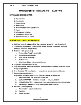
- 1. ARF II MAGDI AWAD SASI 2014 1 DR MAGDI AWAD SASI MANAGEMENT OF PRERENAL ARF-----PART TWO REVERSALBE CAUSES OF ARF: 1. Hypovolemia 2. Hypotension 3. Hypercalemia 4. Heart failure 5. Accelerated phase OF hypertension 6. Sepsis 7. Urinary tract infection 8. Urinary tract obstruction 9. Nephrotoxic drug or poison GENERAL LINES OF ARF MANAGEMENT 1. Fluid and electrolyte balance( I/O chart ,patients weight ,RFT and electrolytes) 2. 500 ml fluid and asses the losses (( urine ,faeces ,vomitus ,aspirations ,ventilator , sweating ,increased temperature)) 3. Nutrition ( N/G ,parenteral ): i. Protein 40gm/day (70 gm if H.D.) ii. Potassium restricted diets iii. Fluid and fat for energy iv. Carbohydrate v. Vitamins and trace elements supplement 4. Treatment of complications of ARF 5. Trial of frusemide ,mannitol ,dopamine ,dobutamine infusion after correction of fluid loss ( using CVP line) 6. If no dieresis or ARF complication-----early start of renal replacement therapy 7. Renal replacement therapy: CONTINOUS ARTERIOVENOUS(CAV), CONTINOUS VENOVENOUS(CVV) I. HAEMODIALYSIS OR PERITONEAL DIALYSIS II. ULTRAFILTRATION ---REMOVAL OF FLUID 0—4KG /H.D. SESSION III. HAEMOFILTRATION-----REMOVAL OF FLUID AND REPLACEMENT FLUID TREATMENT IV. HAEMODIFILTRATION---- FLUID REMOVAL AND HEMODIALYSIS 8. Recovery ( polyuric phase) i. Accurate I/O , electrolyte balance ii. Nutrition iii. Follow up (RFTs)
- 2. ARF II MAGDI AWAD SASI 2014 2 DR MAGDI AWAD SASI SUMMARY OF CAUSES OF PRERENAL ARF: 1. Decrease of blood volume: I. Hemorrhage –from orifices, internal bleeding ,hematemsis, delivery, surgery II. Loss of plasma—burns , crush injuries III. Na and Water depletion from : a. GIT---severe vomiting ,diarrhea , acute intestinal obstruction ,paralytic ileus ,pancreatitis , fistulae b. Urine ------excessive diuretics ,DKA 2. Decrease cardiac output and myocardial failure----cardiogenic shock 3. Increase in the size of vascular bed---septic shock 4. Intravascular hemolysis 5. Rhabdomyolysis-----release of globins----toxic effect on the kidney 6. Disease of the major renal vessels-----decrease of renal blood: A. Thrombosis of arteries B. Occlusive embolus of the aorta or renal arteries C. Aortic dissecting aneurysm MANAGEMENT: 1. Compatible history (risk factor for ARF ) 2. The clinical finding 3. Progressive increase in blood urea and creatinine 4. Examine for: Fluid treatment chart (( input and output )) balance. Blood pressure ---hypotension (postural) in shock states. Look signs of dehydration---- Dry mucous membranes, inelastic skin ,decrease its turgor Postural hypotension and taccychardia ( worse in sitting) Collapsed superficial veins ,sunken eyes Cold cyanosed extremities Decrease JVP and CVP, sharp decrease in body weight Changes in the body weight (( over hydration /dehydration)). Urine ----------decrease volume
- 3. ARF II MAGDI AWAD SASI 2014 3 DR MAGDI AWAD SASI Effect of I.V. treatment (rehydration) Diuresis ----prerenal cause Fluid over load and danger of pulmonary oedema. Treatment : 1. Treat the cause. 2. Treat hypovolemia: A. Restore blood pressure( blood , plasma ,fluids) B. Use of inotropic drugs e.g. after correction of blood volume Body weight x 15 = ----mg of dopamine to be used in 250ccN/S fluid. One drop = 1 micgm 5 –10 drops/min will increase renal perfusion. c. Best treated by monitoring CVP or PCWP. 3. Treatment of septicemia---avoid nephrotoxic antibiotics 4. Aim of Early treatment---diuresis and improvement of RF Otherwise, if not, pt is still oliguric ----- treat as ATN. 5. Frusemide and dopamine for pre renal failure is questionable. They are used if unresponsive to fluid challenge. Frusemide: Don’t give until you are sure that the pt is fully hydrated. Don’t give if aminoglycoside have been used Give 250mg I.V. over one hour by syringe pump. If the urine output remains ˂ 40 ml/hr over the next hours, give further 500mg I.V. over 2 hours by syringe pump. If the urine output increases ,further doses can be given as required up to maximum daily dose of 2 gm or continuous infusion can be used (30--- 60mg/hr) Dopamine : 200---6000microgm/min (has a beneficial effect on renal output.). 400mg in 250ml ( 1600 micrigm/ml) give 1—2 micgm/kg/min (4mg). If dieresis occur , keep dopamine for 24—72hr and taper it.
- 4. ARF II MAGDI AWAD SASI 2014 4 DR MAGDI AWAD SASI MANAGEMENT OF RENAL ARF SUMMARY OF CAUSES: A. Acute parenchymal disorders: I. Glomerular and vascular diseases: 1. Acute glomerular nephritis (primary or part of systemic disease) 2. Vasculitis (including drug induced) 3. Malignant HTN 4. Arterial or venous occlusion(thrombosis , emboli) 5. Microangiopathic haemolysis (TTP,HUS,DIC) II. Tubulo-interstitial diseases: 1. Acute interstitial nephritis—drug induced 2. Pyelonephritis 3. Hypercalcemia ,multiple myeloma ,acute uric acid nephropathy (tumor lysis) B. Acute tubular necrosis((ATN)): 1. POST-ISCHEMIC INJURY: i. Shock ii. Major trauma ,burns ,crush injuries ,eclampsia iii. Surgery ( cardiac ,aortic ,biliary) iv. Pancreatitis v. Prolonged use of vasopressors vi. Obstetric--- eclampsia ,abruption placenta vii. Prolonged pre-renal failure 2. NEPHROTOXIC INJURY: i. Antibiotic( aminoglycoside ,amphotericin ,rifampicin ,sulphonamide) ii. Radioactive contrast media iii. Anesthetics ---methoxyflurane iv. Myoglobalinuria( rabdomylosis) ,Haemoglobulinuria (hemolysis) v. Poisoning (paracetamol ,aspirin , methanol ,ethanol) vi. Heavy meatals platinum ,amantide phalloides
- 5. ARF II MAGDI AWAD SASI 2014 5 DR MAGDI AWAD SASI ATN A. Very poor prognosis 1. Extensive burns 2. BY-pass surgery 3. Sepsis following abdominal surgery 4. Trauma to abdomen and thorax 5. Severe hepatic failure 6. Paraquat poisoning 7. Criminal abortion associated with clostridal infection 8. Any patient requiring ventilation for more than 48hr B. Good prognosis 1. Obstetrical accident without Sepsis 2. Trauma to limbs 3. Mismatched blood transfusion 4. Viral infection( Coxsackie ,influenza ) causing myoglobinuria 5. Some nephrotoxin CLINICAL POINTS TO BE REMMEBERED: 1. ATN is the commonest histology in ARF secondary to pre-renal causes or nephrotoxic drugs. 2. A urine output ˂25-30ml/hour in adult should be investigated and urgently corrected. If oliguria persists ˃2-3 hour, it may be irreversible 3. The simplest screening test for RF in ICU patients are : I. Hourly urine output ( at least 0.5ml/kg/hour) II. Accurate I/O fluid chart III.Daily serum creatinine assessment Prognosis of ARF : DEPENDS ON 1. CAUSE-----------------Nature and severity of precipitating factor 2. DIAGNOSIS----------Early diagnosis and treatment of ARF complication 3. TREATMENT---------Speed and efficiency of ARF treatment.
- 6. ARF II MAGDI AWAD SASI 2014 6 DR MAGDI AWAD SASI Clinical presentation: Symptoms and signs of causative disease. Urine----oliguric Pre renal normal kidney Established ARF CVP ˂ 2CM OF H2O N or high ≥ 12cm Response to 1) Hydration 2) Mannitol or lasix Dieresis ± Dieresis ± No effect or risk of pulmonary Edema URINE volume Specific gravity Sediment Na + Urea Osmolality FE Na% Small ˃ 1020 normal ˂ 10mmol/l ˃ 250 moml/l ˃600 ˂ 1 Small ˂ 1020 Casts ,depris , cells ˃ 20 ˃ 40(ATN) ˂ 150 moml/l 300-400mosm/l ˃ 3 Urine / plasma Urea ratio Creat ratio 0smolality ˃ 10 ˃ 10 ˃ 2 ˂ 10 ˂3(ATN) ˂ 10 ˂ 1.1 Fractional excretion of Na FE Na= (U/P Na) / (U/P CR) X 100 Increase blood urea ,creatinine (if blood urea ˃ 50—100mg/d= hypercatabolic state—trauma ,infection , surgery ,drugs Electrolytes : Hyperkalemia , dilutional hyponatremia (over load + oliguria), ca low Metabolic acidosis: not so marked if excessive vomiting or aspiration of gastric content. UREA is increased in: Steroids , tetracycline, catabolic state ,high protein diet, dehydration ,GIT hemorrhage. CREATININE is increased in: Large muscle bulk, acute rhabdomylsis , high protein meal ,trimethoprium ,low GFR UREA IS DECREASED IN : Liver disease , anabolic state, starvation ,low protein diet ,pregnancy ,increase ADH
- 7. ARF II MAGDI AWAD SASI 2014 7 DR MAGDI AWAD SASI ON EXAMINATION: 1) First feels well----after days ----uremic S&S 2) c/o anorexia ,nauseas ,vomiting ,hiccough 3) Apathy , mental confusion 4) Muscles twitches ,fits ,drowsiness ,coma 5) Bleeding episodes 6) GIT bleeding ( may be serious) 7) Respiratory rate—increase – infection , metabolic acidosis 8) Pulmonary edema S&S A. I.V. fluids and oliguria (over treatment) B. Increase PCWP in uremia – ARDS 9) Anemia -----increased hemolysis ,repeated sampling ,bleeding ----- decrease erythropoiesis 10) Infection –septicemia and shock due to -----( decrease cellular and humoral immunity) 11) Phases -- oliguric then diuretic 12) Diuretic phase: 7 –10 days ( 4 weeks ) of the onset of S&S Urine increase 3 – 5 l/day persist 4 days Urea is usually increased and creatinine slowly decrease Needs accurate supplement of fluid and electrolytes Few days later----normal chemistry and urine volume and pts can eats normal diet 13) PROGNOSIS DEPENDS ON : 1. Speed and efficiency of treatment 2. Early recognition and treatment of complication 3. Nature and severity of causal disorder e.g. I. Simple hemorrhage II. Drugs III. Severe infections IV. Multiorgan failure mortality rate 50n--70% 4. Untreated cases----- MR ˃ 90%
- 8. ARF II MAGDI AWAD SASI 2014 8 DR MAGDI AWAD SASI GENERAL TREATMENT OF THE OLIGURIC PHASE: 1. Get drug history and alter prescriptions appropriately. 2. Fluid and electrolyte balance (( daily weight ,fluid chart )). 3. 500 ml fluid and asses the losses (( urine ,faeces ,vomitus ,aspirations ,ventilator , sweating ,increased temperature—add 500ml for each increase ˃ 37.5)) 4. Skilled nursing care (( mouth , skin )) and senior trained specialists. 5. Establish under lying causes and start prompt medical action 6. Exclude obstruction of the urinary tract. 7. Assess and correct any respiratory or circulatory impairment. 8. Nutrition ( NGT, Parenteral) Protein—40mg/d (( increase to 70gm/d if on dialysis ) K restriction diet Fat and carbohydrate for energy Vitamins supplement 9. Treatment of anemia, hypocalcemia ,DIC ,infections . 10.Manage life threatening complications of ARF . I. HYPERKALEMIA II. SEVERE ACODOSIS III. SALT AND WATER OVERLOAD IV. SEVERE UREMIA 11.Dialysis investigations and accurate I/O . 12.Trial of : I. Furosemide – 1 -2 mg/d and increase to 500-2000mg in 1 hr infusion or ethacryme acid 0.5mg/kg---- If urine output is initiated, it can be maintained by lasix 40—80mg 4 -8 hourly Dopamine drip (200—600microgm/min) 13.If all fail-----------DIALYSIS .
- 9. ARF II MAGDI AWAD SASI 2014 9 DR MAGDI AWAD SASI MANAGEMENT OF POST --RENAL ARF Obstructive nephropathy may be 2ry to upper or lower urinary tract obstruction. Early diagnosis and relief of obstruction are essential to prevent renal damage by ultrasound abdomen and urologist consultation. CLINICAL FEATURES: 1. H/O urinary symptoms—renal colic ,hematuria , difficulty of micturation. 2. May be clinically silent and discovered on investigation 3. Anuria--------suggestive of obstructive lesion. Abdominal USS should be done to R/O any obstructive lesion .If it showed pelvic or uretric dilatation ----urgent percutaneous nephrostomy is indicated to avoid need for dialysis treatment. 4. P.V.---palpable CA cervix P.R.----palpable recto-sigmoid junction carcinoma 5. Management --- I. Investigation aiming to diagnose and relief urinary bladder obstruction II. Supportive and palliative treatment may be needed III. Accurate I.V. fluid treatment after relief of obstruction –postobstuctive dieresis WHAT ARE THE INVESTIGATIONS TO BE DONE? 1. BLOOD----C.B.C. ,Blood glucose , Renal function test ,Na ,K ,Ca , CL , Bicarbonate ,osmolality ,Coagulation screen ,ESR ,Serum &urine electrophoresis , CPK 2. URINE---Na , Urea , Creatinine ,Osmolality . 3. Culture-------blood ,urine ,sputum , wound culture ,tracheostomy. 4. S .complement ,immunological studies –AGN 5. Chest x ray , ABD USS , ECG , IVU ( ABDOMEN CAT OR MRI) 6. Renal biopsy 7. Daily pt weight ---input and out put
- 10. ARF II MAGDI AWAD SASI 2014 10 DR MAGDI AWAD SASI TREATMENT OF ARF 1. Emergency measures : I. RX of hyperkalemia II. Indication of dialysis 2. Determination of ARF cause . 3. General management of the oliguric phase: I. Fluid and electrolyte balance II. Nutrition III. Nursing care IV. Infection V. Anemia ,DIC, GIT hemorrhage 4. General management of recovery (diuretic ) phase : Fluid electrolyte , nutrition. INDICATIONS OF DIALYSIS IN AFR: URGENT DIALYSIS--- 1. Uraemic encephalopathy—impaired conscious level ,coma ,fits 2. Fluid overload----- pulmonary edema 3. Uraemic Pericarditis 4. Uraemic gastritis 5. Hyperkalemia(˃ 6.5)---- not responding to medical therapy 6. Severe metabolic acidosis( HC03 ≤ 13 or PH ˂ 7.1 7. Severe hyeruricemia (tumor lysis syndrome 8. Oxylate toxicity (ethylene glycol overdose) 9. Severe uraemia----200mm0l/d ROUTINE DIALYSIS : 1. Prevention of severe uremia and its complications ( when conservative TR failed) 2. Maintenance of normal fluid ,electrolyte and acid base status ( TR medically failed) 3. Space making for parenteral nutrition ,transfusion of blood and blood products 4. Prophylaxis before surgical procedures
- 11. ARF II MAGDI AWAD SASI 2014 11 DR MAGDI AWAD SASI TREATMENT OF HYPERKALEMIA( K ˃ 6mmol/L ) The first thing is to rule out pseudohyperkalemia: 1. Tourniquet application 2. Haemolysis of drawn blood 3. Increase WBC & platelets Symptoms: Weakness Lethargy Muscle cramps Paresthesias Hypoactive DTRs Dysrhythmias ECG--- K > 5.5 -6 Tall, peaked symmetrical T waves Wide QRS Prolong PR Diminished /absent P Depressed ST Prolonged QT QRS- blends in T ((merge)) – classical sine wave of increase K Ventricular fibrillation asystole
- 12. ARF II MAGDI AWAD SASI 2014 12 DR MAGDI AWAD SASI I.V. access for: Ca gluconate (10ml 10%)diluted over 10 minutes --AIM --protect heart Glucose 50ml 50%--with regular insulin 10 units , may be repeated. Na HCO3 8.4% 50cc for each 0.1 ▼ blood PH to correct metabolic acidosis Ca or Na resonium (30mg) orally or rectally B-agonist (IV /inhaler)-10-20mg albuterol decrease K in half hour for 2 hours Once k ˂ 6mmol/l---dextrose 20% (30ml/hr) to prevent rebound hyperkalemia Dialysis---- definitive treatment if there is ECG changes Summary of hyperkalemia TR: TREATMENT OF BLOOD PRESSURE Hypotension(shock)-----best under C.V.P. 1. I.V. fluids ,blood 2. Dopamine ---IV drip Hypertension:- 1. Ca channel blockers ,clonidine ,prazosine 2. IV Na nitroprusside (may be needed in some cases) 3. Dialysis TO BE CONTINUED