

Empfohlen
Weitere ähnliche Inhalte
Was ist angesagt?
Was ist angesagt? (18)
Andere mochten auch
Andere mochten auch (18)
Ähnlich wie Final thesis pdf (a4)
Ähnlich wie Final thesis pdf (a4) (20)
Kürzlich hochgeladen
Kürzlich hochgeladen (20)
Final thesis pdf (a4)
- 1. IDENTIFICATION OF COMPONENTS IN EXTRACTS OF HERICIUM ERINACEUS (BULL.: FR.) PERS THAT STIMULATE IN VITRO NEURITE OUTGROWTH OF NG108-15 WONG YUIN TENG FACULTY OF SCIENCE UNIVERSITY OF MALAYA KUALA LUMPUR 2012
- 2. IDENTIFICATION OF COMPONENTS IN EXTRACTS OF HERICIUM ERINACEUS (BULL.: FR.) PERS THAT STIMULATE IN VITRO NEURITE OUTGROWTH OF NG108-15 WONG YUIN TENG DISSERTATION SUBMITTED IN FULFILLMENT OF THE REQUIREMENT FOR THE DEGREE OF MASTER OF SCIENCE INSTITUTE OF BIOLOGICAL SCIENCES FACULTY OF SCIENCE UNIVERSITY OF MALAYA KUALA LUMPUR 2012
- 3. ABSTRACT Hericium erinaceus, locally known as cauliflower mushroom, and elsewhere as lion’s mane mushroom, Houtou (monkey head mushroom), Yamabushitake and Harisenbon (balloon fish), is an edible mushroom. It is well known for its medicinal and nutritional values. Hericium erinaceus is reported to have good anti-tumor properties and nerve tonic effects. Although H. erinaceus is a temperate mushroom, it has been successfully cultivated in Malaysia. However, there are very few reported studies on the chemical constituents that stimulate the neurite outgrowth for the locally cultivated species. The crude aqueous ethanol extract of H. erinaceus and its fractionated extracts (hexane, ethyl acetate and water) were evaluated for their effect in stimulating the neurite outgrowth using neural cell line NG108-15 whilst the Nerve Growth Factor (NGF) was used as the positive standard. The crude aqueous ethanol extract of H. erinaceus showed 15.0 % increase in neurite outgrowth at the concentration of 10.0 µg/ml. However, the crude aqueous ethanol extract showed decreased neurite growth as the dose was increased. The hexane, ethyl acetate and water fractions showed an increase in neurite outgrowth when the dose was increased exponentially (10.0, 25.0, 50.0 and 100.0 µg/ml). Maximum stimulation of neurite outgrowth was recorded with ethyl acetate fraction with 68.5 % increase compared to negative control followed by hexane fraction with 65.2 % increase. The combined fraction of hexane and ethyl acetate was further subjected to flash column chromatography. Among the 7 isolated fractions (fraction E1-E7), fraction E1 and fraction E2 show relatively higher neurite stimulation activity compared to other fractions. Maximum stimulation was recorded as 160.6 % increase and 149.1 % ii
- 4. increase compared to negative control at the concentration of 100 µg/ml for fraction E1 and fraction E2 respectively. The chemical compositions of the fraction E1 of H. erinaceus were analayzed by GCMS. Four components were identified from fraction E1 comprising about 80.5 % of the total. Fraction E1 was made up of ethyl palmitate (29.8 %), ethyl stearate (2.3 %), ethyl oleate (18.6 %) and ethyl linoleate (29.9 %). Further isolation of fraction E2 using preparative TLC and HPLC gave subfraction sub4b_4 and subfraction sub4b_6. Subfraction sub4b_4 showed better neurite stimulation activity compared to subfraction sub4b_6 with 187.1 % increase in comparison. The chemical compositions of subfraction sub4b_4 and sub4b_6 were analyzed by NMR and LC/MS/MS. The components identified from subfraction sub4b_4 were hericenone C (and its isomer) and 4-(3’,7’-dimethyl-5’-oxo-2’,6’-octadienyl)-2-formyl- 3-hydroxy-5-methoxylbenzyl oleate (and its isomer). On the other hand, subfraction sub4b_6 comprised of hericenone C, 4-(3’,7’-dimethyl-5’-oxo-2’,6’-octadienyl)-2- formyl-3-hydroxy-5-methoxylbenzyl oleate and a phenolic component attached to the fatty ester side chain contained 26 carbons with 3 double bonds. iii
- 5. ABSTRAK Hericium erinaceus, dikenali sebagai cendawan bunga kobis di Malaysia dan cendawan “lion’s mane”, Houtou (cendawan kepala monyet), Yamabushitake dan Harisenbon di tempat lain. Cendawan ini boleh dimakan dan ia terkenal dari segi nilai perubatan dan nutrisi. Kebelakangan ini, laporan saintifik menunjukkan H. erinaceus mempunyai nilai anti-tumor yang baik dan sebagai tonik terhadap saraf. Walaupun H. erinaceus merupakan cendawan yang ditanam di kawasan sederhana tetapi kini berjaya ditanam di Malaysia yang beriklim tropika. Walaubagaimanapun, tidak banyak terbitan laporan termpatan yang melaporkan tentang komposisi kimia dalam H.erinaceus yang ditanam secara tempatan dalam rangsangan pertumbuhan saraf. Ekstrak mentah akueus etanol dan fraksi-fraksi (heksana, etil asetat dan air) dari H.erinaceus telah diselidik dalam rangsangan pertumbuhan saraf pada sel saraf NG108- 15 dan NGF digunakan sebagai kawalan positif. Ekstrak mentah akueus ethanol menunjukkan peningkatan sebanyak 15.0 % dalam pertumbuhan saraf pada kepekatan 10.0 µg/ml. Walaubagaimanapun, peningkatan kepekatan ekstrak mentah akueus ethanol akan menyebabkan penurunan dalam pertumbuhan saraf. Fraksi hexana, etil asetat dan air akan menyebabkan peningkatan dalam pertumbuhan saraf apabila kepekatan fraksi-fraksi ditingkatkan secara eksponen (10.0, 25.0, 50.0 and 100.0 µg/ml). Pertumbuhan maksimum saraf direkodkan oleh fraksi etil asetat dengan 68.5 % peningkatan berbanding dengan kawalan negatif dan diikuti oleh fraksi hexana dengan 65.2 % peningkatan berbanding dengan kawalan negatif. Pengasingan komponen daripada gabungan fraksi heksana dal etil asetat menggunakan kaedah kromatografi kolum kilat menghasilkan 7 fraksi (fraksi E1-E7). Fraksi E1 dan E2 menunjukkan aktiviti pertumbuhan saraf yang lebih tinggi jika berbanding dengan fraksi-fraksi lain. Peningkatan pertumbuhan maksimum sebanyak iv
- 6. 160.6 % dan 149.1 % direkodkan oleh fraksi E1 dan E2 pada kepekatan 100µg/ml berbanding kawalan negatif. Komposisi kimia fraksi E1 bagi H. erinaceus dianalisis dengan menggunakan GCMS. Empat komponen telah dikenalpasti daripada fraksi E1 dan komponen- komponen tersebut adalah terdiri daripada 80.5 % daripada keseluruhan fraksi E1. Komponen yang terkandung dalam fraksi E1 adalah etil palmitat, etil stearat, etil oleat dan etil linoleat. Subfraksi sub4b_4 dan subfraksi sub4b_6 adalah hasil daripada isolasi fraksi E2 dengan menggunakan preparatif TLC dan HPLC. Subfraksi sub4b_4 menunjukkan aktiviti pertumbuhan saraf yang lebih baik daripada subfraksi sub4b_6 iaitu 187.1 % peningkatan pada kepekatan 100 µg/ml jika dibandingkan dengan kawalan negatif. Komposisi kimia subfraksi sub4b_4 dan sub4b_6 dianalisis dengan menggunakan NMR dan LC/MS/MS. Komponen-komponen yang dikenalpasti daripada subfraksi sub4b_4 termasuk hericenone C (dan isomernya) dan 4-(3’,7’-dimetil-5’-oxo- 2’,6’-octadienil)-2-formil-3-hidroksi-5-metoksibenzil oleat (dan isomernya). Identifikasi subfraksi sub4b_6 menunjukkan kehadiran hericenone C, 4-(3’,7’-dimetil-5’-oxo-2’,6’- octadienil)-2-formil-3-hidroksi-5-metoksibenzil oleat dan satu komponen fenolik yang mengandungi rantai ester asid lemak yang mempunyai 26 karbon dan 3 ikatan dubel. v
- 7. ACKNOWLEDGEMENT Writing a significant scientific thesis is hard work and it would be impossible without support from various people. First of all, I wish to express my greatest appreciation towards my supervisor Professor Datin Dr. Sri Nurestri Abdul Malek, my co-supervisor, Professor Dr. Noorlidah binti Abdullah, the project leader of this research, Professor Dr. Vikineswary Sabaratnam from Mushroom Research Centre and Dr. Murali Naidu from the Faculty of Medicine for the intellectual guidance, valuable advices and help that was given to me during my research. The thesis would not have been written successfully without their continuous supervision and guidance. I would like to thank to University Malaya for the grant (PS191/2009A) and fellowship support. My special appreciation to my labmates and friends’ enthusiasm and support in providing relevant assistance and help to complete this study. Thanks to Wong Kah Hui, Lai Puei-Lene, Priscilla Ann, Joanna Eik Lee Fang, Hong Sok Lai, Lee Guan Serm, Phang Chung Weng, Sujatha Ramasamy, Sharifah Nur Syed Abdul Rahman, Gowri Kanagasabapathy, Jaime Stella Richardson, Ong Kia Ju and Mamalay. A special thank to Madam Chang May Hing for her kind gesture in helping me especially in the laboratory system operation procedures. Last but not least, I would like to express my appreciation to my parents, Mr Wong Wai Yew and Madam Chia Saw Meng, and other members of my family for their emotional, financial support and providing a lovely environment for me. vi
- 8. CONTENTS PAGE ABSTRACT ii ABSTRAK iv ACKNOWLEDGEMENTS vi LIST OF FIGURES x LIST OF TABLES xii LIST OF APPENDICES xiii LIST OF SYMBOLS AND ABBREVIATIONS xv CHAPTER I INTRODUCTION 1 CHAPTER II LITERATURE REVIEW 2.1 Medicinal mushrooms and its usages 5 2.2 Hericium erinaceus 2.2.1 Origin 7 2.2.2 Classification 8 2.2.3 Medicinal properties, nutritional and bioactive components derived 8 from Hericium erinaceus 2.3 Nervous system and neurodegenerative diseases 2.3.1 Neurite 12 2.3.2 Neurodegenerative diseases (factors, therapies to cure and prevent) 12 2.4 Neuroprotective, neurotrophic, neuronal differentiation and neurite 16 stimulation effects of Hericium erinaceus 2.5 Neurite outgrowth bioassay system of neural hybrid cell line NG108-15 2.5.1 Formation of NG108-15 hybrid cell 19 2.5.2 Characteristics of NG108-15 hybrid cell 19 CHAPTER III MATERIALS AND METHODS 3.1 Extraction 3.1.1 Preparations of fruitbodies 21 vii
- 9. 3.1.2 Preparation of aqueous ethanol crude extract 21 3.1.3 Solvent-solvent extraction (fractionation) 21 3.2 Neurite outgrowth activity assay 3.2.1 Preparation of stock solutions 23 3.2.2 Cell culture 23 3.2.3 Preparation of medium and buffer for cell culture 3.2.3.1 Dulbecco’s Modified Eagle’s Medium (DMEM) 23 3.2.3.2 Phosphate buffer saline 24 3.2.4 Cell culture techniques 3.2.4.1 Revival of frozen cells 24 3.2.4.2 Subculture of cells 25 3.2.4.3 Medium renewal 25 3.2.4.4 Cryopreservation of cells 26 3.2.5 Effect of Hericium erinaceus on stimulation of neurite outgrowth 26 activity of NG108-15 3.2.6 Scoring of neurites 26 3.2.7 Statistical analysis 27 3.3 Isolation of bioactive constituents 3.3.1 Column chromatography 28 3.3.2 Analytical thin layer chromatography 28 3.3.3 Preparative thin layer chromatography 29 3.3.4 High performance liquid chromatography (HPLC) 3.3.4.1 HPLC samples and mobile phase preparation 29 3.3.4.2 Analytical HPLC 30 3.3.4.3 Semipreparative HPLC 30 3.4 Identification 3.4.1 Gas chromatography-mass spectrometry (GCMS) 32 3.4.2 Nuclear magnetic resonance spectroscopy (NMR) 32 3.4.3 Liquid chromatography- mass spectrometry (LC/MS/MS) 32 CHAPTER IV RESULTS & DISCUSSION 4.1 Extraction, fractionation and isolation 4.1.1 Extraction, fractionation and isolation of aqueous ethanol extract of 33 Hericium erinaceus viii
- 10. 4.2 Neurite outgrowth activity 4.2.1 Effect of aqueous ethanol extract and fractions of Hericium 37 erinaceus on the neural cell line NG108-15 4.2.2 Effect of the fraction E1-E7 of Hericium erinaceus on the neural 45 cell line NG108-15 4.2.3 Effect of the subfraction sub4b_4 and sub4b_6 of Hericium 57 Erinaceus on the neural cell line NG108-15 4.3 Overall comparison of aqueous ethanol extract, hexane fraction, ethyl 62 acetate fraction, water fraction, fraction E1-E7, subfraction sub4b_4 and subfraction sub4b_6 4.4 Identification of chemical constituents 4.4.1 Identification of chemical constituents of the fraction E1 64 4.4.2 Identification of chemical constituents of the subfraction sub4b_4 66 4.4.3 Identification of chemical constituents of the subfraction sub4b_6 70 4.5 Overall comparison of the identified compounds and the neurite 75 stimulation activity in fraction E1, fraction E2 (subfraction sub4b_4 and subfraction sub4b_6) of Hericium erinaceus CHAPTER V GENERAL DISCUSSION & CONCLUSION 78 REFERENCES 83 APPENDICES 91 ix
- 11. LIST OF FIGURES FIGURE TITLE PAGE 2.1 Hericium erinaceus (Bull.: Fr.) Pers. 7 2.2 HeLa cell growth inhibitory substances isolated from Hericium 10 erinaceus 2.3 Hericenones isolated from fruiting body of Hericium erinaceus 16 which showed NGF synthesis promoting activity 2.4 Erinacines isolated from fruiting body of Hericium erinaceus 18 which showed NGF synthesis promoting activity 3.1 A schematic diagram showing the extraction and fractionation 22 procedures, process of biological investigations and isolation of active fractions of Hericium erinaceus 3.2 A schematic diagram showing the isolation of active fractions, 31 process of biological investigations and identification of the active fractions 4.1 Aqueous ethanol extraction of Hericium erinaceus 33 4.2 Fractionation of aqueous ethanol extract of Hericium erinaceus 34 4.3 Isolation of combined hexane and ethyl acetate extract of 35 Hericium erinaceus obtained through flash column chromatography 4.4 Isolation of fraction E2 of Hericium erinaceus by using 36 preparative thin layer chromatography and high performance liquid chromatography 4.5 Percentage of neurite bearing cells incubated with varying 37 concentrations of aqueous ethanol crude extract, hexane fraction, ethyl acetate fraction and water fraction of Hericium erinaceus 4.6 The morphology of the NG108-15 cells treated with various 39 concentrations of crude aqueous ethanol extract of Hericium erinaceus 4.7 The morphology of the NG108-15 cells treated with various 40 concentrations of hexane fraction of Hericium erinaceus 4.8 The morphology of the NG108-15 cells treated with various 41 concentrations of ethyl acetate fraction of Hericium erinaceus 4.9 The morphology of the NG108-15 cells treated with various 42 concentrations of water fraction of Hericium erinaceus x
- 12. 4.10 The morphology of the NG108-15 cells treated with various 48 concentrations of fraction E1 of Hericium erinaceus 4.11 The morphology of the NG108-15 cells treated with various 49 concentrations of fraction E2 of Hericium erinaceus 4.12 The morphology of the NG108-15 cells treated with various 51 concentrations of fraction E3 of Hericium erinaceus 4.13 The morphology of the NG108-15 cells treated with various 52 concentrations of fraction E4 of Hericium erinaceus 4.14 The morphology of the NG108-15 cells treated with various 53 concentrations of fraction E5 of Hericium erinaceus 4.15 The morphology of the NG108-15 cells treated with various 55 concentrations of fraction E6 of Hericium erinaceus 4.16 The morphology of the NG108-15 cells treated with various 56 concentrations of fraction E7 of Hericium erinaceus 4.17 Percentage of neurite bearing cells incubated with varying 57 concentrations of subfractions sub4b_4 and sub4b_6 of Hericium erinaceus 4.18 The morphology of the NG108-15 cells treated with various 59 concentrations of subfraction sub4b_4 of Hericium erinaceus 4.19 The morphology of the NG108-15 cells treated with various 60 concentrations of subfraction sub4b_6 of Hericium erinaceus 4.20 Compounds (I and II) identified in subfraction sub4b_4 68 4.21 Compounds (I, II and III) identified in subfraction sub4b_6. 73 xi
- 13. LIST OF TABLES TABLE TITLE PAGE 2.1 Neuronal properties of neuroblastoma x glioma hybrid cells 20 NG108-15 4.1 Stimulation of neurite outgrowth activity in the NG108-15 cells 38 with varying concentrations of aqueous ethanol extract and fractions of Hericium erinaceus 4.2 Stimulation of neurite outgrowth activity in the NG108-15 cells 46 with varying concentrations of fractions (E1-E4) of Hericium erinaceus 4.3 Stimulation of neurite outgrowth activity in the NG108-15 cells 47 with varying concentrations of fractions (E5-E7) of Hericium erinaceus 4.4 Stimulation of neurite outgrowth activity of the NG108-15 cells 58 with varying concentrations of sub4b_4 and sub4b_6 of Hericium erinaceus 4.5 Identified constituents of fraction E1 of Hericium erinaceus 64 1 4.6 H- and 13C-NMR for subfraction sub4b_4 in CDCl3 69 1 4.7 H- and 13C-NMR for subfraction sub4b_6 in CDCl3 74 xii
- 14. LIST OF APPENDICES APPENDIX TITLE PAGE 1 Calculation for sample yield 91 2 Statistical analysis for percentage of neurite bearing cells of 92 aqueous ethanol extract by using one way ANOVA 3 Statistical analysis for percentage of neurite bearing cells of 93 hexane fraction by using one way ANOVA 4 Statistical analysis for percentage of neurite bearing cells of 94 ethyl acetate fraction by using one way ANOVA 5 Statistical analysis for percentage of neurite bearing cells of 95 water fraction by using one way ANOVA 6 Statistical analysis for percentage of neurite bearing cells of 96 fraction E1 by using one way ANOVA 7 Statistical analysis for percentage of neurite bearing cells of 97 fraction E2 by using one way ANOVA 8 Statistical analysis for percentage of neurite bearing cells of 98 fraction E3 by using one way ANOVA 9 Statistical analysis for percentage of neurite bearing cells of 99 fraction E4 by using one way ANOVA 10 Statistical analysis for percentage of neurite bearing cells of 100 fraction E5 by using one way ANOVA 11 Statistical analysis for percentage of neurite bearing cells of 101 fraction E6 by using one way ANOVA 12 Statistical analysis for percentage of neurite bearing cells of 102 fraction E7 by using one way ANOVA 13 Statistical analysis for percentage of neurite bearing cells of 103 subfraction sub4b_4 by using one way ANOVA 14 Statistical analysis for percentage of neurite bearing cells of 104 subfraction sub4b_6 by using one way ANOVA 15 The total ion chromatogram (TIC) of fraction E1 of 105 Hericium erinaceus 16 Mass spectrum of fraction E1 of Hericium erinaceus 106-107 1 17 H-NMR spectrum of subfraction sub4b_4 108- 110 13 18 C NMR spectrum of subfraction sub4b_4 111-113 xiii
- 15. 19 DEPT NMR spectrum of subfraction sub4b_4 114-116 1 20 H-NMR spectrum of subfraction sub4b_6 117-119 13 21 C NMR spectrum of subfraction sub4b_6 120-122 22 DEPT NMR spectrum of subfraction sub4b_6 123-126 23 Chromatogram and mass spectrum data of peak in 127 LC/MS/MS at retention time 5.76 min in subfraction sub4b_4 with [M+H]+ of 148.8 24 Chromatogram and mass spectrum data of peak in 128 LC/MS/MS at retention time 7.66 min in subfraction sub4b_4 with [M+H]+ of 571.3 25 Chromatogram and mass spectrum data of peak in 129 LC/MS/MS at retention time 7.92 min in subfraction sub4b_4 with [M+H]+ of 597.3 26 Chromatogram and mass spectrum data of peak in 130 LC/MS/MS at retention time 8.72 min in subfraction sub4b_4 with [M+H]+ of 571.3 27 Chromatogram and mass spectrum data of peak in 131 LC/MS/MS at retention time 9.01 min in subfraction sub4b_4 with [M+H]+ of 597.3 28 Chromatogram and mass spectrum data of peak in 132 LC/MS/MS at retention time 6.24 min in subfraction sub4b_6 with [M+H]+ of 162.9 29 Chromatogram and mass spectrum data of peak in 133 LC/MS/MS at retention time 8.02 min in subfraction sub4b_6 with [M+H]+ of 569.3 30 Chromatogram and mass spectrum data of peak in 134 LC/MS/MS at retention time 9.11 min in subfraction sub4b_6 with [M+H]+ of 571.3 31 Chromatogram and mass spectrum data of peak in 135 LC/MS/MS at retention time 9.31 min in subfraction sub4b_6 with [M+H]+ of 597.3 xiv
- 16. LIST OF SYMBOLS AND ABBREVIATIONS Ac Acetone AD Alzheimer’s disease ADFM Alzheimer’s Disease Foundation Malaysia ADI Alzheimer's Disease International α Alpha ANOVA Analysis of variance ApoE4 Apolipoprotein E4 ATCC American Tissue Culture Collection β beta Ca2+ Calcium ion CO2 Carbon dioxide CHCl3 Chloroform cm Centimeter °C Degree celcius CDCl3 Deuterated chloroform Da Dalton DLPE Dilinoleoyl-phosphatidylethanolamine DMSO Dimethyl sulfoxide DMEM Dulbecco’s Modified Eagle’s Medium EDTA Ethylenediaminetetraacetic acid ER Endoplasmic reticulum GC-MS Gas Chromatography-Mass Spectroscopy g Gram g/l Gram per litre HMG-CoA 3-hydroxy-3-methyl-glutaryl-CoA xv
- 17. HPLC High-performance liquid chromatography hr Hour HIV Human immunodeficiency virus HCl Hydrochloric acid HAT Hypoxanthine- aminopterine- thymidine kg Kilogram λ Lambda < Less than LC/MS/MS Liquid chromatography-mass spectrometry L Litre LDL Low-density lipoprotein m/z Mass-to-charge ratio MHz Megahertz mRNA Messenger RNA MeOH Methanol µg/ml Microgram per mililitre µM Micromolar mg/ml Miligram per mililitre ml Mililitre mm Milimetre min Minute ng/ml Nanogram per mililitre nm Nanometer NGF Nerve Growth Factor NO Nitric oxide N Normality xvi
- 18. NMR Nuclear magnetic resonance spectroscopy % Percentage PTFE Polytetrafluoroethylene KH2PO4 Potassium hydrogen phosphate psi Pounds per square inch ± Plus-minus RP Reverse phase rpm Rotation per minute Na2HPO4 Disodium hydrogen orthophosphate NaHCO3 Sodium bicarbonate NaCl Sodium chloride NaOH Sodium hydroxide Na+ Sodium ion TMS Tetramethylsilane TLC Thin layer chromatography USP-NF The United States Pharmacopeia–National Formulary UPLC Ultra pure liquid chromatography UV Ultraviolet v/v Volume per volume w/v weight per volume xvii
- 19. CHAPTER I INTRODUCTION Neurodegenerative diseases can be defined as hereditary and sporadic conditions which are characterized by progressive nervous system dysfunction. Alzheimer’s disease (AD) is one of the major neurodegenerative diseases. According to a report from Alzheimer's Disease International (ADI), it is estimated that there are currently about 18 million people with AD worldwide. According to Alzheimer’s Disease Foundation Malaysia (ADFM), approximately 50,000 Malaysians are currently diagnosed with the illness. The production of reactive oxygen species during oxidative stress is speculated to be pathologically important in neurodegenerative diseases. Degeneration of cholinergic neurons and concomitant impairment of cortical and hippocampal neurotransmission lead to cognitive and memory deficits (Schorderet, 1995). Therefore, the characterization of neurite formation, maturation and collapse/ resorption is an area of interest because these cellular processes are essential for the interconnection of neuronal cell bodies. Choline supplementation (lecithins) and/ or acetylcholinesterase inhibitors (Tacrine) have been used to attenuate the cognitive and memory deficits. However, these agents have showed several side effects such as gastrointestinal troubles, hepatitis and reversible hepatotoxicity. The use of neurotrophin NGF (nerve growth factor) has been initiated to treat neurodegenerative diseases. However, NGF cannot pass through the blood-brain barrier. Therefore, it needs to be injected directly into the brain to be effective (Kawagishi et al., 2002). If a substance can permeate the membrane and stimulates the NGF synthesis in brain, this may result in the repair of the damaged nervous functions. 1
- 20. Mushrooms, belonging to the kingdom fungi, are well-known for their medicinal and therapeutic values for centuries, since every culture has a written or oral tradition of using mushroom for their healing powers (Hobbs, 1995). There are over 1.5 million species of fungi on earth, but mushrooms only constitute 14,000 species (Hawksworth, 2001). However, the well investigated known species of mushrooms are still very low. Only 700 species are eaten as food and 50 species are poisonous (Halpern, 2007). In recent years, studies on the medicinal values of the edible mushrooms have gained a great deal of interests from researchers, as there is demand for more natural remedies for life's ailments. Mushrooms are valuable health food - low in calories, high in vegetable proteins, chitin, iron, zinc, fiber, essential amino acids, vitamins and minerals. Besides that, mushrooms have been used as bioengineering resources in the development of food materials (functional foods) as well as starting materials in the production of drugs. For example, the hot water extracts from dried fruitbodies of H. erinaceus are used as health drink (Yang and Jong, 1989). It has been pickled in brewed wine to give a health drink (Mizuno, 1999). Mushrooms possess many medicinal properties, pharmacological effects and physiological properties such as bioregulation, maintenance of homeostasis, regulation of biorhythm, prevention and improvement in cancer, cerebral stroke and heart diseases, decreasing blood cholesterol, antifungal, anti-inflammatory, antiviral, antibacterial and antiseptic, antidiabetic, serve as kidney and nerve tonic, hepatoprotective and sexual potentiator (Wasser and Weis,1999). Hericium erinaceus, belonging to the Basidiomycetes class, is an edible mushroom occurring widely in Japan and China. These mushrooms grow on dead or dying wood. Hericium erinaceus, known as Yamabushitake (mountain hidden mushroom), Jokotake (drinker fungus), Usagitake (rabbit fungus) and Harisenbon 2
- 21. (balloon fish) in Japan; Houtou (monkey head mushroom) and Hedgehog mushroom in China and cauliflower mushroom (cendawan ‘bunga kobis’) in Malaysia. As a culinary delicacy, H. erinaceus is one of the few mushrooms imparting the flavor of lobster and shrimp when cooked. Hericium erinaceus has served as traditional medicines in many regions. In China, it is prescribed for stomach disorders, ulcers and gastrointestinal ailments. In North American, native Americans used H. erinaceus as a styptic, applied as a dried powder to cuts and scratches to stop them from bleeding. Some compounds have been successfully isolated from the fruiting bodies and mycelia of H. erinaceus which showed NGF stimulation. Hericenones isolated from the fruiting bodies of H. erinaceus have been shown to promote NGF synthesis (Kawagishi et al., 1991). Erinacines isolated from mycelium of H. erinaceus have been identified as stimulators of nerve growth factor (NGF) synthesis (Kawagishi et al., 1996; Shimbo et al., 2005). Dilinoleoyl-phosphatidylethanolamine (DLPE) isolated from the fruiting bodies of H. erinaceus may reduce the risk of neurodegenerative diseases by reducing the endoplasmic reticulum (ER) stress (Nagai et al., 2006). The screening for neurite outgrowth activity by H. erinaceus in an in vitro model provides important preliminary data to select mushroom extracts for isolation purposes. The neural hybrid clone, NG108-15 cell line is most widely used as an in vitro model of neuronal differentiation because of its high proliferative activity and rapid elaboration of neurites (Smalheiser, 1991). The advantages of this bioassay is that it uses a continuous cell line, thus avoiding the need for dissection. Hericium erinaceus is a temperate mushroom reported to fruit in cool temperature. Currently, it is successfully cultivated in Malaysia. The mushroom now grown in tropical climate, may have bioactive profiles different from temperate grown 3
- 22. H. erinacius. However, Wong et al. (2007, 2009) have shown that the cultivation temperature did not affect this. Both the ethanol and water extract of H. erinaceus grown locally displayed stimulation of the neurite outgrowth using an in vitro model. Further, antioxidant and antimicrobial activities have been reported (Wong et al., 2009). It was reported that extracts of H. erinaceus enhanced nerve regeneration (Wong et al., 2009; 2011). It was therefore of interest to identify the chemical constituents in the mushroom extract which may be responsible for stimulating neurite outgrowth. Objectives of study The objectives of the study were to: (a) evaluate the crude and fractionated extracts of H. erinaceus for their effects in stimulating the neurite outgrowth using the neural cell line NG 108-15. (b) identify the most active fraction. (c) identify the components present in the most active fraction . 4
- 23. CHAPTER II LITERATURE REVIEW 2.1 MEDICINAL MUSHROOMS AND ITS USAGES Fleshy mushrooms (members of the class basidiomycetes) have long been used for their medicinal and therapeutic values. The term ‘medicinal mushroom’ is now increasingly gaining worldwide recognition due to its value in the prevention and treatment of diseases. Furthermore, it can be easily obtained from the natural environment. Medicinal properties of mushrooms have been widely studied. It was recorded that mushrooms can exert a number of beneficial physiological effects. Auricularia auricula-judae has been identified as a mushroom with reducing effect on the risk factors of cardiovascular diseases. It has been reported to lower down the total cholesterol and low density lipoprotein (LDL) level in hypercholesterolemic rats (Cheung, 1996; Chen et al., 2008) and reduced blood platelet binding which will cause arterial thromboses (Fan et al., 1989). Cordyceps sinensis, Grifola frondosa and Lentinus edodes were effective in reduce the triglyceride level (Francia et al., 1999). Besides that, Ganoderma lucidum and G. frondosa reduced blood pressure in spontaneously hypertensive rats (Kabir et al., 1988; 1989). There were few species of mushrooms which possessed hypoglycemic action such as Agaricus bisporus (Swanston-Flatt et al., 1989), Agrocybe aegerita (Kiho et al., 1994), C. sinensis (Kiho et al., 1996) and G. frondosa (Kubo et al., 1994). Ergosterol, an antitumor compound which has been isolated out from the mushroom Agaricus blazei, reduced the tumor growth with no side effects (Takaku et al., 2001). Mushrooms are also good candidates for promoting neuronal differentiation and survival. For example, polysaccharides in aqueous extract of G. lucidum induce 5
- 24. neuronal differentiation of rat pheochromocytoma PC12 cells and prevent NGF- dependent PC12 neurons from undergoing apoptosis (Cheung et al., 2000; Silva, 2004). Cyathane diterpenoid, termed scabronines, have been isolated from Sarcodon scabrosus, a bitter mushroom (Ohta et al., 1998), and have been reported to stimulate neurite outgrowth in rat pheochromocytoma cells (PC12) cultivated with the conditioned medium of human astrocytoma cells (1221N1) (Obara et al., 1999). Water extract of Tremella fuciformis induced neurite outgrowth in PC12 cells and improved the memory deficit in rats by increasing the central cholinergic activity (Kim et al., 2007). Wu Ri, a famous Chinese physician from the Ming Dynasty (A.D. 1368- 1644), claimed that L. edodes contain the ability to increase energy, cure colds, eliminate worms and improve blood circulation. In “Shen Nong Ben Cao Jing”, G. lucidum, is ranked under the superior medicine reported to be effective for multiple diseases and mostly responsible for maintaining and restoring the body balance with no unfavorable side effects. In the Taoist tradition, G. lucidum is said to enhance spiritual receptivity and it was used by monks to calm the spirit and mind. It is also considered a symbol of feminine sexuality as it refines the beauty and complexion. 6
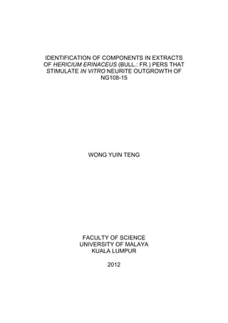
- 25. 2.2 HERICIUM ERINACEUS 2.2.1 Origin Hericium erinaceus (Bull.: Fr.) Pers. (Figure 2.1), a member of the basidiomycetous fungus, is well known as a traditional medicine or food in Japan and China. In Japan, H. erinacues is called Yamabushitake because it resembles the ornamental cloth worn by Yamabushi. It is also called Jokotake (drinker fungus), Usagitake (rabbit fungus), Harisenbon (balloon fish) due to its shape. Figure 2.1: Hericium erinaceus (Bull.: Fr.) Pers. This mushroom is called Houtou (monkey head mushroom) in China due to the close resemblance of fruiting body to the head of a baby monkey. It also known as Hedgehog mushroom according to its shape. A Chinese traditional drug prepared by drying this mushroom is also called Houtou. The hot water extracts from dried fruitbodies are used as health drink (Yang and Jong, 1989). It can be pickled in brewed wine to give a health drink (Mizuno, 1999). Hericium erinaceus is a wood destroying fungus and grows in standing and decayed broadleaf trees such as oak, beech, and walnut. The cultivation of H. erinaceus 7
- 26. has been established using artificial logs made with agricultural residues in either bottles or polypropylene bags (Mizuno, 1999; Chang and Miles, 2004). 2.2.2 Classification Kingdom : Fungi Phylum : Basidiomycota Class : Basidiomycetes Order : Russulales Family : Hericiaceae Genus : Hericium Species : erinaceus 2.2.3 Medicinal properties, nutritional and bioactive components derived from Hericium erinaceus Medicinal properties of H. erinaceus have been widely studied. Both the fruiting bodies and mycelia of H. erinaceus contain bioactive polysaccharides which exhibit various pharmacological activities including immunomodulatory effect, as well as anti- tumor, hypoglycemic and anti-aging properties (Zhang et al., 2007). Fifteen polysaccharides have been successively extracted out with hot water. Five types of polysaccharides which showed relatively strong antitumor activity and a good life prolongation effect were glucoxylan, xylan, heteroxyloglucan, glucoxylan protein complex and galactoxyloglucan protein complex (Zhang et al., 2007). Besides the polysaccharides, an ergosterol derivative was also isolated from H. erinaceus. This compound showed cytotoxic effects on the cervical carcinoma HeLa cells (Mizuno, 1999), antitiumor activity against Walker carcinosarcoma and human mammary adenocarcinoma cell lines in vitro (Jong and Donovick, 1989), human gastric 8
- 27. tumor cell line, human hepatoma cell line, human colorectal tumor cell line and murine sarcoma-180. It also showed antivenom, anti-inflammatory (Keyzers and Davies- Coleman, 2005) and antimicrobial activity (Lu et al., 2000). Hericenones, erinacines, hericerin and hericenes, the aromatic compounds that identified in H. erinaceus, showed a wide range of in vitro and in vivo bioactivities (Shang et al., 2012). The novel oxyketo acid, Y-A-2, cytotoxic phenols, hericenone A and hericenone B (Kawagishi et al., 1990), two novel γ- pyrones, erinapyrone A and erinapyrone B (Figure 2.2) (Kawagishi et al., 1992) extracted from the fruiting body of H. erinaceus using ethanol or acetone, showed inhibition against the proliferative activity of HeLa cells. Besides that, various acidic phenol-like and neutral fatty acid-like compounds such as hericenones and hericerins found in H. erinaceus (Kim et al., 2000) were effective against pathogenic microorganisms and showed antibacterial activity at low concentrations against S. aureus, B. subtilis and E. coli respectively. Two novel and a known chlorinated orcinol derivaties were also isolated from the mycelium of H. erinaceus. These three compounds exhibited antimicrobial activities against Bacillus subtilis, Saccharomyces cerevisiae, Vetticillium dahlia and Aspergillus niger (Okamato et al., 1993). Ethanol extract of mycelia or fruitbodies promoted better antimutagenic effects than water extract examined with the Ames test (Wang et al., 2001). On the other hand, methanol extract of fruitbodies was found to have hypoglycemic effect and reduce elevation rates of serum triglyceride and total cholesterol levels when administered to streptozotocin-included diabetic rats (Wang et al., 2005). Yang et al., (2003) investigated the hypolipidemic effect of an exo-biopolymer produced from a submerged culture of H. erinaceus in dietary-included hyperlipidemic rats. The exo-biopolymer reduced the level of plasma total cholesterol, low density lipoprotein cholesterol, 9
- 28. triglyceride, phospholipids, atherogenic index and hepatic HMG-CoA reductase activity; and preserving the high density lipoprotein at relatively high level. These effects would help to reduce the risk of atherosclerosis. OH O COOH OH Y-A-2 (Novel oxyketo acid) O OH O O H3CO Hericenone A O OH O N H3CO Hericenone B O O HO OH O O Erinapyrone A Erinapyrone B Figure 2.2: HeLa cell growth inhibitory substances isolated from Hericium erinaceus 10
- 29. This mushroom has been reported to exhibit significant antioxidant activity which might help to reduce the oxidative damage caused by the uncontrolled production of oxygen-derived free radicals (Mau et al., 2001). The reduction of free radicals might lower the risk in the onset of many diseases such as cancer, rheumatoid arthritis, artherosclerosis, degenerative processes and deterioration of physiological functions associated with aging. Besides that, total polyphenols were the major natural antioxidant components found in the methanol extract from dried H. erinaceus fruit bodies (Mau et al., 2002). In Chinese traditional medicine, it is used for the treatment for neurasthenic gastritis and gastroduodenal ulcer. In recent year, cultures or their extracts processed in tablets have been produced in large scale for curing gastric ulcer and chronic gastricism. Nitric oxide (NO) is a pleiotropic biological molecule involved in a myriad of physiological and pathological processes such as regulation of blood pressure, neurotransmission, signal transduction, anti-microbial defense, immunomodulation, cellular redox regulation and apoptosis. The water extract of H. erinaceus activated the macrophages and induce NO production in peritoneal macrophages and RAW 264.7 cell line through the activation of transcription factor NF-KB (Son et al., 2006). A 63kDa laccase, with a novel N-terminal sequence isolated from the water extract of H. erinaceus dried fruiting bodies showed inhibitory effect towards HIV-1 reverse transcriptase (Wang and Ng, 2004). HIV-1 reverse transcriptase was involved in HIV replication; inhibitors of this enzyme are potential therapeutic agents in the battle against HIV (Sarafianos et al., 2009). 11
- 30. 2.3 NERVOUS SYSTEM AND NEURODEGENERATIVE DISEASES 2.3.1 Neurite The characterization of neurite formation, maturation and collapse/ resorption is an area of interest because these cellular processes are essential for the interconnection of neuronal cell bodies. Neurites are particularly interesting in relation to neuropathological disorders, neuronal injury/ regeneration and neuropharmacological research and screening (Smit et al., 2003). Neurites emerging from cloned neural cell lines have been studied extensively over the past 15 years (Smalheiser & Schwartz, 1987). It was appreciated very early that some clones can express neurites spontaneously, even without inducing them to differentiate, but most neurobiologists have ignored this class of neurites in favor of studying clones such as the PC12 cell line, whose neurites are under inducible control and contain characteristics of axons in differentiated neurons. Bioassay which uses the PC12 cell line of rat pheochromocytoma was described by Greene (1977) and Greene & Tischler (1982). The matured neurite, called neuron, is responsible for receiving stimuli, producing and transmiting electrical signal called nerve impulses, or action potentials. It also synthesizes and releases neurotransmitters. 2.3.2 Neurodegenerative diseases (factors, therapies to cure and prevent) Neurodegenerative diseases can be defined as hereditary and sporadic conditions which are characterized by progressive nervous system dysfunction. These disorders are often associated with atrophy of the affected central or peripheral nervous system structures. Neurological disorders are quite diverse, chronic, challenging to treat, and often disabling. They can be caused by many different factors, including (but not limited to): inherited genetic abnormalities, problems in the immune system, injury to 12
- 31. the brain or nervous system, or diabetes. Many mental illnesses are believed to be neurological disorders of the central nervous system, but they are classified separately. The production of reactive oxygen species during oxidative stress is speculated to be pathologically important in neurodegenerative diseases which include Alzheimer’s disease, Parkinson’s disease, amyotrophic lateral sclerosis, and Huntington’s disease (Halliwell and Gutteridge, 1999). Alzheimer’s disease is the most common form of senile dementia. Alzheimer’s disease could be caused by both environmental and genetic factors. This has been proved by the genetic linkage studies on the chromosomes 14, 21 (early-onset) and 19 (late-onset). Trisomy and mutations of the β- amyloid precursor protein gene on chromosome 21 are the causes that lead to the early- onset of the Alzheimer’s disease (Goate et al., 1991). Early-onset familial forms of Alzheimer’s disease could be caused by genetic mutations which may affect chromosome 14 (Mullan et al., 1993). Mutations on chromosome 19 and the concomitant expression of variant apolipoprotein E4 (ApoE4) from ApoE gene (ε4 allele) are associated with sporadic and late-onset familial forms of Alzheimer’s disease (Uterman, 1994). However, less than 1% of patients who suffer from the disease are due to these genetic causes. It is probable that the majority of the cases is caused by a variety of environmental factors which may be either sufficient to trigger disease by themselves, or sufficient when acting synergistically with the patient genotype. A direct or indirect role has been attributed to normal or structurally altered amyloid β-protein (concentrated in senile plaques) and/or excessively phosphorylated tau protein (located in neurofibrillary tangles) (Schorderet, 1995). Degeneration of cholinergic neurons and concomitant impairment of cortical and hippocampal neurotransmission lead to cognitive and memory deficits (Schorderet, 1995). Neuronal cell death is an essential feature of neurodegenerative disease. Many types of neuronal cell death, for example, these which are associated with amyloid-β, 13
- 32. glutamate and nitric oxide are thought to be caused by endoplasmic reticulum stress. Glutamate toxicity is a major contributor to pathological cell death within the nervous system and appears to be mediated by reactive oxygen species (Lee et al., 2003). Thus, it is reasonable to suspect that molecules which are able to attenuate endoplasmic reticulum stress might reduce both the risk for and the extent of the damage in neurodegenerative disease (Nagai et al., 2006). Treatments for reducing neuronal cell death are important for preventing as well treating neurodegenerative disease, including dementia and motor dysfunction. However, because neurodegenerative diseases have, typically, a long incubation period prior to diagnosis and are symptom-free; hence, there is a late diagnosis of the disease. This is a severe problem because once neurons are dead or neuronal circuits destroyed, lost of brain function associated with the neurons or neuronal circuits is almost impossible to restore. Attenuation of the cognitive deficits by using choline supplementation (lecithins) and/ or acetylcholinesterase inhibitors might enhance the cholinergic activity if the cognitive deficits are due to the loss of cholinergic activity. Tacrine (tetrahydroaminoacridine, Cognex®), a potent, centrally active and reversible acetylcholinesterase inhibitor, was used together with morphine to lessen respiratory depression without affecting analgesia in the mid 1940s. Tacrine has been used alone, or in combination with lecithin, to treat symptoms of the Alzheimer’s disease (Chatellier et al., 1990; Farlow et al., 1992). It showed a slight but statistically significant improvement in the physician's score on the visual analogue scale (Chatellier et al., 1990). Only a small percentage of patients, moderately affected or treated at an early stage of Alzheimer’s disease, seem to benefit from the drug (Farlow et al., 1992). However, it showed several side effects such as gastrointestinal troubles, hepatitis and reversible hepatotoxicity. 14
- 33. Investigation on the projection of neurotrophin NGF (Nerve Growth Factor) which could counter the degeneration of cholinergic neurons to the hippocampus, a recognized memory center, were recently initiated. NGF is a protein that is essential for supporting the growth and maintenance of peripheral sympathetic neurons as well as facilitating the development of some sensory neurons for a brief period during early development (Shimbo et al., 2005). Infusions of NGF into the brain of a patient can improve performance in memory test and prevent or stabilize the processes of cholinergic pathway degeneration (Schorderet, 1995). Alternatively, antioxidants, free redical scavengers and/ or non-steroidal anti- inflammatory agents such as α-tocopherol (vitamin E), ubiquinols (coenzyme Q), retinoic acid (vitamin A), and ascorbic acid (vitamin C), may be screened as potential therapies for neurodegenerative disease induced by multiple endogenous and/ or exogenous factors (Schorderet, 1995). 15
- 34. 2.4 NEUROPROTECTIVE, NEUROTROPHIC, NEURONAL DIFFERENTIATION AND NEURITE STIMULATION EFFECTS OF HERICIUM ERINACEUS Hericenone C, D, E, F, G, H have been successively isolated from H. erinaceus (Kawagishi et al., 1991, 1993). Among them, hericenone C, D, E (Figure 2.3) have been proven to show NGF synthesis promoting activity (Kawagishi et al., 1991). O OH CHO 16" O H3CO O Hericenone C O OH CHO 18" O H3CO O Hericenone D O OH CHO 18" O H3CO 9" 12" O Hericenone E Figure 2.3: Hericenones isolated from fruiting body of Hericium erinaceus which showed NGF synthesis promoting activity. 16
- 35. This mushroom also produces erinacines A (Shimbo et al., 2005), B, C (Kawagishi et al., 1994), D (Kawagishi et al., 1996a), E, F, G (Kawagishi et al., 1996b) which have been identified as stimulators of nerve growth factor (NGF) synthesis (Figure 2.4). Stimulators of NGF synthesis have been used as medicines for degenerative neuronal disorders such as Alzheimer’s disease and peripheral nerve regeneration. NGF is a protein that is essential for supporting the growth and maintenance of peripheral sympathetic neurons as well as facilitating the development of some sensory neurons for a brief period during early development (Shimbo et al., 2005). NGF cannot pass through the blood-brain barrier, the semi-permeable membrane between the blood and brain. Only small and lipid soluble molecules can pass through the membrane. NGF is too large to permeate it. Therefore, it needs to be injected directly into the brain to be effective (Kawagishi et al., 2002). If a substance can permeate the membrane and stimulates the NGF synthesis in brain, this may result in repairing the damaged nervous functions. Dilinoleoyl-phosphatidylethanolamine (DLPE), an endoplasmic reticulum (ER) stress- attenuating molecule which might reduce the ER-stress, has been isolated from the fruit bodies of H. erinaceus and these may reduce the risk of getting neurodegenerative diseases (Nagai et al., 2006). ER stress is the major cause of the neuronal cell death which leads to the neurodegenerative diseases. 17
- 36. O O O O H OH OH O HO OH OH CHO CHO Erinacine A Erinacine B O O O O H H OH OH O HO OH OH CH2OH C2H5O CHO Erinacine C Erinacine D H O O O O H H OH H H OH H H OH OH HO OH HO OH Eriancine E Erinacine F O O O O H H OH H OH HO OH Erinacine G Figure 2.4: Erinacines isolated from mycelium of Hericium erinaceus which showed NGF synthesis promoting activity. 18
- 37. 2.5 NEURITE OUTGROWTH BIOASSAY SYSTEM OF NEURAL HYBRID CELL LINE NG108-15 2.5.1 Formation of NG108-15 hybrid cell 6-thioguanine-resistant clonal mouse neuroblastoma cells N18TG2 and the bromodeoxyuridine-resistant rat glioma cells C6-BU-1 were fused with the aid of inactivated Sendai virus to generate the neuroblastoma x glioma hybrid cell clone, NG108-15 (Hamprecht et al., 1985). Cells were grown in selective hypoxanthine- aminopterin-thymidine (HAT) medium, which was known to select for the wild-type hybrid cells and against the parental cell lines and their corresponding homokaryocytes (Littlefield, 1964). 2.5.2 Characteristics of NG108-15 hybrid cell The hybrid cell is used as model neurons because every characteristic generally ascribed to neurons has been observed with the hybrid cell. The properties of NG108-15 are summarized in Table 2.1 (Hamprecht et al., 1985). Due to the complexity of the mammalian nervous system, it is hard to assign a certain effect observed to a certain cell type. Problems that are difficult to solve with animal or tissue experiments can be tackled with the aid of cultured cells. Then, it is like having at one’s disposal the numerous cell types as homogenous cell populations for studying their individual differentiated functions and their mechanisms of intercellular communication. 19
- 38. Table 2.1: Neuronal properties of neuroblastoma x glioma hybrid cells (NG108-15) Neuronal Properties Extension of long processes Clear and dense core vesicles Excitable membranes (inward current of action potentials carried by Na+ or Ca2+ Formation of functional synapses Neurotransmitter enzymes - Choline acetyltransferase - Dopamine-β-hydroxylase Synthesis of neurohormones - Acetylcholine - Leu- and Met- enkephalin - Dynorphine-(1-8), α-neoendorphine - β-Endorphine - Vasoactive intestinal peptide - Angiotensin - Hydra head activator- like activity Uptake system for - Catecholamines - Taurine Depolarization- induced Ca2+- dependent release of acetylcholine Receptors for neurohormones - Acetylcholine - Noradrenaline - Opioids (Morphine, enkephaline) - Prostaglandin E1 - Adenosine 20
- 39. CHAPTER III MATERIALS AND METHODS 3.1 EXTRACTION 3.1.1 Preparation of fruitbodies Hericium erinaceus fresh fruitbodies were obtained from Ganofarm Sdn. Bhd. in Tanjung Sepat, Selangor. The fruitbodies were freeze-dried at 50± 2 C for 48 hours. The dried fruit bodies were blended in the commercial Waring blender and stored in airtight containers prior to assay. 3.1.2 Preparation of aqueous ethanol crude extract The powdered freeze-dried fruit bodies of H. erinaceus was soaked in 80 % (v/v) aqueous ethanol for 3 days. The residue was then resoaked in 80 % (v/v) aqueous ethanol and the extraction and filtration process was repeated three times. The solvent containing extract was then concentrated under vacuum using a rotary evaporator. 3.1.3 Solvent- solvent extraction (fractionation) The crude extract obtained was successively fractionated with hexane, ethyl acetate and water using a separating funnel. All the fractions (hexane, ethyl acetate and water) were filtered and concentrated under vacuum using a rotary evaporator to give hexane, ethyl acetate and water fractions Figure 3.1 shows the flow chart of the extraction and fractionation procedures, process of biological investigations and isolation of active fractions of H. erinaceus. 21
- 40. Fresh Hericium erinaceus Freeze-dried and ground to fine powder Dried and ground H. erinaceus i. Extraction with 80% ethanol (3 times) ii. Concentration under reduced pressure Aqueous ethanol crude extract i. Extraction with hexane ii. Concentration under reduced pressure Hexane soluble fraction Hexane insoluble fraction i. Partition (v/v) between ethyl acetate and water (Ratio 1:2) ii. Concentration under reduced pressure Ethyl acetate fraction Water fraction In vitro neurite outgrowth assay by using NG108-15 hybrid clone Isolation of active fractions Figure 3.1: A schematic diagram showing the extraction and fractionation procedures, process of biological investigations and isolation of active fractions of Hericium erinaceus 22
- 41. 3.2 NEURITE OUTGROWTH ACTIVITY ASSAY 3.2.1 Preparation of stock solutions Each extract and fractions were dissolved in dimethylsulfoxide (DMSO) to form stock solutions 20 mg/ml for neurite outgrowth assay and kept at -20 ºC for future use. The concentration of samples was prepared according to the requirements for the assay by serial dilutions using the media or media with Tween 80. 3.2.2 Cell culture The neural hybrid clone NG108-15 was chosen for this purpose. NG108-15 cells were purchased from American Type Culture Collection (ATCC) and cultured in Dulbecco’s Modified Eagle’s Medium (DMEM). 3.2.3 Preparation of medium and buffer for cell culture 3.2.3.1 Dulbecco’s Modified Eagle’s Medium (DMEM) Basic medium Final volume cell culture grade water (80 - 90 %) was measured. Water temperature should be 15-30 C. Dry powder medium (13.38 g/l) was added slowly and allowing mixing time between additions (original package was rinsed with small amount of water to remove all traces of powder and added to solution). The solution was mixed for 30 minutes. 3.7 g/l of sodium bicarbonate (NaHCO3) and hypoxanthine- aminopterine- thymidine (HAT) were added and stirred until dissolved. While mixing, the pH of the medium was adjusted to 6.9-7.1 using 1N NaOH or 1N HCl. Additional water was added to bring the solution to final volume and continue mixing for at least 30 minutes. The medium was sterilized by filtration using a membrane with a pore size of 0.22 microns and aseptically dispensed into sterile container. 23
- 42. Complete growth medium Basic medium described above supplemented with 10 % (v/v) of fetal bovine serum, 100units/ml penicillin and 100 µg/ml streptomycin. Revival medium Complete growth medium described above supplemented with 20 % (v/v) instead of 10 % (v/v) fetal bovine serum. Cryoprotectant medium Basic growth medium described above supplemented with 10 % (v/v) dimethyl sulfoxide (DMSO) and 50 % (v/v) fetal bovine serum. 3.2.3.2 Phosphate Buffer Saline 1.52 g of sodium hydrogen phosphate (Na2HPO4), 0.58 g of potassium hydrogen phosphate (KH2PO4) and 8.5 g of sodium chloride (NaCl) were dissolved in 1L distilled water and pH was adjusted to 7.2. The solution was filtered with filter paper and autoclaved for 15 minutes at 121 C, 15 psi. The solution was stored at room temperature. 3.2.4 Cell culture techniques 3.2.4.1 Revival of frozen cells The vial containing frozen cells was thawed by gentle agitation in a 37 C water bath. To reduce the loss of viability, the vial was thawed rapidly. Once the contents were thawed, the contents were transferred aseptically into a centrifuge tube by pipetting. 1 ml of revival medium was added and centrifuged at 1000 rpm for 5 minutes. 24
- 43. Supernatant was discarded and pellet was resuspended in 1 ml of revival medium. The suspended cells were pipetted into a cell culture flask containing 5- 10 ml revival medium. The culture was incubated at 37C in a 5% CO2 incubator at atmospheric pressure. 3.2.4.2 Subculture of cells The culture was examined with an inverted microscope to check for any evidence of microbial contamination and to determine whether the majority of the cells were attached to the bottom of the flask. When the color of the medium changed from reddish to yellowish (the medium become acidic) or every 2- 3 days, the culture medium was removed and discarded. Trypsin-EDTA (1 ml) and phosphate buffer saline solution (3 ml) were added to the flask and cells were observed under inverted microscope until cell layer was detached from the bottom of the flask. The contents of the flask were transferred aseptically into a centrifuge tube containing 2 ml of complete growth medium by pipetting. The cells were centrifuged at 1000 rpm for 5 minutes. Supernatant was discarded and pellet was resuspended in 3- 4 ml of complete growth medium. Appropriate amounts of suspended cell were added to a new culture flask containing complete growth medium. Cultures were incubated at 37 C in a 5 % CO2 incubator at air atmosphere. 3.2.4.3 Medium renewal The medium was changed 3 to 4 times weekly. 25
- 44. 3.2.4.4 Cryopreservation of cells Cryopreservation of cells was carried out in accordance with subculturing procedure except that the pellet obtained after centrifugation was suspended in cryoprotectant medium instead of complete growth medium. The suspended cells were then transferred to a cryogenic vial and stored in liquid nitrogen vapor phase. 3.2.5 Effect of Hericium erinaceus on stimulation of neurite outgrowth activity of NG108-15 The neural hybrid cell NG 108-15 was cultured until 60-70 % confluent prior to assay. Cells were detached from the flask with 0.25 % (w/v) solution of trypsin in phosphate buffer saline solution and washed in culture medium. The cell pellet was obtained by centrifugation at 1000 rpm for 5 minutes. The density of the cells was counted by 0.4 % (w/v) of tryphan blue exclusion method in a haemocytometer. The cells were plated into 6 well plates coated with 2 x 10⁻⁵ % (w/v) of poly-D-lysine at a cell density of 10000 cells per well in medium containing 4 various concentrations (10, 25, 50, 100 µg/ml) of extract and fractions and subfractions of fruit bodies. Negative control is the well with the untreated cells whilst positive control is the well with the cells treated with Nerve Growth Factor (NGF). Plates were incubated at 37 C in a 5 % CO2 humidified incubator. Cells were observed for neurite outgrowth, branching of neurites after 24 hours. 3.2.6 Scoring of neurites A cell was considered as positive for bearing neurites if it had at least one thin extension longer than one full diameter of its cell body. Specifically excluded were extensions associated with clearly different patterns of cell responses, such as broad, 26
- 45. sheet-like spreading of cells or the rare radially oriented processes apparently arising by “shrinkage” (Smalheiser and Schwartz, 1987). Cell clumps containing more than five cells were also not included in the results. If more relaxed criteria had been used, many short extensions would have been counted as neurites, and the assay would not have been useful to detect stimulatory effects upon neurite formation (Smalheiser and Schwartz, 1987). Duplicates were set up for each concentrations tested. Approximately 300 cells in each well were evaluated. Neurite formation was quantified by scoring the number of cells processing neurites and expressed as a percentage of the total number of cells counted. Neurite bearing cells (%) = number of cells processing neurites x 100 % total number of cells counted The result was also expressed in percentage increase in neurite bearing cells in comparison to negative control. Percentage increase compared to negative control = neurite bearing cells of extract – neurite bearing cells of negative control x 100 % neurite bearing cells of negative control 3.2.7 Statistical analysis The means of data were subjected to a one way analysis of variance (ANOVA) and the significance of the difference between means was determined by the Duncan’s multiple range test at 95 % least significance difference (P<0.05). 27
- 46. 3.3 ISOLATION OF BIOACTIVE CONSTITUENTS 3.3.1 Column chromatography The combined fraction of hexane and ethyl acetate was subjected to silica gel flash column chromatography. Column chromatography was performed by using Merck silica gel. The gel was packed onto the column. After the sample was introduced to the column, solvent with increasing polarity gradient was used to elute the column [developing solvent: chloroform (100 % chloroform) → chloroform-acetone mixtures (20 % Ac/CHCl3, 40 % Ac/CHCl3) → chloroform-methanol mixtures (10 % MeOH/CHCl3, 30 % MeOH/CHCl3) → methanol (100 % MeOH)]. Fractions were monitored by thin layer chromatography (TLC) and fractions that possess the same spots/bands on the TLC were combined and where necessary subjected to further separation. Nine fractions were obtained (E1-E9) from the flash column chromatography which monitored by TLC. Figure 3.2 shows the flow chart of the isolation of active fractions of H. erinaceus.by using different types of chromatographic technique, process of biological investigations and identification of the active fractions. 3.3.2 Analytical thin layer chromatography TLC was routinely used to detect and separate the various compounds. The fractions from column chromatography were examined by TLC using precoated glass plates, 0.25 mm thickness, silica gel F254 (Merck, Darmstadt, G.F.R). The TLC plates were spotted with a piece of fine glass capillary tube and then developed in saturated chromatography tanks with various solvent systems at room temperature. The spots were visualized by examination of the TLC plates under UV light, followed by applying iodine vapor. 28
- 47. 3.3.3 Preparative thin layer chromatography Subfraction E2 was successively subjected to preparative thin layer chromatography. The sample was dissolved by using chloroform. The TLC precoated (silica gel F254) glass plates (Merck, Darmstadt, G.F.R) with 0.25 mm thickness, 10 cm (width) x 10 cm (height) were used. A line was drawed (about 1 cm) from the bottom of the plate. ). The line on the TLC plates was spotted with a piece of fine glass capillary tube and then developed in saturated chromatography tanks (100 % CHCl3) at room temperature. After the solvent reach the solvent front, the plates were taken out from the chromatography tanks. When the plates were dried enough, the bands were visualized by using UV light. The bands were marked lightly by using pencil. The bands were scrapped off onto a clean, white paper by using the edge of a spatula. The compounds were washed off from the silica gel by using chloroform. The solvent containing samples were filtered and concentrated under vacuum using a rotary evaporator. 3.3.4 High performance liquid chromatography (HPLC) 3.3.4.1 HPLC samples and mobile phase preparation The samples were dissolved in methanol/acetonitrile mixture. The samples were filtered through 0.45 µm Sartorius minisart PTFE-membrane syringe filter to remove any particular matter that might clog the column. The mobile phase that used was acetonitrile. The mobile phase was filtered by using 0.45 µm Sartorius PTFE-membrane and degassed before introduced to the system. All the solvents that used were in HPLC grade. 29
- 48. 3.3.4.2 Analytical HPLC Analytical HPLC analysis were carried out by using the instrument Waters Delta Prep consists of water Prep LC controller, quaternary pump, vacuum degasser, UV detector (water 2487, Dual λ Absorbance Detector). The separation profiles of the samples can be improved by changing the solvent system of the mobile phase, flow rate and column. The column that used for analytical HPLC was performance RP-18 encapped column 100-4.6 mm purchased from Merck. The analysis was carried out in isocratic mode at a flow rate of 1 ml/min, with column effluent being monitored at the wavelength of 214 nm and 254 nm. 3.3.4.3 Semipreparative HPLC The samples were then further isolated out by using Chromolith Semiprep RP- 18 column encapped 100-10 mm purchased from Merck. The separation was carried out in isocratic mode by using 100 % acetonitrile as mobile phase at a flow rate of 3 ml/min, with column effluent being monitored at the wavelength of 214 nm and 254 nm. The separated subfraction was collected manually. 30
- 49. Combined fraction of hexane and ethyl acetate Flash column chromatograhy E1 E2 E3 E4 E5 E6 E7 In vitro neurite outgrowth assay by using NG108-15 hybrid clone Identification of the Preparative thin layer chemical constituents chromatography by GC-MS sub4b HPLC sub4b_4 sub4b_6 Identification of the chemical constituents by NMR Identification of the chemical constituents by LC/MS/MS In vitro neurite outgrowth assay by using NG108-15 hybrid clone Figure 3.2: A schematic diagram showing the isolation of active fractions, process of biological investigations and identification of the active fractions. 31
- 50. 3.4 IDENTIFICATION 3.4.1 Gas chromatography-mass spectrometry (GCMS) GCMS analysis was performed on fraction E1 using Network Gas Chromatography System (Agilent Technologies 6890) and Inert Mass Selective Detector (Agilent Technologies 5975) (70eV direct inlet) on a HP-5MS (5 % phenyl methyl siloxane) capillary column (30 m x 250 µm x 0.25 µm) initially set at 150 ºC, then programmed to 300 ºC at 5 ºC min-1 and held for 10 minutes at 300 ºC using helium as the carrier gas. The total ion chromatogram obtained was autointegrated by chemstation and the constituents were identified by comparison with the accompanying mass-spectra database (NIST 05 Mass Prectral Library, USA) wherever possible. 3.4.2 Nuclear magnetic resonance spectroscopy (NMR) All the NMR experiments were performed on a JEOL 400MHz NMR spectrometer that install with the JEOL Delta software. Subfractions sub4b_4 and sub4b_6 were dissolved in deuterated chloroform and 1H, 13C and 1H-1H-COSY spectra were collected. The internal standard for 1H NMR was TMS (δ: 0.00) and 13 C was CDCl3 (δ: 77.00). 3.4.3 Liquid chromatography-mass spectrometry (LC/MS/MS) LC/MS/MS analysis were carried out on subfractions sub4b_4 and sub4b_6 by using the instrument Applied Biosystems 3200Q Trap LCMS/MS with Shimadzu ultra pure liquid chromatography (UPLC) system. Full scan with MS/MS data collection was used. Positive ionization mode was set. The column that was used is Phenomenex Aqua C-18 with dimension 50.0 mm x 2.0 mm x 5.0 µM. Rapid screening was performed with 10 min run time. 32
- 51. CHAPTER IV RESULTS & DISCUSSION 4.1 EXTRACTION, FRACTIONATION AND ISOLATION 4.1.1 Extraction, fractionation and isolation of aqueous ethanol extract of Hericium erinaceus The flow chart shows the extraction and fractionation procedures for H. erinaceus (Figure 4.1 and 4.2) Fresh H. erinaceus (1.33 kg) Dried and ground to fine powder Dried and ground H. erinaceus (200.00 g) i. Extraction with 80 % ethanol (3 times) ii. Concentration under reduced pressure Aqueous ethanol extract (52.29 g) Figure 4.1: Aqueous ethanol extraction of Hericium erinaceus. Fresh H. erinaceus (1.33 kg) yielded 200.00 g of dried and ground H.erinaceus. The dried H. erinaceus sample (200.00 g) yielded 52.29 g of crude aqueous ethanol extract. 33
- 52. Aqueous ethanol extract i. Extraction with hexane ii. Concentration under reduced pressure Hexane soluble fraction Hexane insoluble fraction (3.85 g, 7.36 %) i. Partition (v/v) between ethyl acetate and water (Ratio 1:2) ii. Concentration under reduced pressure Ethyl acetate fraction Water fraction (0.77 g, 1.47 %) (44. 34 g, 84.80 %) Figure 4.2: Fractionation of aqueous ethanol extract of Hericium erinaceus. The aqueous ethanol extract was further extracted with hexane to give hexane- soluble fraction (3.85 g, 7.36 %) and hexane-insoluble residues. The hexane-insoluble residues were further partitioned between ethyl acetate-water (ratio 1:2) to give the ethyl acetate-soluble fraction (0.77 g, 1.47 %) and water fraction (44. 34 g, 84.80 %). 34
- 53. The combined hexane and ethyl acetate fraction were subjected to flash column chromatography to yield 7 fractions, which were E1 (384.0 mg, 0.73 %), E2 (780.8 mg, 1.49 %), E3 (438.2 mg, 0.84 %), E4 (62.4 mg, 0.12 %), E5 (39.7 mg, 0.08 %), E6 (183.1 mg, 0.35 %), E7 (1068.2 mg, 2.04 %) (Figure 4.3). The percentage yields were calculated based on the crude aqueous ethanol extract. Combined fraction of hexane and ethyl acetate Flash column chromatography (developing solvent: CHCl3 → CHCl3/Ac → CHCl3/MeOH →MeOH) E1 (384.0mg, E3 (438.2mg, E5 (39.7mg, E7 (1068.2mg, 0.73 %) 0.84 %) 0.08 %) 2.04 %) E2 (780.8mg, E4 (62.4mg E6 (183.1mg, 1.49 %) 0.12 %) 0.35 %) Figure 4.3: Isolation of combined hexane and ethyl acetate extract of Hericium erinaceus obtained through flash column chromatography. 35
- 54. Fraction E2 obtained from flash column chromatography were further subjected to preparative thin layer chromatography to yield subfraction sub4b (187.7 mg, 0.36 %) (Figure 4.4). Sub4b was then subjected to high performance liquid chromatography (HPLC) to give sub4b_4 (68.5 mg, 0.13 %) and sub4b_6 (38.5 mg, 0.07 %) (Figure 4.4). E2 Preparative thin layer chromatography Sub4b (187.7 mg, 0.36 %) High perfomance liquid chromatography (HPLC) Sub4b_4 (68.5 mg, Sub4b_6 (38.5 mg, 0.13 %) 0.07 %) Figure 4.4: Isolation of fraction E2 of Hericium erinaceus using preparative thin layer chromatography and high performance liquid chromatography. 36
- 55. 4.2 NEURITE OUTGROWTH ACTIVITY 4.2.1 Effect of aqueous ethanol extract and fractions of Hericium erinaceus on the neural cell line NG108-15 Aqueous ethanol extract and fractions of H. erinaceus were screened for the in vitro neurite outgrowth activity on the neural hybrid cell line NG108-15 at various concentrations (µg/ml) (Figure 4.5; Table 4.1). Cells were observed under a phase contrast microscope for the neurite outgrowth and branching of neurites. The effect of the various extracts on the morphology and neurite extension of the NG108-15 cells are given in Figure 4.6 (crude aqueous ethanol extract), Figure 4.7 (hexane fraction), Figure 4.8 (ethyl acetate fraction) and Figure 4.9 (water fraction). 50 45 Neurite- bearing cells (%) 40 35 ethanol crude extract 30 Hexane fraction 25 Ethyl acetate fraction 40.6 20 Water fraction 34.5 34.1 32.3 27.8 27.4 15 26.5 25.6 25.4 24.6 24.2 24.1 24.0 23.7 23.5 23.0 22.9 22.5 21.9 20.9 20.5 19.9 19.0 18.6 10 5 0 Negative NGF 10 25 50 100 control (20ng/ml) Concentrations (µg/ml) Figure 4.5: Percentage of neurite bearing cells incubated with varying concentrations of aqueous ethanol crude extract, hexane fraction, ethyl acetate fraction and water fraction of Hericium erinaceus (nerve growth factor, 20 ng/ml, used as positive control). 37
- 56. Table 4.1: Stimulation of neurite outgrowth activity in the NG108-15 cells with varying concentrations of aqueous ethanol extract and fractions of Hericium erinaceus. NG108-15 cells without extract was negative control. 20 ng/ml of nerve growth factor (NGF) was used as positive control. Ethanol crude extract Hexane fraction Ethyl acetate fraction Water fraction Treatment Neurite Increase Neurite Increase Neurite Increase Neurite Increase concentration bearing cells compared to bearing cells compared to bearing cells compared to bearing cells compared to (µg/ml) (%) control (%) (%) control (%) (%) control (%) (%) control (%) Negative control 19.9±1.5ab - 24.6±1.5a - 20.5±1.5a - 19.0±1.7a - Positive control (NGF) 23.0±1.8c 15.5 28.9±0.9bc 11.3 24.1±0.2b 17.8 24.2±0.2b 27.1 10 22.9±0.5c 15.0 25.4±1.3ab 3.3 23.7±0.8b 15.9 21.9±1.8ab 14.9 25 22.5±0.7bc 13.2 32.3±2.6bc 31.4 26.5±2.0bc 29.4 24.0±1.6b 26.3 50 20.9±0.9abc 4.8 34.1±0.1c 38.7 27.8±1.6c 35.4 23.5±1.9b 23.3 100 18.6±1.3a -6.4 40.6±2.5d 65.2 34.5±0.9d 68.5 25.6±1.3b 34.4 Note: Data are expressed as means ± standard deviation (n = 2). Means with different letters in the same column are significantly different (P < 0.05), one-way analysis of variance/ANOVA) 38
- 57. A B neurite C D E F neurite Figure 4.6: The morphology of the NG108-15 cells treated with various concentrations of crude aqueous ethanol extract of Hericium erinaceus [24hr of incubation at 37 ºC in a 5 % CO2 humidified incubator. NG108-15 cells without extract or treated with NGF (20 ng/ml) was negative and positive control, respectively.] A: negative control (cells without extract); B: positive control - NGF (20 ng/ml); C: 10 µg/ml of crude aqueous ethanol extract; D: 25 µg/ml of crude aqueous ethanol extract; E: 50 µg/ml of crude aqueous ethanol extract; F: 100µg/ml of crude aqueous ethanol extract 39
- 58. A B neurite C D neurite neurite E F neurite neurite Figure 4.7: The morphology of the NG108-15 cells treated with various concentrations of hexane fraction of Hericium erinaceus [24 hr of incubation at 37 ºC in a 5 % CO2 humidified incubator. NG108-15 cells without extract or treated with NGF (20 ng/ml) was negative and positive control, respectively.] A: negative control (cells without extract); B: positive control - NGF (20 ng/ml); C: 10 µg/ml of hexane fraction; D: 25 µg/ml of hexane fraction; E: 50 µg/ml of hexane fraction; F: 100 µg/ml of hexane fraction 40
- 59. A B neurite neurite C D neurite neurite E F neurite neurite neurite Figure 4.8: The morphology of the NG108-15 cells treated with various concentrations of ethyl acetate fraction of Hericium erinaceus [24 hr of incubation at 37 ºC in a 5 % CO2 humidified incubator. NG108-15 cells without extract or treated with NGF (20 ng/ml) was negative and positive control, respectively.] A: negative control (cells without extract); B: positive control - NGF (20 ng/ml); C: 10 µg/ml of ethyl acetate fraction; D: 25 µg/ml of ethyl acetate fraction; E: 50 µg/ml of ethyl acetate fraction; F: 100 µg/ml of ethyl acetate fraction 41
- 60. A B neurite C D neurite E F neurite neurite Figure 4.9: The morphology of the NG108-15 cells treated with various concentrations of water fraction of Hericium erinaceus [24hr of incubation at 37 ºC in a 5 % CO2 humidified incubator. NG108-15 cells without extract or treated with NGF (20 ng/ml) was negative and positive control, respectively.] A: negative control (cells without extract); B: positive control - NGF (20 ng/ml); C: 10 µg/ml of water fraction; D: 25 µg/ml of water fraction; E: 50 µg/ml of water fraction; F: 100 µg/ml of water fraction 42
- 61. Table 4.1 and Figure 4.2 showed the effects of various concentrations of crude aqueous ethanol extract and fractions of H. erinaceus on neurite outgrowth of the NG108-15 cells after 24 hr of incubation. Aqueous ethanol extract at 10 µg/ml cause maximal stimulation of neurite outgrowth. The percentage of neurite bearing cells was significantly higher in extract concentration of 10 µg/ml compared to negative control. Furthermore, increasing the concentration of the extract has a minimal effect on the number of neurite bearing cells. Extract concentration of 100 µg/ml showed 6.4 % decreased of neurite bearing cells when compared to negative control. When the aqueous ethanol extract was further fractionated, three fractions were obtained (hexane, ethyl acetate and water fraction). Hexane fraction showed significant stimulation of neurite outgrowth at the concentration of 25 µg/ml. When the concentration was increased (10, 25, 50 and 100 µg/ml), there was an increase in the percentage of neurite bearing cells (25.4 %, 32.3 %, 34.1 % and 40.6 % respectively) for the hexane fraction. Hexane fraction showed 65.2 % increased in neurite bearing cells when compared to negative control at the highest tested concentration of 100 µg/ml. Ethyl acetate fraction showed significant stimulation (p ˂ 0.05) of neurite outgrowth at the concentration as low as 10 µg/ml. There was an increase in the percentage of neurite bearing cells for the ethyl acetate fraction when the concentration is increased (23.7 %, 26.5 %, 27.8 %, 34.5 % for 10, 25, 50 and 100 µg/ml respectively). Ethyl acetate fraction showed 68.5 % increased in neurite bearing cells compared to negative control at the highest tested concentration of 100 µg/ml. At 25 µg/ml, water fractions caused significant stimulation (p ˂ 0.05) of neurite- bearing cells compared to negative control. However, water fraction did not show a significant difference of neurite bearing cells at concentration above 25 µg/ml. 43