SlideShare ist ein Scribd-Unternehmen logo
1 von 83
Downloaden Sie, um offline zu lesen
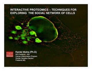
INTERACTIVE PROTEOMICS – TECHNIQUES FOR
EXPLORING THE SOCIAL NETWORK OF CELLS


   Karobi Moitra (Ph.D)
   NCI Frederick , NIH
   Cancer Inflammation Program
   Human Genetics Section
   Frederick MD.
Proteome: the entire protein complement of a cell , tissue, or organism


                      The proteome is DYNAMIC !
Why is the proteome dynamic ?


Proteins can be:

Synthesized

Modified by post-translational modifications

Undergo translocations within the cell

Degraded
Examination of the proteome of a cell is like
taking a “snapshot” of the protein environment
              at any given time
FREEZE FRAME………
Proteome: the entire protein complement of a cell , tissue, or organism


 Proteomics: is the large scale characterization of this
                       proteome
Why do we need to characterize the proteome?

• To obtain a more global and integrated view of biology by
studying all the proteins of a cell rather than each one
individually


• To create a complete three-dimensional (3-D) map of the cell
indicating where proteins are located
The Google Earth Analogy


                           Global Protein
                           Landscapes
Different areas of study are now grouped under
the rubric of proteomics include:


Protein modifications

Protein function

Protein localization

Protein-protein interactions
(Interactive proteomics)
WHAT IS INTERACTIVE PROTEOMICS OR PROTEIN –
              PROTEIN INTERACTIONS?


THE ABILITY OF A PROTEIN TO BIND OR INTERACT WITH ANOTHER PROTEIN
OR PROTEINS
 Types of protein interactions :

 Permanent interactions

 Transient interactions
From the need for more -omics came the term
                interactome
THE INTERACTOME
  COMPLETE PROTEIN INTERACTION NETWORK OF A CELL OR
  AN ORGANISM


Just as humans don’t thrive when isolated from other humans - the same can be
                             said for proteins !
WHY ???
Proteins interact with other proteins to provide :


Structural integrity to the cell (e.g., actin filaments)
Transport molecules (e.g.,Transporters)
Propagate signals (e.g., kinases)
Transcribe DNA, translate other proteins etc.
….there is no protein discovered yet that acts on its own
       without interacting with any other entity !
The entire protein complement of a cell, tissue, or organism is called
the PROTEOME


The proteome is DYNAMIC


Proteins can interact or bind with other protein(s)


This ‘social’ network is called the INTERACTOME
….in the real world you have to interact with people to learn the
                             ‘social dynamic’


       …. in the protein world you would have to know how proteins
(and other components of a cell) interact with each other in order to explore
                           the ‘cellular’ dynamic
You have been asked to find the interacting partners of
a protein named ‘C3PO’. Your first task is to find out
everything you need to know about this protein in order to
undertake this study.

Your tool is the internet, which sites you would go to and
what information might you obtain from these sites to get
the relevant background knowledge you would need to
carry out the study?
Partial List of potential websites:

www.google.com

www.ncbi.nlm.nih.gov/

http://www.ensembl.org/index.html

www.expasy.ch

http://www.expasy.org/links.html
And a lot more links from this page………
1.Clone and express the protein (C3PO) in an expression
  system of your choice

2. Optimize protein expression

3. Decide which techniques you would use to study
   protein-protein interactions
TECHNIQUES USED TO STUDY
PROTEIN-PROTEIN INTERACTIONS
A. Standard techniques to probe protein-protein interactions
Affinity purification
Mass Spectrometry
Two-Hybrid Assay
Phage Display

B. In Vivo Imaging
Fluorescence Microscopy

C. Biophysical Approaches
Protein Co-crystallization

D. Microarrays
High Density Protein Microarray

E. Computational/Bioinformatics Methods
Computer programs that simulate protein-protein interactions
Prediction of co-evolved protein pairs based on similar
phylogenetic trees
A. Standard techniques to probe protein-protein interactions


Affinity purification
Basic Principle:
Historically affinity purification
was based on a specific biological
interaction such as
enzyme-substrate.
In a broader sense it may mean
Chemical/biological affinity.

Stationary/solid phase
Dynamic/liquid phase


 Immunoprecipitation

 Immunoprecipitation (IP) is the technique of precipitating a protein antigen
 out of solution using an antibody that specifically binds to that particular
 protein.
Immunoprecipitation/coimmunoprecipitation

Basic Principle:
                                                                              B                    lysate

                                                          X        A
                                                              Y                                        B
                                                                                                                A
                                                                                                  X
                                                                                                       Y

                         B
                                    Cell lysis
           X         A              Freeze thaw
               Y                    Lysis buffer
                                    Hypotonic
                                    Mild detergent                                        ProteinA/G beads
                                    (Ripa, NP40)                          X               (binds to Fc of Ab)
                                                                  A           Y

                     Post- ip
 Run gel
 Visualize protein
 Excise band                                          elute               wash
 Digest
                                                                       Lysis buffer   A
                                X                    Low pH(change pH)
 MS                                                  SDS loading buffer
                                    Y
Disadvantage: An antibody to the specific protein of interest is required


Solution: We can tag our protein of interest with an epitope tag
Epitope Tagging :
 Antibody recognizes a specific portion of the protein - epitope.


                      Target protein    Flag tag
                                                            Anti-Flag Ab
                                                            coupled to
                                                            beads
Associated proteins


  (i)   Single Tag

         FLAG tag , c-Myc tag, GST tag, His tag etc.

  (ii) Tandem affinity purification
       TAP tag
(i) Single Tag
Attaching the Tag :

                                                           Note:
                                                           Tags can be N
                                                           terminal or C terminal
                                                           depending on where the
                                                           functional region of the
                                                           protein is located. Tagging
                                                           close to the functional region
                                                           may interfere with binding
                                                           sites.


                                  Clone into vector


                      Transfect into cells to express the protein
Histidine Tag
                                            Imidazole groups
                Imidazole                   can form a coordinate
                                            covalent bond with metals
                groups


                            Nickel column


Or
Imidazole
Transfect cells
                                                                            Controls :
 48-72hrs
                                                                            Transfection:
                                               (for peptide purification-
                                                                            Untransfected cells
  (Complex pulldown)                             antibody production        Vector transfected
                                               Also complex pulldown)       Known positive control

                                                                            Pulldown:
                                                                            Vector transfected cells
                                                                            Known positive control
  Or
  Mechanical lysis
  Freeze-thaw method


                              His-tagged protein
                              binds to Ni column


 (Low conc. to wash
  out non-specific binding)


(Compete off His tagged
Protein)
(ii) Tandem Affinity Purification :


    2 step purification :

    1.   Purify through Protein A tag on a IgG-
         sephrose column

    2.   Purify through Calmodulin binding domain
         on a Calmodulin-sepharose column


2 step purification removes a lot of the background / non-specific protein binding
Tandem affinity purification (TAP)


        TAP   TAG


                      (tobacco etch virus)
IgG-sepharose
                                                                        bead


Step 1                                              TEV cleavage
Purify protein by passing through
IgG column and elute with TEV


                                                                    IgG-sepharose
                                                                         bead

Step 2
Purify protein by passing
Through Ca + calmodulin column
And elute with EDTA


                                                                             Elute
                                                                             (EGTA)
                                                                 IgG-sepharose
                                                                        Target protein
                                                                      bead


                                    Calmodulin-sepharose beads
The TAP strategy
Evaluation of a Co-IP Captured interaction


1.Confirm that the co-precipitated protein is obtained only by the antibody against
  the target , try and use monoclonal antibodies , if using polyclonals purify
  the antibody using an affinity column containing pure target


2. Use an antibody against the co-precipitated protein to co-IP the same complex


3. Determine that the interaction takes place in the cell and not as a consequence
   of cell lysis, use co-localizatiion or mutation studies to confirm interaction.

4. Run a negative control IP with unrelated antibodies.
(i) Single Tag 
(ii) Tap tag   
(iii)Photochemical/chemical crosslinking
Photochemical / Chemical Crosslinking of Proteins


The interactions or proximity of proteins can be studied by the clever use
of crosslinking agents. Protein A and B may be quite close to each other
 in a cell and a chemical crosslinker can be used to probe the protein-protein
interaction by linking them together, disrupting the cell and detecting the
crosslinked proteins.


                                        B
                        A
Diazirine based photo crosslinking

Cells grown with photoreactive diazirine compounds

Diazirine incorporated into protein

 UV light                       A        B

 Diazirines activated and bind to interacting proteins (within a few angstroms)
Chemical Crosslinking
 • Covalently links distinct chemical functional groups & can detect both stable and
   transient interactions


 • If 2 proteins physically interact with each other they can be covalently crosslinked


 Crude cellular extract + crosslinking agent (maleimides -SH reactive groups
              would form disulphide bonds between proteins )


           IP
                                              A        B

  Recover complexes


  Cleave with DTT, BME which would break disulphide bonds

Example : SMCC, succinimidal trans -4 (maleimidemethyl) cyclohexane-1- carboxylate
(a)   Epitope tagging
(b)   Chemical cross-linking
(c)   TAP tag approach.
You have your putative protein -complex of interest

how would you identify the individual proteins that make up

                      this complex ?
SCHEMATIC DIAGRAM OF PROTEIN IDENTIFICATION


(can ID only 50-60 aa)
MASS SPECTROMETERY


Protein structural information : peptide mass
                                 amino acid sequences

Type and location of post-translational modifications
Experimental Design


Immuno-precipitation


Examine complexes by SDS-PAGE


Mass Spectrometry
LC- TANDEM MS


In-gel digestion
with trypsin (K/R)


Extract peptides


                     MALDI-TOF/TOF
PROTEIN IDENTIFICATION BY PEPTIDE MAPPING
   (MALDI-TOF)


MALDI-TOF
Matrix assisted laser desorption ionisation- time of flight

Soft ionisation technique suitable for fragile biomolecules like peptides
Basic Principle of Mass Spectrometry


How it works :


                                       The amount of deflection for a
                                       sideways force depends on the
                                       Mass of the ball (acceleration constant)

                                        Acceleration - known
                                        Force - known
                                        Mass - can be calculated
                                        Force= mass x acceleration
Peptides + matrix (matrix protects peptides from the direct laser beam
                    and help absorption of laser energy)
Spotted onto a target plate

Ionised by laser beam (charge needed for deflection by electric field)
Ionised particles enter flight tube

Charged peptides move to other side of tube according to mass

Peptides hit the detector and time of flight (TOF) is recorded (to calculate speed)


                                                 Opposite charge


                                                  (speed)

                                                  (known force)
The computer generates a mass spectrum, with each peak representing the
mass to charge ratio (m/z) as a function of the % relative intensity (abundance)
of the detected peptide
The list of experimental peptide masses is compared against the theoretical
tryptic digest of every protein in a protein database.
When the experimental data matches the theoretical, the protein is identified.
A probability based scoring system is used for the search, indicating the
‘hit’ is not a random event.
PROTEIN IDENTIFICATION BY TANDEM MASS SPECTOMETRY (MS/MS)


   If the protein cannot be identifed via the peptide mass profile (eg the
   protein may not be listed in any database) then Tandem (MS/MS) may
   be used to obtain an amino acid sequence.


Q1- 1st mass analyser (quadrupole) isolates peptide ion of interest

Q2- Collision chamber peptide ion collides with neutral gas molecules (helium,nitrogen
    or argon) and fragments into smaller pieces

Q3- 2nd analyser (TOF) leads to detector which gives a product profile (aa sequence)
  Fragments the peptides into the smallest length to ID short sequences
A. Standard techniques to probe protein-protein interactions
Affinity purification 
Mass Spectrometry 
Two-Hybrid Assay
Phage Display

B. In Vivo Imaging
Fluorescence Microscopy

C. Biophysical Approaches
Protein Co-crystallization

D. Microarrays
High Density Protein Microarray

E. Computational/Bioinformatics Methods
Computer programs that simulate protein-protein interactions
Prediction of co-evolved protein pairs based on similar
phylogenetic trees
Yeast 2-hybrid assay


• Test the association of two specific proteins that
  are believed to interact on the basis of other
  criteria.
• Define domains or amino acids that are critical for the
  interactions of two proteins that are known to interact
• Screen libraries for proteins that interact with a
  specific protein.
Activating
Basic Principle of 2- Hybrid Assays                 domain
                                          Binding
                                          domain
The basic premise of a 2- hybrid assay
is that a prey protein is detected with
the help of a bait protein.

A transcription factor is split into 2
parts a DNA binding domain - BD and
an activation domain AD.


The BD is engineered to bind to the
bait and the AD is engineered to
bind to the prey.


Only if the bait and the prey protein
interact will the transcription factor
come together and transcribe a reporter
gene.
Probing Protein-Protein Interactions
           with the Yeast 2-Hybrid Assay


DNA Binding Domain   Protein X   +   Activation Domain          Protein Y


                                                                        YES    NO
                                                         HIS3           HIS+   his-

                                                         lacZ           Blue   white

                                                         ADE2           Whit    red
Split-Ubiquitin Membrane Yeast Two-Hybrid System


Drawbacks of typical Y2H necessitated the split-ubiquitin Y2H

1.Hybrid proteins are directed towards the nucleus so proteins that fold
  incorrectly in nucleus are excluded from the method (integral membrane
  proteins).

2. Interactions dependent on post-translational modifications ( in ER) won’t
   take place.

3. Interactions mediated by the amino-terminus may not work because the
   transcription factor domain blocks accessibility.
Split-Ubiquitin Membrane Yeast Two-Hybrid System
1.   Contains 2 fragments of ubiquitin brought
     together upon interaction of the
     2 proteins.
                                                     Prey   Bait
2.   The bait protein X is fused to the C-term of
     ubiquitin (Cub) followed by a TF

3.   The prey protein Y is fused to N-term of          Y    X
     ubiquitin (NubG)

4.   The 2 plasmids are introduced into yeast
     L40 strain.                                                   Transcription factor

5.   Interaction of X and Y leads to the
     assembly of ubiquitin and the proteolytic
     release of transcription factor (by ubiquitin
     proteases).

6.   The transcription factor activates the 2
     reporter genes lacZ and His3 so
     the interactions can be monitored by
     growing yeast in histidine deficient
     media or by performing an X-gal test for
     the expression of beta galactosidase.
A. Standard techniques to probe protein-protein interactions
Affinity purification 
Mass Spectrometry 
Two-Hybrid Assay 
Phage Display

B. In Vivo Imaging
Fluorescence Microscopy

C. Biophysical Approaches
Protein Co-crystallization

D. Microarrays
High Density Protein Microarray

E. Computational/Bioinformatics Methods
Computer programs that simulate protein-protein interactions
Prediction of co-evolved protein pairs based on similar
phylogenetic trees
PHAGE DISPLAY

Basic Principle:
In phage display new genetic material is inserted into a phage gene and
the bacteria process the new gene so that a protein/peptide is made and
exposed on the phage surface (due to a tag which only expresses on the cell surface).


A population of bacteriophages display hundreds/millions of protein -
one protein per phage.This is called a phage display library.
This library can be exposed to an immobilized target protein and some members will
bind to the target. The immobilized target is then washed to remove non/loose binding
phages. The DNA of phages that bind can be sequenced to identify the gene/protein.
B. IN VIVO IMAGING

Fluorescence Microscopy
Basic Principle

Fluorescent molecules are irradiated
with high intensity light.

When these molecules absorb a photon of
light an electron is boosted up to a higher
energy orbit creating an excited state

When this electron returns to the ground state
a photon of light may be emitted- this is
called fluorescence.

Fluorophores have distinct excitation and
emission spectra.
How can we use fluorescence microscopy to study
protein-protein interactions?
1. FRET (Fluorescent Resonance Energy Transfer)


2. BRET (Bioluminescence Resonance Energy Transfer)
1. FRET (Fluorescent Resonance Energy Transfer)

Normally an excited photon returns to the ground state when a photon is emitted.
FRET results in the excitation of a nearby acceptor fluorophore which will emit a
photon when it goes back to the ground state.

The occurrence of FRET thus results in decreased donor emission and increased
acceptor emission.


  Distance is everything !
 FRET is extremely sensitive to the distance among fluorophores
 For CFP and YFP the half maximum distance or Forster radius is 49-52 angstroms
Basic Principle of FRET


                                                            475


  CFP                               YFP


One probable interaction partner is tagged with CFP the other with YFP.
If the 2 proteins interact emission will be observed at 530nm instead of 475nm
Problems of FRET

1. Tissues and cells may be damaged by excitation light

2. Some tissues like the retina and most plant tissues are photoresponsive

3. Photobleaching, autofluorescence or diect excitation of the acceptor
   fluorophore may occur.
2. BRET (Bioluminescence Resonance Energy Transfer)

 In BRET the excitation light is replaced by bioluminescent light from
 Renilla luciferase (RLUC)
 The luciferase is activated by its substrate coelenterazine.


     Bioluminescent light
C. Biophysical Approach

  1. Protein Co-crystallization
Protein Co-crystallization


 -grow crystal

 -collect diffraction data

 -calculate electron density

 -trace chain & generate structure
SNL1 & YPD1 co-crystals


                          SNL1 and YPD1 are part of a phosphorelay
                          signal transduction pathway in yeast.
                          these protein can be co-crystalized by using
                          a phosphate analog (BeF3) which bind
                          covalently and activates respose regulator
                          proteins.

                                                     (Chooback . L 2003)
Co-Crystal structure of YPD1 and SLN1


YPD - Yellow
SLN1- Cyan


                                                       (Xu et. al 2003)
D. Microarrays

High Density Protein Microarray

Microspots of the captured molecules
are immobilized in rows and columns
on a solid support

They are exposed to samples containing
the corresponding binding molecules.


                                         Proteins interact
Readout systems based on fluorescence, chemiluminescence,mass
spectometry, radioactivity etc. can be used to detect
complex formation
E. Computational/Bioinformatics Methods

1.Computer programs that simulate protein-protein interactions
  ie Docking programs like Autodock.


2. Prediction of co-evolved protein pairs based on similar phylogenetic trees

This method involves using a sequence search tool such as BLAST for finding
homologues of a pair of proteins, then building multiple sequence alignments with
alignment tools such as Clustal. From these multiple sequence alignments,
phylogenetic distance matrices are calculated for each protein in the hypothesized
interacting pair. If the matrices are sufficiently similar they are deemed likely to
interact.
A. Standard techniques to probe protein-protein interactions
Affinity purification   
Mass Spectrometry       
Two-Hybrid Assay        
Phage Display           

B. In Vivo Imaging
Fluorescence Microscopy 

C. Biophysical Approaches
Protein Co-crystallization 

D. Microarrays
High Density Protein Microarray 

E. Computational/Bioinformatics Methods
Computer programs that simulate protein-protein interactions 
Prediction of co-evolved protein pairs based on similar 
phylogenetic trees
Humans do not thrive when isolated from others - the same can be
said for proteins !
TO MARGUERITE

 by: Matthew Arnold (1822-1888)

‘Yes in the sea of life enisled,
With echoing straits between us thrown.
Dotting the shoreless watery wild,
We mortal millions live alone.
The islands feel the enclasping flow,
And then their endless bounds they know…’
Think-Pair- Share Activity


In light of what you have learnt about proteins today do you think
that a protein can function on its own isolated from other proteins?

For or Against
‘O then a longing like despair
Is to their farthest caverns sent!
For surely once, they feel, we were
Parts of a single continent.
Now round us spreads the watery plain--
O might our marges meet again!’

Weitere ähnliche Inhalte

Was ist angesagt?

Immunoprecipitation Presentation
Immunoprecipitation PresentationImmunoprecipitation Presentation
Immunoprecipitation PresentationVibhutiSardana1
 
Protein 3 d structure prediction
Protein 3 d structure predictionProtein 3 d structure prediction
Protein 3 d structure predictionSamvartika Majumdar
 
Chou fasman algorithm for protein structure prediction
Chou fasman algorithm for protein structure predictionChou fasman algorithm for protein structure prediction
Chou fasman algorithm for protein structure predictionRoshan Karunarathna
 
BITS: Basics of sequence analysis
BITS: Basics of sequence analysisBITS: Basics of sequence analysis
BITS: Basics of sequence analysisBITS
 
Comparative genomics @ sid 2003 format
Comparative genomics @ sid 2003 formatComparative genomics @ sid 2003 format
Comparative genomics @ sid 2003 formatsidjena70
 
Protein Microarrays
Protein MicroarraysProtein Microarrays
Protein MicroarraysRomilMistry
 
Immunoprecipitation - Overview and technical tips
Immunoprecipitation - Overview and technical tipsImmunoprecipitation - Overview and technical tips
Immunoprecipitation - Overview and technical tipsProteintech Group
 
Whole genome sequencing of bacteria & analysis
Whole genome sequencing of bacteria & analysisWhole genome sequencing of bacteria & analysis
Whole genome sequencing of bacteria & analysisdrelamuruganvet
 
Uni prot presentation
Uni prot presentationUni prot presentation
Uni prot presentationRida Khalid
 
Proteomics 2 d gel, mass spectrometry, maldi tof
Proteomics 2 d gel, mass spectrometry, maldi tofProteomics 2 d gel, mass spectrometry, maldi tof
Proteomics 2 d gel, mass spectrometry, maldi tofnirvarna gr
 
De novo genome assembly - T.Seemann - IMB winter school 2016 - brisbane, au ...
De novo genome assembly  - T.Seemann - IMB winter school 2016 - brisbane, au ...De novo genome assembly  - T.Seemann - IMB winter school 2016 - brisbane, au ...
De novo genome assembly - T.Seemann - IMB winter school 2016 - brisbane, au ...Torsten Seemann
 
Pathways and genomes databases in bioinformatics
Pathways and genomes databases in bioinformaticsPathways and genomes databases in bioinformatics
Pathways and genomes databases in bioinformaticssarwat bashir
 

Was ist angesagt? (20)

Immunoprecipitation Presentation
Immunoprecipitation PresentationImmunoprecipitation Presentation
Immunoprecipitation Presentation
 
Protein 3 d structure prediction
Protein 3 d structure predictionProtein 3 d structure prediction
Protein 3 d structure prediction
 
IMMUNOPRECIPITATION
IMMUNOPRECIPITATIONIMMUNOPRECIPITATION
IMMUNOPRECIPITATION
 
Chou fasman algorithm for protein structure prediction
Chou fasman algorithm for protein structure predictionChou fasman algorithm for protein structure prediction
Chou fasman algorithm for protein structure prediction
 
Protein protein interaction
Protein protein interactionProtein protein interaction
Protein protein interaction
 
BITS: Basics of sequence analysis
BITS: Basics of sequence analysisBITS: Basics of sequence analysis
BITS: Basics of sequence analysis
 
Comparative genomics @ sid 2003 format
Comparative genomics @ sid 2003 formatComparative genomics @ sid 2003 format
Comparative genomics @ sid 2003 format
 
2D-PAGE & DIGE
2D-PAGE & DIGE2D-PAGE & DIGE
2D-PAGE & DIGE
 
PROTEIN MICROARRAYS
PROTEIN MICROARRAYSPROTEIN MICROARRAYS
PROTEIN MICROARRAYS
 
IMMUNOTECHNIQUES (immunoprecipitation)
IMMUNOTECHNIQUES (immunoprecipitation)IMMUNOTECHNIQUES (immunoprecipitation)
IMMUNOTECHNIQUES (immunoprecipitation)
 
Protein Microarrays
Protein MicroarraysProtein Microarrays
Protein Microarrays
 
Immunoprecipitation - Overview and technical tips
Immunoprecipitation - Overview and technical tipsImmunoprecipitation - Overview and technical tips
Immunoprecipitation - Overview and technical tips
 
Whole genome sequencing of bacteria & analysis
Whole genome sequencing of bacteria & analysisWhole genome sequencing of bacteria & analysis
Whole genome sequencing of bacteria & analysis
 
Prosite
PrositeProsite
Prosite
 
blast bioinformatics
blast bioinformaticsblast bioinformatics
blast bioinformatics
 
Uni prot presentation
Uni prot presentationUni prot presentation
Uni prot presentation
 
Proteomics 2 d gel, mass spectrometry, maldi tof
Proteomics 2 d gel, mass spectrometry, maldi tofProteomics 2 d gel, mass spectrometry, maldi tof
Proteomics 2 d gel, mass spectrometry, maldi tof
 
De novo genome assembly - T.Seemann - IMB winter school 2016 - brisbane, au ...
De novo genome assembly  - T.Seemann - IMB winter school 2016 - brisbane, au ...De novo genome assembly  - T.Seemann - IMB winter school 2016 - brisbane, au ...
De novo genome assembly - T.Seemann - IMB winter school 2016 - brisbane, au ...
 
Pathways and genomes databases in bioinformatics
Pathways and genomes databases in bioinformaticsPathways and genomes databases in bioinformatics
Pathways and genomes databases in bioinformatics
 
Genome assembly
Genome assemblyGenome assembly
Genome assembly
 

Ähnlich wie Km Usuhs Class Improved1.2 Copy

Ag presentation & role of mhc somu
Ag presentation & role of mhc somuAg presentation & role of mhc somu
Ag presentation & role of mhc somuSomshekhar Hogtapur
 
(050407)protein chip
(050407)protein chip(050407)protein chip
(050407)protein chipnamvgta
 
Publications citing schott_nexterion_microarray_products_20100929
Publications citing schott_nexterion_microarray_products_20100929Publications citing schott_nexterion_microarray_products_20100929
Publications citing schott_nexterion_microarray_products_20100929SCHOTT
 
Bls 107 general microbiology
Bls 107 general microbiologyBls 107 general microbiology
Bls 107 general microbiologyBruno Mmassy
 
Bls 107 general microbiology
Bls 107 general microbiologyBls 107 general microbiology
Bls 107 general microbiologyBruno Mmassy
 
Antigen Processing
Antigen ProcessingAntigen Processing
Antigen Processingraj kumar
 
Ibidi membrane fusion
Ibidi membrane fusion Ibidi membrane fusion
Ibidi membrane fusion Sasha Thomas
 
Recombinant Antibody Overview I - Creative Biolabs
Recombinant Antibody Overview I - Creative BiolabsRecombinant Antibody Overview I - Creative Biolabs
Recombinant Antibody Overview I - Creative BiolabsCreative-Biolabs
 
L08 protein metabolism
L08 protein metabolismL08 protein metabolism
L08 protein metabolismMUBOSScz
 
Stephen Friend Fanconi Anemia Research Fund 2012-01-21
Stephen Friend Fanconi Anemia Research Fund 2012-01-21Stephen Friend Fanconi Anemia Research Fund 2012-01-21
Stephen Friend Fanconi Anemia Research Fund 2012-01-21Sage Base
 
67 biotechnology2008 3
67 biotechnology2008 367 biotechnology2008 3
67 biotechnology2008 3sbarkanic
 
27 ch06cellmembranediffusion2008
27 ch06cellmembranediffusion200827 ch06cellmembranediffusion2008
27 ch06cellmembranediffusion2008sbarkanic
 
Cho Hcp Immunogenicity Iciw Bailey Kellog
Cho Hcp Immunogenicity Iciw Bailey KellogCho Hcp Immunogenicity Iciw Bailey Kellog
Cho Hcp Immunogenicity Iciw Bailey KellogAnnie De Groot
 
Computational Synthetic Biology
Computational Synthetic BiologyComputational Synthetic Biology
Computational Synthetic BiologyNatalio Krasnogor
 
Biopharma Solution
Biopharma SolutionBiopharma Solution
Biopharma SolutionSujin Prabhu
 

Ähnlich wie Km Usuhs Class Improved1.2 Copy (20)

Ag presentation & role of mhc somu
Ag presentation & role of mhc somuAg presentation & role of mhc somu
Ag presentation & role of mhc somu
 
(050407)protein chip
(050407)protein chip(050407)protein chip
(050407)protein chip
 
Publications citing schott_nexterion_microarray_products_20100929
Publications citing schott_nexterion_microarray_products_20100929Publications citing schott_nexterion_microarray_products_20100929
Publications citing schott_nexterion_microarray_products_20100929
 
Bls 107 general microbiology
Bls 107 general microbiologyBls 107 general microbiology
Bls 107 general microbiology
 
Bls 107 general microbiology
Bls 107 general microbiologyBls 107 general microbiology
Bls 107 general microbiology
 
Antigen Processing
Antigen ProcessingAntigen Processing
Antigen Processing
 
Ibidi membrane fusion
Ibidi membrane fusion Ibidi membrane fusion
Ibidi membrane fusion
 
Protein protein interactions
Protein protein interactionsProtein protein interactions
Protein protein interactions
 
Recombinant Antibody Overview I - Creative Biolabs
Recombinant Antibody Overview I - Creative BiolabsRecombinant Antibody Overview I - Creative Biolabs
Recombinant Antibody Overview I - Creative Biolabs
 
L08 protein metabolism
L08 protein metabolismL08 protein metabolism
L08 protein metabolism
 
Stephen Friend Fanconi Anemia Research Fund 2012-01-21
Stephen Friend Fanconi Anemia Research Fund 2012-01-21Stephen Friend Fanconi Anemia Research Fund 2012-01-21
Stephen Friend Fanconi Anemia Research Fund 2012-01-21
 
67 biotechnology2008 3
67 biotechnology2008 367 biotechnology2008 3
67 biotechnology2008 3
 
27 ch06cellmembranediffusion2008
27 ch06cellmembranediffusion200827 ch06cellmembranediffusion2008
27 ch06cellmembranediffusion2008
 
Bioinformatica t7-protein structure
Bioinformatica t7-protein structureBioinformatica t7-protein structure
Bioinformatica t7-protein structure
 
15 arrays
15 arrays15 arrays
15 arrays
 
Cho Hcp Immunogenicity Iciw Bailey Kellog
Cho Hcp Immunogenicity Iciw Bailey KellogCho Hcp Immunogenicity Iciw Bailey Kellog
Cho Hcp Immunogenicity Iciw Bailey Kellog
 
Computational Synthetic Biology
Computational Synthetic BiologyComputational Synthetic Biology
Computational Synthetic Biology
 
Biopharmaceutical
BiopharmaceuticalBiopharmaceutical
Biopharmaceutical
 
Biopharma Solution
Biopharma SolutionBiopharma Solution
Biopharma Solution
 
Protein purification
Protein purificationProtein purification
Protein purification
 

Mehr von Karobi Moitra CFD, MS, PhD

The story of us: The journey of man by karobi moitra
The story of us:  The journey of man by karobi moitra The story of us:  The journey of man by karobi moitra
The story of us: The journey of man by karobi moitra Karobi Moitra CFD, MS, PhD
 
Storytelling in STEM poster lily conference 2014 Karobi Moitra
Storytelling in STEM poster lily conference 2014 Karobi MoitraStorytelling in STEM poster lily conference 2014 Karobi Moitra
Storytelling in STEM poster lily conference 2014 Karobi MoitraKarobi Moitra CFD, MS, PhD
 
Story telling in the classroom lilly conference presentation 2014 1hr session...
Story telling in the classroom lilly conference presentation 2014 1hr session...Story telling in the classroom lilly conference presentation 2014 1hr session...
Story telling in the classroom lilly conference presentation 2014 1hr session...Karobi Moitra CFD, MS, PhD
 
I teach, what's your superpower? ccr fyi 2013
I teach, what's your superpower? ccr fyi 2013 I teach, what's your superpower? ccr fyi 2013
I teach, what's your superpower? ccr fyi 2013 Karobi Moitra CFD, MS, PhD
 
Biology of cancer lectures 1 to 5 revision Karobi Moitra
Biology of cancer lectures 1 to 5 revision Karobi MoitraBiology of cancer lectures 1 to 5 revision Karobi Moitra
Biology of cancer lectures 1 to 5 revision Karobi MoitraKarobi Moitra CFD, MS, PhD
 
Biology of cancer lecture 5 cell cycle Karobi Moitra
Biology of cancer lecture 5 cell cycle Karobi MoitraBiology of cancer lecture 5 cell cycle Karobi Moitra
Biology of cancer lecture 5 cell cycle Karobi MoitraKarobi Moitra CFD, MS, PhD
 
Boc Lecture 3 the multistep nature of cancer, Karobi Moitra
Boc Lecture 3 the multistep nature of cancer, Karobi MoitraBoc Lecture 3 the multistep nature of cancer, Karobi Moitra
Boc Lecture 3 the multistep nature of cancer, Karobi MoitraKarobi Moitra CFD, MS, PhD
 
Biology of cancer, lecture 2 tumor viruses,oncogenes,tsgs
Biology of cancer, lecture 2 tumor viruses,oncogenes,tsgs Biology of cancer, lecture 2 tumor viruses,oncogenes,tsgs
Biology of cancer, lecture 2 tumor viruses,oncogenes,tsgs Karobi Moitra CFD, MS, PhD
 

Mehr von Karobi Moitra CFD, MS, PhD (18)

The story of DNA modified by Karobi Moitra
The story of DNA modified by Karobi MoitraThe story of DNA modified by Karobi Moitra
The story of DNA modified by Karobi Moitra
 
The story of us: The journey of man by karobi moitra
The story of us:  The journey of man by karobi moitra The story of us:  The journey of man by karobi moitra
The story of us: The journey of man by karobi moitra
 
Storytelling in STEM poster lily conference 2014 Karobi Moitra
Storytelling in STEM poster lily conference 2014 Karobi MoitraStorytelling in STEM poster lily conference 2014 Karobi Moitra
Storytelling in STEM poster lily conference 2014 Karobi Moitra
 
Story telling in the classroom lilly conference presentation 2014 1hr session...
Story telling in the classroom lilly conference presentation 2014 1hr session...Story telling in the classroom lilly conference presentation 2014 1hr session...
Story telling in the classroom lilly conference presentation 2014 1hr session...
 
I teach, what's your superpower? ccr fyi 2013
I teach, what's your superpower? ccr fyi 2013 I teach, what's your superpower? ccr fyi 2013
I teach, what's your superpower? ccr fyi 2013
 
Evolutionary Game Theory
Evolutionary Game TheoryEvolutionary Game Theory
Evolutionary Game Theory
 
The panda's roots
The panda's roots The panda's roots
The panda's roots
 
Darwin's voyage final
Darwin's  voyage finalDarwin's  voyage final
Darwin's voyage final
 
BOC lecture 7 cell death
BOC lecture 7 cell deathBOC lecture 7 cell death
BOC lecture 7 cell death
 
BOC 134 lecture - 7 Cell Death
BOC 134 lecture - 7 Cell DeathBOC 134 lecture - 7 Cell Death
BOC 134 lecture - 7 Cell Death
 
Biology of cancer lectures 1 to 5 revision Karobi Moitra
Biology of cancer lectures 1 to 5 revision Karobi MoitraBiology of cancer lectures 1 to 5 revision Karobi Moitra
Biology of cancer lectures 1 to 5 revision Karobi Moitra
 
Biology of cancer lecture 5 cell cycle Karobi Moitra
Biology of cancer lecture 5 cell cycle Karobi MoitraBiology of cancer lecture 5 cell cycle Karobi Moitra
Biology of cancer lecture 5 cell cycle Karobi Moitra
 
Boc Lecture 4 the hallmarks of cancer km
Boc Lecture 4 the hallmarks of cancer  kmBoc Lecture 4 the hallmarks of cancer  km
Boc Lecture 4 the hallmarks of cancer km
 
Boc Lecture 3 the multistep nature of cancer, Karobi Moitra
Boc Lecture 3 the multistep nature of cancer, Karobi MoitraBoc Lecture 3 the multistep nature of cancer, Karobi Moitra
Boc Lecture 3 the multistep nature of cancer, Karobi Moitra
 
Biology of cancer, lecture 2 tumor viruses,oncogenes,tsgs
Biology of cancer, lecture 2 tumor viruses,oncogenes,tsgs Biology of cancer, lecture 2 tumor viruses,oncogenes,tsgs
Biology of cancer, lecture 2 tumor viruses,oncogenes,tsgs
 
Biology of cancer 134 L1 karobi moitra
Biology of cancer 134 L1  karobi moitraBiology of cancer 134 L1  karobi moitra
Biology of cancer 134 L1 karobi moitra
 
Karobi Moitra 4 H Class Whale Of A Tale
Karobi Moitra  4 H Class Whale Of A TaleKarobi Moitra  4 H Class Whale Of A Tale
Karobi Moitra 4 H Class Whale Of A Tale
 
Tears Of The Cheetah
Tears Of The CheetahTears Of The Cheetah
Tears Of The Cheetah
 

Km Usuhs Class Improved1.2 Copy

  • 1. INTERACTIVE PROTEOMICS – TECHNIQUES FOR EXPLORING THE SOCIAL NETWORK OF CELLS Karobi Moitra (Ph.D) NCI Frederick , NIH Cancer Inflammation Program Human Genetics Section Frederick MD.
  • 2. Proteome: the entire protein complement of a cell , tissue, or organism The proteome is DYNAMIC !
  • 3. Why is the proteome dynamic ? Proteins can be: Synthesized Modified by post-translational modifications Undergo translocations within the cell Degraded
  • 4. Examination of the proteome of a cell is like taking a “snapshot” of the protein environment at any given time
  • 6. Proteome: the entire protein complement of a cell , tissue, or organism Proteomics: is the large scale characterization of this proteome
  • 7. Why do we need to characterize the proteome? • To obtain a more global and integrated view of biology by studying all the proteins of a cell rather than each one individually • To create a complete three-dimensional (3-D) map of the cell indicating where proteins are located
  • 8. The Google Earth Analogy Global Protein Landscapes
  • 9.
  • 10. Different areas of study are now grouped under the rubric of proteomics include: Protein modifications Protein function Protein localization Protein-protein interactions (Interactive proteomics)
  • 11. WHAT IS INTERACTIVE PROTEOMICS OR PROTEIN – PROTEIN INTERACTIONS? THE ABILITY OF A PROTEIN TO BIND OR INTERACT WITH ANOTHER PROTEIN OR PROTEINS Types of protein interactions : Permanent interactions Transient interactions
  • 12. From the need for more -omics came the term interactome
  • 13. THE INTERACTOME COMPLETE PROTEIN INTERACTION NETWORK OF A CELL OR AN ORGANISM Just as humans don’t thrive when isolated from other humans - the same can be said for proteins !
  • 15. Proteins interact with other proteins to provide : Structural integrity to the cell (e.g., actin filaments) Transport molecules (e.g.,Transporters) Propagate signals (e.g., kinases) Transcribe DNA, translate other proteins etc.
  • 16. ….there is no protein discovered yet that acts on its own without interacting with any other entity !
  • 17. The entire protein complement of a cell, tissue, or organism is called the PROTEOME The proteome is DYNAMIC Proteins can interact or bind with other protein(s) This ‘social’ network is called the INTERACTOME
  • 18. ….in the real world you have to interact with people to learn the ‘social dynamic’ …. in the protein world you would have to know how proteins (and other components of a cell) interact with each other in order to explore the ‘cellular’ dynamic
  • 19. You have been asked to find the interacting partners of a protein named ‘C3PO’. Your first task is to find out everything you need to know about this protein in order to undertake this study. Your tool is the internet, which sites you would go to and what information might you obtain from these sites to get the relevant background knowledge you would need to carry out the study?
  • 20. Partial List of potential websites: www.google.com www.ncbi.nlm.nih.gov/ http://www.ensembl.org/index.html www.expasy.ch http://www.expasy.org/links.html
  • 21. And a lot more links from this page………
  • 22. 1.Clone and express the protein (C3PO) in an expression system of your choice 2. Optimize protein expression 3. Decide which techniques you would use to study protein-protein interactions
  • 23. TECHNIQUES USED TO STUDY PROTEIN-PROTEIN INTERACTIONS
  • 24. A. Standard techniques to probe protein-protein interactions Affinity purification Mass Spectrometry Two-Hybrid Assay Phage Display B. In Vivo Imaging Fluorescence Microscopy C. Biophysical Approaches Protein Co-crystallization D. Microarrays High Density Protein Microarray E. Computational/Bioinformatics Methods Computer programs that simulate protein-protein interactions Prediction of co-evolved protein pairs based on similar phylogenetic trees
  • 25. A. Standard techniques to probe protein-protein interactions Affinity purification Basic Principle: Historically affinity purification was based on a specific biological interaction such as enzyme-substrate. In a broader sense it may mean Chemical/biological affinity. Stationary/solid phase Dynamic/liquid phase Immunoprecipitation Immunoprecipitation (IP) is the technique of precipitating a protein antigen out of solution using an antibody that specifically binds to that particular protein.
  • 26. Immunoprecipitation/coimmunoprecipitation Basic Principle: B lysate X A Y B A X Y B Cell lysis X A Freeze thaw Y Lysis buffer Hypotonic Mild detergent ProteinA/G beads (Ripa, NP40) X (binds to Fc of Ab) A Y Post- ip Run gel Visualize protein Excise band elute wash Digest Lysis buffer A X Low pH(change pH) MS SDS loading buffer Y
  • 27. Disadvantage: An antibody to the specific protein of interest is required Solution: We can tag our protein of interest with an epitope tag
  • 28. Epitope Tagging : Antibody recognizes a specific portion of the protein - epitope. Target protein Flag tag Anti-Flag Ab coupled to beads Associated proteins (i) Single Tag FLAG tag , c-Myc tag, GST tag, His tag etc. (ii) Tandem affinity purification TAP tag
  • 30. Attaching the Tag : Note: Tags can be N terminal or C terminal depending on where the functional region of the protein is located. Tagging close to the functional region may interfere with binding sites. Clone into vector Transfect into cells to express the protein
  • 31. Histidine Tag Imidazole groups Imidazole can form a coordinate covalent bond with metals groups Nickel column Or Imidazole
  • 32. Transfect cells Controls : 48-72hrs Transfection: (for peptide purification- Untransfected cells (Complex pulldown) antibody production Vector transfected Also complex pulldown) Known positive control Pulldown: Vector transfected cells Known positive control Or Mechanical lysis Freeze-thaw method His-tagged protein binds to Ni column (Low conc. to wash out non-specific binding) (Compete off His tagged Protein)
  • 33.
  • 34. (ii) Tandem Affinity Purification : 2 step purification : 1. Purify through Protein A tag on a IgG- sephrose column 2. Purify through Calmodulin binding domain on a Calmodulin-sepharose column 2 step purification removes a lot of the background / non-specific protein binding
  • 35. Tandem affinity purification (TAP) TAP TAG (tobacco etch virus)
  • 36. IgG-sepharose bead Step 1 TEV cleavage Purify protein by passing through IgG column and elute with TEV IgG-sepharose bead Step 2 Purify protein by passing Through Ca + calmodulin column And elute with EDTA Elute (EGTA) IgG-sepharose Target protein bead Calmodulin-sepharose beads
  • 38. Evaluation of a Co-IP Captured interaction 1.Confirm that the co-precipitated protein is obtained only by the antibody against the target , try and use monoclonal antibodies , if using polyclonals purify the antibody using an affinity column containing pure target 2. Use an antibody against the co-precipitated protein to co-IP the same complex 3. Determine that the interaction takes place in the cell and not as a consequence of cell lysis, use co-localizatiion or mutation studies to confirm interaction. 4. Run a negative control IP with unrelated antibodies.
  • 39. (i) Single Tag  (ii) Tap tag  (iii)Photochemical/chemical crosslinking
  • 40. Photochemical / Chemical Crosslinking of Proteins The interactions or proximity of proteins can be studied by the clever use of crosslinking agents. Protein A and B may be quite close to each other in a cell and a chemical crosslinker can be used to probe the protein-protein interaction by linking them together, disrupting the cell and detecting the crosslinked proteins. B A
  • 41. Diazirine based photo crosslinking Cells grown with photoreactive diazirine compounds Diazirine incorporated into protein UV light A B Diazirines activated and bind to interacting proteins (within a few angstroms)
  • 42. Chemical Crosslinking • Covalently links distinct chemical functional groups & can detect both stable and transient interactions • If 2 proteins physically interact with each other they can be covalently crosslinked Crude cellular extract + crosslinking agent (maleimides -SH reactive groups would form disulphide bonds between proteins ) IP A B Recover complexes Cleave with DTT, BME which would break disulphide bonds Example : SMCC, succinimidal trans -4 (maleimidemethyl) cyclohexane-1- carboxylate
  • 43. (a) Epitope tagging (b) Chemical cross-linking (c) TAP tag approach.
  • 44. You have your putative protein -complex of interest how would you identify the individual proteins that make up this complex ?
  • 45. SCHEMATIC DIAGRAM OF PROTEIN IDENTIFICATION (can ID only 50-60 aa)
  • 46. MASS SPECTROMETERY Protein structural information : peptide mass amino acid sequences Type and location of post-translational modifications
  • 48. LC- TANDEM MS In-gel digestion with trypsin (K/R) Extract peptides MALDI-TOF/TOF
  • 49. PROTEIN IDENTIFICATION BY PEPTIDE MAPPING (MALDI-TOF) MALDI-TOF Matrix assisted laser desorption ionisation- time of flight Soft ionisation technique suitable for fragile biomolecules like peptides
  • 50. Basic Principle of Mass Spectrometry How it works : The amount of deflection for a sideways force depends on the Mass of the ball (acceleration constant) Acceleration - known Force - known Mass - can be calculated Force= mass x acceleration
  • 51. Peptides + matrix (matrix protects peptides from the direct laser beam and help absorption of laser energy) Spotted onto a target plate Ionised by laser beam (charge needed for deflection by electric field) Ionised particles enter flight tube Charged peptides move to other side of tube according to mass Peptides hit the detector and time of flight (TOF) is recorded (to calculate speed) Opposite charge (speed) (known force)
  • 52.
  • 53. The computer generates a mass spectrum, with each peak representing the mass to charge ratio (m/z) as a function of the % relative intensity (abundance) of the detected peptide The list of experimental peptide masses is compared against the theoretical tryptic digest of every protein in a protein database. When the experimental data matches the theoretical, the protein is identified. A probability based scoring system is used for the search, indicating the ‘hit’ is not a random event.
  • 54. PROTEIN IDENTIFICATION BY TANDEM MASS SPECTOMETRY (MS/MS) If the protein cannot be identifed via the peptide mass profile (eg the protein may not be listed in any database) then Tandem (MS/MS) may be used to obtain an amino acid sequence. Q1- 1st mass analyser (quadrupole) isolates peptide ion of interest Q2- Collision chamber peptide ion collides with neutral gas molecules (helium,nitrogen or argon) and fragments into smaller pieces Q3- 2nd analyser (TOF) leads to detector which gives a product profile (aa sequence) Fragments the peptides into the smallest length to ID short sequences
  • 55. A. Standard techniques to probe protein-protein interactions Affinity purification  Mass Spectrometry  Two-Hybrid Assay Phage Display B. In Vivo Imaging Fluorescence Microscopy C. Biophysical Approaches Protein Co-crystallization D. Microarrays High Density Protein Microarray E. Computational/Bioinformatics Methods Computer programs that simulate protein-protein interactions Prediction of co-evolved protein pairs based on similar phylogenetic trees
  • 56. Yeast 2-hybrid assay • Test the association of two specific proteins that are believed to interact on the basis of other criteria. • Define domains or amino acids that are critical for the interactions of two proteins that are known to interact • Screen libraries for proteins that interact with a specific protein.
  • 57.
  • 58. Activating Basic Principle of 2- Hybrid Assays domain Binding domain The basic premise of a 2- hybrid assay is that a prey protein is detected with the help of a bait protein. A transcription factor is split into 2 parts a DNA binding domain - BD and an activation domain AD. The BD is engineered to bind to the bait and the AD is engineered to bind to the prey. Only if the bait and the prey protein interact will the transcription factor come together and transcribe a reporter gene.
  • 59. Probing Protein-Protein Interactions with the Yeast 2-Hybrid Assay DNA Binding Domain Protein X + Activation Domain Protein Y YES NO HIS3 HIS+ his- lacZ Blue white ADE2 Whit red
  • 60. Split-Ubiquitin Membrane Yeast Two-Hybrid System Drawbacks of typical Y2H necessitated the split-ubiquitin Y2H 1.Hybrid proteins are directed towards the nucleus so proteins that fold incorrectly in nucleus are excluded from the method (integral membrane proteins). 2. Interactions dependent on post-translational modifications ( in ER) won’t take place. 3. Interactions mediated by the amino-terminus may not work because the transcription factor domain blocks accessibility.
  • 61. Split-Ubiquitin Membrane Yeast Two-Hybrid System 1. Contains 2 fragments of ubiquitin brought together upon interaction of the 2 proteins. Prey Bait 2. The bait protein X is fused to the C-term of ubiquitin (Cub) followed by a TF 3. The prey protein Y is fused to N-term of Y X ubiquitin (NubG) 4. The 2 plasmids are introduced into yeast L40 strain. Transcription factor 5. Interaction of X and Y leads to the assembly of ubiquitin and the proteolytic release of transcription factor (by ubiquitin proteases). 6. The transcription factor activates the 2 reporter genes lacZ and His3 so the interactions can be monitored by growing yeast in histidine deficient media or by performing an X-gal test for the expression of beta galactosidase.
  • 62. A. Standard techniques to probe protein-protein interactions Affinity purification  Mass Spectrometry  Two-Hybrid Assay  Phage Display B. In Vivo Imaging Fluorescence Microscopy C. Biophysical Approaches Protein Co-crystallization D. Microarrays High Density Protein Microarray E. Computational/Bioinformatics Methods Computer programs that simulate protein-protein interactions Prediction of co-evolved protein pairs based on similar phylogenetic trees
  • 63. PHAGE DISPLAY Basic Principle: In phage display new genetic material is inserted into a phage gene and the bacteria process the new gene so that a protein/peptide is made and exposed on the phage surface (due to a tag which only expresses on the cell surface). A population of bacteriophages display hundreds/millions of protein - one protein per phage.This is called a phage display library.
  • 64. This library can be exposed to an immobilized target protein and some members will bind to the target. The immobilized target is then washed to remove non/loose binding phages. The DNA of phages that bind can be sequenced to identify the gene/protein.
  • 65. B. IN VIVO IMAGING Fluorescence Microscopy Basic Principle Fluorescent molecules are irradiated with high intensity light. When these molecules absorb a photon of light an electron is boosted up to a higher energy orbit creating an excited state When this electron returns to the ground state a photon of light may be emitted- this is called fluorescence. Fluorophores have distinct excitation and emission spectra.
  • 66. How can we use fluorescence microscopy to study protein-protein interactions?
  • 67. 1. FRET (Fluorescent Resonance Energy Transfer) 2. BRET (Bioluminescence Resonance Energy Transfer)
  • 68. 1. FRET (Fluorescent Resonance Energy Transfer) Normally an excited photon returns to the ground state when a photon is emitted. FRET results in the excitation of a nearby acceptor fluorophore which will emit a photon when it goes back to the ground state. The occurrence of FRET thus results in decreased donor emission and increased acceptor emission. Distance is everything ! FRET is extremely sensitive to the distance among fluorophores For CFP and YFP the half maximum distance or Forster radius is 49-52 angstroms
  • 69. Basic Principle of FRET 475 CFP YFP One probable interaction partner is tagged with CFP the other with YFP. If the 2 proteins interact emission will be observed at 530nm instead of 475nm
  • 70. Problems of FRET 1. Tissues and cells may be damaged by excitation light 2. Some tissues like the retina and most plant tissues are photoresponsive 3. Photobleaching, autofluorescence or diect excitation of the acceptor fluorophore may occur.
  • 71. 2. BRET (Bioluminescence Resonance Energy Transfer) In BRET the excitation light is replaced by bioluminescent light from Renilla luciferase (RLUC) The luciferase is activated by its substrate coelenterazine. Bioluminescent light
  • 72. C. Biophysical Approach 1. Protein Co-crystallization
  • 73. Protein Co-crystallization -grow crystal -collect diffraction data -calculate electron density -trace chain & generate structure
  • 74. SNL1 & YPD1 co-crystals SNL1 and YPD1 are part of a phosphorelay signal transduction pathway in yeast. these protein can be co-crystalized by using a phosphate analog (BeF3) which bind covalently and activates respose regulator proteins. (Chooback . L 2003)
  • 75. Co-Crystal structure of YPD1 and SLN1 YPD - Yellow SLN1- Cyan (Xu et. al 2003)
  • 76. D. Microarrays High Density Protein Microarray Microspots of the captured molecules are immobilized in rows and columns on a solid support They are exposed to samples containing the corresponding binding molecules. Proteins interact
  • 77. Readout systems based on fluorescence, chemiluminescence,mass spectometry, radioactivity etc. can be used to detect complex formation
  • 78. E. Computational/Bioinformatics Methods 1.Computer programs that simulate protein-protein interactions ie Docking programs like Autodock. 2. Prediction of co-evolved protein pairs based on similar phylogenetic trees This method involves using a sequence search tool such as BLAST for finding homologues of a pair of proteins, then building multiple sequence alignments with alignment tools such as Clustal. From these multiple sequence alignments, phylogenetic distance matrices are calculated for each protein in the hypothesized interacting pair. If the matrices are sufficiently similar they are deemed likely to interact.
  • 79. A. Standard techniques to probe protein-protein interactions Affinity purification  Mass Spectrometry  Two-Hybrid Assay  Phage Display  B. In Vivo Imaging Fluorescence Microscopy  C. Biophysical Approaches Protein Co-crystallization  D. Microarrays High Density Protein Microarray  E. Computational/Bioinformatics Methods Computer programs that simulate protein-protein interactions  Prediction of co-evolved protein pairs based on similar  phylogenetic trees
  • 80. Humans do not thrive when isolated from others - the same can be said for proteins !
  • 81. TO MARGUERITE by: Matthew Arnold (1822-1888) ‘Yes in the sea of life enisled, With echoing straits between us thrown. Dotting the shoreless watery wild, We mortal millions live alone. The islands feel the enclasping flow, And then their endless bounds they know…’
  • 82. Think-Pair- Share Activity In light of what you have learnt about proteins today do you think that a protein can function on its own isolated from other proteins? For or Against
  • 83. ‘O then a longing like despair Is to their farthest caverns sent! For surely once, they feel, we were Parts of a single continent. Now round us spreads the watery plain-- O might our marges meet again!’