SlideShare ist ein Scribd-Unternehmen logo
1 von 11
Downloaden Sie, um offline zu lesen
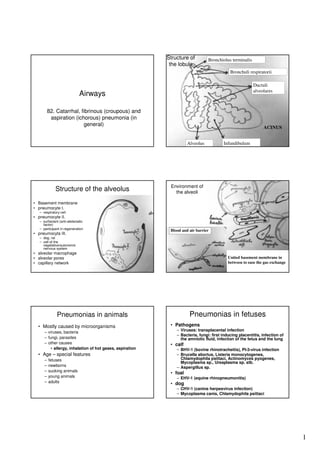
Structure of
the lobule

Bronchiolus terminalis
Bronchuli respiratorii
Ductuli
alveolares

Airways
82. Catarrhal, fibrinous (croupous) and
aspiration (ichorous) pneumonia (in
general)

ACINUS
Alveolus

Structure of the alveolus

Infundibulum

Environment of
the alveoli

• Basement membrane
• pneumocyte I.
– respiratory cell

• pneumocyte II.
– surfactant (anti-atelectatic
factor)
– participant in regeneration

• pneumocyta III.

Blood and air barrier

– dog, rat
– cell of the
vegetative/autonomic
nervous system

• alveolar macrophage
• alveolar pores
• capillary network

United basement membrane in
between to ease the gas exchange

Pneumonias in fetuses

Pneumonias in animals
• Mostly caused by microorganisms
– viruses, bacteria
– fungi, parasites
– other causes
• allergy, inhalation of hot gases, aspiration

• Age – special features
–
–
–
–
–

fetuses
newborns
sucking animals
young animals
adults

• Pathogens
– Viruses: transplacental infection
– Bacteria, fungi: first inducing placentitis, infection of
the amniotic fluid, infection of the fetus and the lung

• calf
– BHV-1 (bovine rhinotracheitis), PI-3-virus infection
– Brucella abortus, Listeria monocytogenes,
Chlamydophila psittaci, Actinomyces pyogenes,
Mycoplasma sp., Ureaplasma sp. stb.
– Aspergillus sp.

• foal
– EHV-1 (equine rhinopneumonitis)

• dog
– CHV-1 (canine herpesvirus infection)
– Mycoplasma canis, Chlamydophila psittaci

1
Pneumonia in newborns

Forms of pneumonias

(perinatal pneumonias)

• neonatal pneumonia (collective category)
– Aerogenous infection before the newborn got the
colostrum (in state of hypogammaglobulinaemia)
– inhalation of different bacteria
• calf, foal, goat
Streptococcus sp.
• piglet
Streptococcus sp., E. coli
• birds
E. coli
– Local process continues often to septicemia

• meconium aspiration syndrome
– aspiration during parturition, typical
– or obturation, or pneumonia due to pathogens

Bronchopneumonias:
Inflammation →
hyperaemia, edema →
neutrophils, macrophages
→ epithelial gaps↑,
↑
vascular permeability↑ →
↑
plasma fluid, proteins →
airspace↓
↓
Serous bronchopneumonia

Desquamative bronchopneumonia

• definition
– inflammatory process in the parenchyma of the lung often
accompanied with the lesions of the smaller or larger
bronchi

• Forms
– bronchoalveolar
• serous, desquamative, catarrhal, suppurative,
fibrinous (croupous)
– interstitial
– granulomatous
– embolic-metastatic
– aspiration
– special

Desquamative bronchopneumonia

Desquamative bronchopneumonia

2
Serous
bronchopneumonia

Normal shape, slightly enlarged,
darker, gland-like by palpation,
doesn’t crepitate when cut, clear
liquid oozes to the cut surface

Desquamative pneumonia

Catarrhal
bronchitis

Bronchoalveolar pneumonia

Fibrinous (croupous) pneumonia

(bronchopneumonia)
•
•

spreads quickly → entire lobes are affected - “Lobar pneumonia”
Development

•

Appearance

– Congestion, hemorrhage, massive exudation, fibrin accumulation
– Fibrin → pleural surface = sero-fibrinous pleuritis
– intense redness, firmer texture, “consolidation”, hepatization (red~, gray~).
– Dilation and thrombosis of lymph vessels → edema → thick septae → marbled
appearance
– Focal coagulative necrosis. “Sequestra”.
– Histology

• massive exudation → fluid obliterates airspace. Neutrophils,
macrophages. Fibroblasts
•

Stages
– initial phase

Extension:

• stadium incrementi s. hyperaemicum
– phase of hepatisation (std. hepatisationis)

lobules - lobular
lobes - lobal
focal - pseudolobular

• red hepatisation (std. h. rubrae)
• grey hepatisation (std. h. griseae)
• yellow hepatisation (std. h. flavae)
– phase of resolution (std. resolutionis)

•

Croupous (fibrinous) pneumonia

nasal discharge: mucoid or mucopurulent (P. m., B. b., Str.)

Croupous (fibrinous) pneumonia

3
Croupous pneumonia

Croupous pneumonia – stadium incrementi

Croupous pneumonia - hepatisation

Grey hepatisation

Croupous pneumonia – hemorrhagic character

Healing of the croupous pneumonia
• restitutio ad integrum
– if the integrity of the tissue structure is maintained
– freed enzymes of the degenerated neutrophils cause
– fibrinolysis
• partially absorbtion (lymph vessels)
• partially expectoration
– epithel layer of the acini regenerates

• restitutio cum defectu
– connective tissue proliferation from the wall of the
respiratory ducts
– carnificatio pulmonum

4
Croupous pneumonia

Croupous pneumonia

• Due to direct (thrombotic) effect of the pathogen
– Smaller or larger necrotic foci
– Sometime hemorrhagic character
– Secondary ichorous pneumonia on the affected areas

• Cause of death:
– Suffocation or heart failure
– Following croupous pneumonia
• Metastatic foci – focal necrotizing-purulent-ichorous
pneumonia
• Allergic reaction in horses
– Hemorrhagic purpura, septic pododermatitis,
uveitis, urticaria

Pathology of the croupous pneumonia
•
•
•
•
•
•

shape maintained
enlarged
marbled
liver-like (in stages of hepatisation)
No crepitation on cutting!
cut surface
– mottled, homogenous
– dry, uneven, granular

Aspiration pneumonia

Aspiration pneumonia
• Different aspirated substances
– amniotic fluid
– milk, saliva, food, drinking water, blood
– fluid used for gastric lavage, contrast medium used for
filling the GI tract
– eructated or vomited gastric content

• Consequences are different according to
– what type of material and what amount is aspirated
– where did the substance go
– if there where any putrefactive bacteria present

Aspiration pneumonia

5
Aspiration pneumonia

Aspiration pneumonia
• Putrid bronchitis
– Putrefactive bacteria colonize in the mucous membrane
of the bronchi
– the mucous membrane necrotizes
– accompanied with putrid smell, the tissue debris
becomes soft fluid
– all the layers of the bronchi are affected
– if there is enough time, the lungs also become affected
• usually the animal dies because of the absorbed
toxins

Ichorous pneumonia

Ichorous pneumonia

• Way of infection:
– Aerogenous infection (aspiration)
• putrid bronchitis first, than spreading to the lungs
• starts from the airways
– lympho-haematogenous infection may occur

• disseminated foci
• from the blood vessels of the interstitium
• Due to putrefactive bacteria
– coagulation necrosis
– secondary softening in the necrotized areas
• putrid smell, histolytic tissue debris causing intoxication
(sapraemia)
– acute ichorous abscess, ichorous cavern (cavity)
• Slow, delayed demarcation

Interstitial pneumonia
• Majority of the lesions
– In the interstitium

83. Interstitial, suppurative
and embolic pneumonias
(in general)

• Most typical lesions:
– acute, subacute, chronic proliferative inflammation
– besides proliferative processes immunpathological
processes, accumulation of immune cells

• Appearance:
– intralobular (interalveolar)
– interlobular – between the lobules
– peribronchial – around the small bronchi

6
Intralobular (interalveolar) interstitial pneumonia

Mild proliferation of the alveolar interstitium
Intralobular (interalveolar) interstitial pneumonia

Severe proliferation of the alveolar interstitium

Immunpathologic process

Intralobular (interalveolar) interstitial pneumonia

Intralobular (interalveolar) interstitial pneumonia

Peribronchial interstitial pneumonia

Interstitial pneumonia
• Proliferative processes in the alveoli
– Epithelisation
• pneumocyta II.
– Pseudoepithelisation
• alveolar macrophages

• Age of the process
– acute – histiocyte and macrophage proliferation
– subacute – pronounced cell proliferation
– chronic – severe fibrosis

7
Pathogen has an effect inside the alveoli
Intralobular (interalveolar) interstitial pneumonia

Epithelisation

Pseudoepithelisation

Chronic interstitial pneumonia

Suppurative pneumonia
(pneumonia purulenta)

Purulent bronchopneumonia

• Typically embolic or metastatic
• If hematogenous:
– Purulent process somewhere in the body → distributed
abscesses in the lung
(no surrounding inflammation)

• If bronchiogenous:
– First catarrhal or croupous pneumonia occurs and
secondary purulent foci are formed

• Pathogens:
– cattle/pig → Arcanobacterium (Actinomyces) pyogenes
– rabbit → Staphylococcus aureus
– horse → Streptococcus equi, Rhodococcus equi

8
Purulent bronchopneumonia

Pneumonia with granulome formation
• Accompaying other disease
– Lympho-hematogenous metastasis

• Embolic-metastatic pneumonia
• Possible nodules
– necrotic foci
– purulent (ichorous) abcesses
– ichorous foci

• Appearance:
– solitaer focus (smaller - larger)
– evenly disseminated foci
– foci of the same age / or prolonged form

Necrotic pneumonia

Sequestrum formation after necrotic pneumonia

Mass of necrotic lung parenchyma
seprated from viable lung tissue by
purulent exudate and usually
encased in a fibrous capsule

9
Embolic-metastatic pneumonia

Chronic purulent abscess formation
(cut surface)

Port of entry

Purulent-ichorous pneumonia

Granulomatous pneumonia

Granulomatous pneumonia

10
Granulomatous pneumonia

Locus minoris resistanciae

Special pneumonias
• Actinobacillus pleuropneumonia
– serous-hemorrhagic-proliferative-necrotizing
• in hours, death in 1-2 days
• pronounced macrophage proliferation
• extended demarcation
– sero-fibrinous pleuritis
• perifocal edema

• Anthrax
– Aerogenous infection (piglet inhales the spores)
– Serous, hemorrhagic, necrotizing pneumonia
– Spore reaches the deep areas of the lung, starts to
proliferate → rapid course

11

Weitere ähnliche Inhalte

Was ist angesagt?

Adeno tonsillitis dr.p.k arthikeyan, 11.07.16
Adeno tonsillitis dr.p.k arthikeyan, 11.07.16Adeno tonsillitis dr.p.k arthikeyan, 11.07.16
Adeno tonsillitis dr.p.k arthikeyan, 11.07.16ophthalmgmcri
 
Respiratory diseases of horses
Respiratory diseases of horsesRespiratory diseases of horses
Respiratory diseases of horsesAjith Y
 
TUNDIKERI -ACUTE AND CHRONIC TONSILITIS
TUNDIKERI -ACUTE AND CHRONIC TONSILITISTUNDIKERI -ACUTE AND CHRONIC TONSILITIS
TUNDIKERI -ACUTE AND CHRONIC TONSILITISDr. Raju
 
6 bronchiectasis
6 bronchiectasis6 bronchiectasis
6 bronchiectasisPNK SINGH
 
Chronic tonsillitis
Chronic tonsillitisChronic tonsillitis
Chronic tonsillitisKapil Dhital
 
Tonsillitis slideshare for medical students
Tonsillitis slideshare for medical students Tonsillitis slideshare for medical students
Tonsillitis slideshare for medical students NehaNupur8
 
FlashPath - Lung - Hypersensitivity Pneumonitis - Extrinsic Allergic Alveolitis
FlashPath - Lung - Hypersensitivity Pneumonitis - Extrinsic Allergic AlveolitisFlashPath - Lung - Hypersensitivity Pneumonitis - Extrinsic Allergic Alveolitis
FlashPath - Lung - Hypersensitivity Pneumonitis - Extrinsic Allergic AlveolitisHazem Ali
 

Was ist angesagt? (19)

Acute tonsillitis
Acute tonsillitisAcute tonsillitis
Acute tonsillitis
 
Chronic tonsillitis
Chronic  tonsillitisChronic  tonsillitis
Chronic tonsillitis
 
Tonsillitis
TonsillitisTonsillitis
Tonsillitis
 
Pathology of respiratory system
Pathology of respiratory systemPathology of respiratory system
Pathology of respiratory system
 
Adeno tonsillitis dr.p.k arthikeyan, 11.07.16
Adeno tonsillitis dr.p.k arthikeyan, 11.07.16Adeno tonsillitis dr.p.k arthikeyan, 11.07.16
Adeno tonsillitis dr.p.k arthikeyan, 11.07.16
 
Acute tonsillitis
Acute tonsillitisAcute tonsillitis
Acute tonsillitis
 
Tonsils And Adenoids
Tonsils And  AdenoidsTonsils And  Adenoids
Tonsils And Adenoids
 
Acute tonsilitis
Acute tonsilitis Acute tonsilitis
Acute tonsilitis
 
Respiratory diseases of horses
Respiratory diseases of horsesRespiratory diseases of horses
Respiratory diseases of horses
 
Bronchitis
BronchitisBronchitis
Bronchitis
 
TUNDIKERI -ACUTE AND CHRONIC TONSILITIS
TUNDIKERI -ACUTE AND CHRONIC TONSILITISTUNDIKERI -ACUTE AND CHRONIC TONSILITIS
TUNDIKERI -ACUTE AND CHRONIC TONSILITIS
 
Mcqs on lung abscess
Mcqs on lung abscessMcqs on lung abscess
Mcqs on lung abscess
 
6 bronchiectasis
6 bronchiectasis6 bronchiectasis
6 bronchiectasis
 
Chronic tonsillitis
Chronic tonsillitisChronic tonsillitis
Chronic tonsillitis
 
Tonsillitis slideshare for medical students
Tonsillitis slideshare for medical students Tonsillitis slideshare for medical students
Tonsillitis slideshare for medical students
 
Lung pathology
Lung pathologyLung pathology
Lung pathology
 
Tonsil
TonsilTonsil
Tonsil
 
FlashPath - Lung - Hypersensitivity Pneumonitis - Extrinsic Allergic Alveolitis
FlashPath - Lung - Hypersensitivity Pneumonitis - Extrinsic Allergic AlveolitisFlashPath - Lung - Hypersensitivity Pneumonitis - Extrinsic Allergic Alveolitis
FlashPath - Lung - Hypersensitivity Pneumonitis - Extrinsic Allergic Alveolitis
 
Tonsillitis
TonsillitisTonsillitis
Tonsillitis
 

Ähnlich wie Structure and types of pneumonia

FlashPath - Lung - Pulmonary Interstitial Emphysema
FlashPath - Lung - Pulmonary Interstitial  EmphysemaFlashPath - Lung - Pulmonary Interstitial  Emphysema
FlashPath - Lung - Pulmonary Interstitial EmphysemaHazem Ali
 
diseases of pharynx.pptx
diseases of pharynx.pptxdiseases of pharynx.pptx
diseases of pharynx.pptxEmanZayed17
 
Pulmonary Infections
Pulmonary InfectionsPulmonary Infections
Pulmonary InfectionsPrasad CSBR
 
Pulmonary pathology
Pulmonary pathologyPulmonary pathology
Pulmonary pathologyGhie Santos
 
Worst is best good no one new..............
Worst is best good no one new..............Worst is best good no one new..............
Worst is best good no one new..............HarishankarSharma27
 
Lungs abscess and bronchitis
Lungs abscess and bronchitisLungs abscess and bronchitis
Lungs abscess and bronchitisChanak Trikhatri
 
Wheezing and noisy breathing seminar
Wheezing and noisy breathing seminarWheezing and noisy breathing seminar
Wheezing and noisy breathing seminarNaqib Bajuri
 
16. Gen Path of Helminthic and Protozoal Infections -II .pptx
16. Gen Path of Helminthic and Protozoal Infections -II .pptx16. Gen Path of Helminthic and Protozoal Infections -II .pptx
16. Gen Path of Helminthic and Protozoal Infections -II .pptxVasan37
 
Yersinia pasteurella fransicella
Yersinia pasteurella fransicellaYersinia pasteurella fransicella
Yersinia pasteurella fransicellaRiyaz Sheriff
 
Helminthic (Parasitic) Lung infections
Helminthic (Parasitic)  Lung infections Helminthic (Parasitic)  Lung infections
Helminthic (Parasitic) Lung infections Nanditha Mukesh
 
Group a presentation 20th feb 2012
Group a presentation 20th feb 2012Group a presentation 20th feb 2012
Group a presentation 20th feb 2012Sana Anwari
 

Ähnlich wie Structure and types of pneumonia (20)

FlashPath - Lung - Pulmonary Interstitial Emphysema
FlashPath - Lung - Pulmonary Interstitial  EmphysemaFlashPath - Lung - Pulmonary Interstitial  Emphysema
FlashPath - Lung - Pulmonary Interstitial Emphysema
 
Pneumonia in ruminants.pptx
Pneumonia in ruminants.pptxPneumonia in ruminants.pptx
Pneumonia in ruminants.pptx
 
Rsd
RsdRsd
Rsd
 
diseases of pharynx.pptx
diseases of pharynx.pptxdiseases of pharynx.pptx
diseases of pharynx.pptx
 
Nasal polyp
Nasal polypNasal polyp
Nasal polyp
 
Nasal polyposis
Nasal polyposisNasal polyposis
Nasal polyposis
 
Pulmonary Infections
Pulmonary InfectionsPulmonary Infections
Pulmonary Infections
 
Pulmonary pathology
Pulmonary pathologyPulmonary pathology
Pulmonary pathology
 
Worst is best good no one new..............
Worst is best good no one new..............Worst is best good no one new..............
Worst is best good no one new..............
 
Diseases of tonsils and adenoids
Diseases of tonsils and adenoidsDiseases of tonsils and adenoids
Diseases of tonsils and adenoids
 
Lungs abscess and bronchitis
Lungs abscess and bronchitisLungs abscess and bronchitis
Lungs abscess and bronchitis
 
Lower respiratory disorder
Lower respiratory disorderLower respiratory disorder
Lower respiratory disorder
 
Wheezing and noisy breathing seminar
Wheezing and noisy breathing seminarWheezing and noisy breathing seminar
Wheezing and noisy breathing seminar
 
Sinusitis.pptx
Sinusitis.pptxSinusitis.pptx
Sinusitis.pptx
 
16. Gen Path of Helminthic and Protozoal Infections -II .pptx
16. Gen Path of Helminthic and Protozoal Infections -II .pptx16. Gen Path of Helminthic and Protozoal Infections -II .pptx
16. Gen Path of Helminthic and Protozoal Infections -II .pptx
 
Yersinia pasteurella fransicella
Yersinia pasteurella fransicellaYersinia pasteurella fransicella
Yersinia pasteurella fransicella
 
PNEUMONIA.pptx
PNEUMONIA.pptxPNEUMONIA.pptx
PNEUMONIA.pptx
 
Helminthic (Parasitic) Lung infections
Helminthic (Parasitic)  Lung infections Helminthic (Parasitic)  Lung infections
Helminthic (Parasitic) Lung infections
 
Nasal polyposis
Nasal polyposis Nasal polyposis
Nasal polyposis
 
Group a presentation 20th feb 2012
Group a presentation 20th feb 2012Group a presentation 20th feb 2012
Group a presentation 20th feb 2012
 

Mehr von kantemur

Tumor cell morphology
Tumor cell morphologyTumor cell morphology
Tumor cell morphologykantemur
 
Inflammation adel-1
Inflammation adel-1Inflammation adel-1
Inflammation adel-1kantemur
 
History taking
History takingHistory taking
History takingkantemur
 
Introduction to auscultation
Introduction to auscultationIntroduction to auscultation
Introduction to auscultationkantemur
 
Lung exambates
Lung exambatesLung exambates
Lung exambateskantemur
 
Spinal cord reflexes
Spinal cord reflexesSpinal cord reflexes
Spinal cord reflexeskantemur
 

Mehr von kantemur (7)

Tumor cell morphology
Tumor cell morphologyTumor cell morphology
Tumor cell morphology
 
Inflammation adel-1
Inflammation adel-1Inflammation adel-1
Inflammation adel-1
 
Neoplasia
NeoplasiaNeoplasia
Neoplasia
 
History taking
History takingHistory taking
History taking
 
Introduction to auscultation
Introduction to auscultationIntroduction to auscultation
Introduction to auscultation
 
Lung exambates
Lung exambatesLung exambates
Lung exambates
 
Spinal cord reflexes
Spinal cord reflexesSpinal cord reflexes
Spinal cord reflexes
 

Structure and types of pneumonia

  • 1. Structure of the lobule Bronchiolus terminalis Bronchuli respiratorii Ductuli alveolares Airways 82. Catarrhal, fibrinous (croupous) and aspiration (ichorous) pneumonia (in general) ACINUS Alveolus Structure of the alveolus Infundibulum Environment of the alveoli • Basement membrane • pneumocyte I. – respiratory cell • pneumocyte II. – surfactant (anti-atelectatic factor) – participant in regeneration • pneumocyta III. Blood and air barrier – dog, rat – cell of the vegetative/autonomic nervous system • alveolar macrophage • alveolar pores • capillary network United basement membrane in between to ease the gas exchange Pneumonias in fetuses Pneumonias in animals • Mostly caused by microorganisms – viruses, bacteria – fungi, parasites – other causes • allergy, inhalation of hot gases, aspiration • Age – special features – – – – – fetuses newborns sucking animals young animals adults • Pathogens – Viruses: transplacental infection – Bacteria, fungi: first inducing placentitis, infection of the amniotic fluid, infection of the fetus and the lung • calf – BHV-1 (bovine rhinotracheitis), PI-3-virus infection – Brucella abortus, Listeria monocytogenes, Chlamydophila psittaci, Actinomyces pyogenes, Mycoplasma sp., Ureaplasma sp. stb. – Aspergillus sp. • foal – EHV-1 (equine rhinopneumonitis) • dog – CHV-1 (canine herpesvirus infection) – Mycoplasma canis, Chlamydophila psittaci 1
  • 2. Pneumonia in newborns Forms of pneumonias (perinatal pneumonias) • neonatal pneumonia (collective category) – Aerogenous infection before the newborn got the colostrum (in state of hypogammaglobulinaemia) – inhalation of different bacteria • calf, foal, goat Streptococcus sp. • piglet Streptococcus sp., E. coli • birds E. coli – Local process continues often to septicemia • meconium aspiration syndrome – aspiration during parturition, typical – or obturation, or pneumonia due to pathogens Bronchopneumonias: Inflammation → hyperaemia, edema → neutrophils, macrophages → epithelial gaps↑, ↑ vascular permeability↑ → ↑ plasma fluid, proteins → airspace↓ ↓ Serous bronchopneumonia Desquamative bronchopneumonia • definition – inflammatory process in the parenchyma of the lung often accompanied with the lesions of the smaller or larger bronchi • Forms – bronchoalveolar • serous, desquamative, catarrhal, suppurative, fibrinous (croupous) – interstitial – granulomatous – embolic-metastatic – aspiration – special Desquamative bronchopneumonia Desquamative bronchopneumonia 2
  • 3. Serous bronchopneumonia Normal shape, slightly enlarged, darker, gland-like by palpation, doesn’t crepitate when cut, clear liquid oozes to the cut surface Desquamative pneumonia Catarrhal bronchitis Bronchoalveolar pneumonia Fibrinous (croupous) pneumonia (bronchopneumonia) • • spreads quickly → entire lobes are affected - “Lobar pneumonia” Development • Appearance – Congestion, hemorrhage, massive exudation, fibrin accumulation – Fibrin → pleural surface = sero-fibrinous pleuritis – intense redness, firmer texture, “consolidation”, hepatization (red~, gray~). – Dilation and thrombosis of lymph vessels → edema → thick septae → marbled appearance – Focal coagulative necrosis. “Sequestra”. – Histology • massive exudation → fluid obliterates airspace. Neutrophils, macrophages. Fibroblasts • Stages – initial phase Extension: • stadium incrementi s. hyperaemicum – phase of hepatisation (std. hepatisationis) lobules - lobular lobes - lobal focal - pseudolobular • red hepatisation (std. h. rubrae) • grey hepatisation (std. h. griseae) • yellow hepatisation (std. h. flavae) – phase of resolution (std. resolutionis) • Croupous (fibrinous) pneumonia nasal discharge: mucoid or mucopurulent (P. m., B. b., Str.) Croupous (fibrinous) pneumonia 3
  • 4. Croupous pneumonia Croupous pneumonia – stadium incrementi Croupous pneumonia - hepatisation Grey hepatisation Croupous pneumonia – hemorrhagic character Healing of the croupous pneumonia • restitutio ad integrum – if the integrity of the tissue structure is maintained – freed enzymes of the degenerated neutrophils cause – fibrinolysis • partially absorbtion (lymph vessels) • partially expectoration – epithel layer of the acini regenerates • restitutio cum defectu – connective tissue proliferation from the wall of the respiratory ducts – carnificatio pulmonum 4
  • 5. Croupous pneumonia Croupous pneumonia • Due to direct (thrombotic) effect of the pathogen – Smaller or larger necrotic foci – Sometime hemorrhagic character – Secondary ichorous pneumonia on the affected areas • Cause of death: – Suffocation or heart failure – Following croupous pneumonia • Metastatic foci – focal necrotizing-purulent-ichorous pneumonia • Allergic reaction in horses – Hemorrhagic purpura, septic pododermatitis, uveitis, urticaria Pathology of the croupous pneumonia • • • • • • shape maintained enlarged marbled liver-like (in stages of hepatisation) No crepitation on cutting! cut surface – mottled, homogenous – dry, uneven, granular Aspiration pneumonia Aspiration pneumonia • Different aspirated substances – amniotic fluid – milk, saliva, food, drinking water, blood – fluid used for gastric lavage, contrast medium used for filling the GI tract – eructated or vomited gastric content • Consequences are different according to – what type of material and what amount is aspirated – where did the substance go – if there where any putrefactive bacteria present Aspiration pneumonia 5
  • 6. Aspiration pneumonia Aspiration pneumonia • Putrid bronchitis – Putrefactive bacteria colonize in the mucous membrane of the bronchi – the mucous membrane necrotizes – accompanied with putrid smell, the tissue debris becomes soft fluid – all the layers of the bronchi are affected – if there is enough time, the lungs also become affected • usually the animal dies because of the absorbed toxins Ichorous pneumonia Ichorous pneumonia • Way of infection: – Aerogenous infection (aspiration) • putrid bronchitis first, than spreading to the lungs • starts from the airways – lympho-haematogenous infection may occur • disseminated foci • from the blood vessels of the interstitium • Due to putrefactive bacteria – coagulation necrosis – secondary softening in the necrotized areas • putrid smell, histolytic tissue debris causing intoxication (sapraemia) – acute ichorous abscess, ichorous cavern (cavity) • Slow, delayed demarcation Interstitial pneumonia • Majority of the lesions – In the interstitium 83. Interstitial, suppurative and embolic pneumonias (in general) • Most typical lesions: – acute, subacute, chronic proliferative inflammation – besides proliferative processes immunpathological processes, accumulation of immune cells • Appearance: – intralobular (interalveolar) – interlobular – between the lobules – peribronchial – around the small bronchi 6
  • 7. Intralobular (interalveolar) interstitial pneumonia Mild proliferation of the alveolar interstitium Intralobular (interalveolar) interstitial pneumonia Severe proliferation of the alveolar interstitium Immunpathologic process Intralobular (interalveolar) interstitial pneumonia Intralobular (interalveolar) interstitial pneumonia Peribronchial interstitial pneumonia Interstitial pneumonia • Proliferative processes in the alveoli – Epithelisation • pneumocyta II. – Pseudoepithelisation • alveolar macrophages • Age of the process – acute – histiocyte and macrophage proliferation – subacute – pronounced cell proliferation – chronic – severe fibrosis 7
  • 8. Pathogen has an effect inside the alveoli Intralobular (interalveolar) interstitial pneumonia Epithelisation Pseudoepithelisation Chronic interstitial pneumonia Suppurative pneumonia (pneumonia purulenta) Purulent bronchopneumonia • Typically embolic or metastatic • If hematogenous: – Purulent process somewhere in the body → distributed abscesses in the lung (no surrounding inflammation) • If bronchiogenous: – First catarrhal or croupous pneumonia occurs and secondary purulent foci are formed • Pathogens: – cattle/pig → Arcanobacterium (Actinomyces) pyogenes – rabbit → Staphylococcus aureus – horse → Streptococcus equi, Rhodococcus equi 8
  • 9. Purulent bronchopneumonia Pneumonia with granulome formation • Accompaying other disease – Lympho-hematogenous metastasis • Embolic-metastatic pneumonia • Possible nodules – necrotic foci – purulent (ichorous) abcesses – ichorous foci • Appearance: – solitaer focus (smaller - larger) – evenly disseminated foci – foci of the same age / or prolonged form Necrotic pneumonia Sequestrum formation after necrotic pneumonia Mass of necrotic lung parenchyma seprated from viable lung tissue by purulent exudate and usually encased in a fibrous capsule 9
  • 10. Embolic-metastatic pneumonia Chronic purulent abscess formation (cut surface) Port of entry Purulent-ichorous pneumonia Granulomatous pneumonia Granulomatous pneumonia 10
  • 11. Granulomatous pneumonia Locus minoris resistanciae Special pneumonias • Actinobacillus pleuropneumonia – serous-hemorrhagic-proliferative-necrotizing • in hours, death in 1-2 days • pronounced macrophage proliferation • extended demarcation – sero-fibrinous pleuritis • perifocal edema • Anthrax – Aerogenous infection (piglet inhales the spores) – Serous, hemorrhagic, necrotizing pneumonia – Spore reaches the deep areas of the lung, starts to proliferate → rapid course 11