SlideShare ist ein Scribd-Unternehmen logo
1 von 8
Downloaden Sie, um offline zu lesen
Cell Reports
Resource
Detection of Misfolded Ab Oligomers
for Sensitive Biochemical Diagnosis
of Alzheimer’s Disease
Natalia Salvadores,1,4 Mohammad Shahnawaz,1,4 Elio Scarpini,2 Fabrizio Tagliavini,3 and Claudio Soto1,*
1Mitchell Center for Alzheimer’s Disease and Related Brain Disorders, Department of Neurology, University of Texas Medical School
at Houston, 6431 Fannin Street, Houston, TX 77030, USA
2Neurology Unit, Universita` di Milano, Centro Dino Ferrari, Fondazione Ca’ Granda, IRCC Ospedale Policlinico, via F. Sforza 35, 20122 Milan,
Italy
3IRCCS Foundation ‘‘Carlo Besta’’ Neurological Institute, Via Celoria 11, 20133 Milan, Italy
4These authors contributed equally to this work
*Correspondence: claudio.soto@uth.tmc.edu
http://dx.doi.org/10.1016/j.celrep.2014.02.031
This is an open access article under the CC BY-NC-ND license (http://creativecommons.org/licenses/by-nc-nd/3.0/).
SUMMARY
Alzheimer’s disease (AD) diagnosis is hampered by
the lack of early, sensitive, and objective laboratory
tests. We describe a sensitive method for biochem-
ical diagnosis of AD based on specific detection of
misfolded Ab oligomers, which play a central role in
AD pathogenesis. The protein misfolding cyclic
amplification assay (Ab-PMCA), exploits the func-
tional property of Ab oligomers to seed the poly-
merization of monomeric Ab. Ab-PMCA allowed
detection of as little as 3 fmol of Ab oligomers.
Most importantly, using cerebrospinal fluid, we
were able to distinguish AD patients from control
individuals affected by a variety of other neurode-
generative disorders or nondegenerative neurolog-
ical diseases with overall sensitivity of 90% and
specificity of 92%. These findings provide the
proof-of-principle basis for developing a highly sen-
sitive and specific biochemical test for AD diagnosis.
INTRODUCTION
Alzheimer’s disease (AD) is the most common cause of dementia
in the elderly population and one of the leading causes of death
in the developed world (Hebert et al., 2003). The disease is typi-
cally characterized by a progressive amnestic disorder followed
by impairment of other cognitive functions and behavioral abnor-
malities associated with specific neuropathological changes, in
particular accumulation of protein aggregates in the form of
amyloid plaques and neurofibrillary tangles (Terry, 1994).
Although the etiology of the disease is not yet clear, compelling
evidence suggests that misfolding, oligomerization, and accu-
mulation of amyloid aggregates in the brain is the triggering fac-
tor of the pathology (Selkoe, 2000; Haass and Selkoe, 2007;
Soto, 2003). Amyloid aggregates are composed predominantly
of a 42-residue peptide called amyloid-b (Ab), which is the
product of the enzymatic processing of a larger amyloid precur-
sor protein (Selkoe, 2000). Ab misfolding and fibrillar aggregation
follow a seeding-nucleation mechanism that involves the forma-
tion of several intermediates, including soluble oligomers and
protofibrils (Caughey and Lansbury, 2003; Soto et al., 2006; Jar-
rett and Lansbury, 1993). Recent findings have shown that Ab
oligomers, rather than large amyloid fibrils, might be the culprit
of neurodegeneration in AD (Walsh and Selkoe, 2007; Haass
and Selkoe, 2007; Glabe and Kayed, 2006; Klein et al., 2004).
AD belongs to a large group of diseases associated with mis-
folding, aggregation and tissue accumulation of proteins (Soto,
2003). These diseases, termed protein misfolding disorders
(PMDs), include Parkinson’s disease, type 2 diabetes, Hunting-
ton’s disease, amyotrophic lateral sclerosis, systemic amyloid-
osis, prion diseases, and many others (Soto, 2003; Luheshi
and Dobson, 2009). In all these diseases, misfolded aggregates
composed of different proteins are formed by a similar mecha-
nism resulting in the accumulation of toxic structures that induce
cellular dysfunction and tissue damage (Caughey and Lansbury,
2003; Soto et al., 2006; Jarrett and Lansbury, 1993).
One of the major problems in AD is the lack of a widely
accepted early, sensitive, and objective laboratory diagnosis to
support neuropsychological evaluation, monitor disease pro-
gression, and identify affected individuals before they display
the clinical symptoms (Parnetti and Chiasserini, 2011; Urbanelli
et al., 2009). For diseases affecting the brain, a tissue with low
regeneration capacity, it is crucial to intervene before irreversible
neuropathological changes occur. Therefore, early diagnosis of
AD is of utmost importance. Several lines of evidence point
that the process of Ab misfolding and oligomerization begins
years or decades before the onset of clinical symptoms and sub-
stantial brain damage (Braak et al., 1999; Buchhave et al., 2012).
Recent studies have shown that Ab oligomers are naturally
secreted by cells and circulate in AD biological fluids (Gao
et al., 2010; Head et al., 2010; Walsh et al., 2002; Klyubin
et al., 2008; Georganopoulou et al., 2005; Fukumoto et al.,
2010). Thus, detection of soluble Ab oligomers might represent
the best strategy for early and specific biochemical diagnosis
of AD. The challenge of this approach is that the quantity of Ab
Cell Reports 7, 1–8, April 10, 2014 ª2014 The Authors 1
Please cite this article in press as: Salvadores et al., Detection of Misfolded Ab Oligomers for Sensitive Biochemical Diagnosis of Alzheimer’s Disease,
Cell Reports (2014), http://dx.doi.org/10.1016/j.celrep.2014.02.031
oligomers is likely very small in tissues other than the brain. An
additional difficulty for specific detection of Ab oligomers is
that their sequence and chemical structure is the same as the
native Ab protein.
Our strategy to detect misfolded oligomers is to use their
functional property of being capable of catalyzing the polymeri-
zation of the monomeric protein. For this purpose, we invented
the protein misfolding cyclic amplification (PMCA) technology in
order to achieve the ultrasensitive detection of misfolded aggre-
gates through amplification of the misfolding and aggregation
process in vitro (Saborio et al., 2001). So far, PMCA has been
applied to detect minute quantities of oligomeric misfolded prion
protein (PrPSc
) implicated in prion diseases (Morales et al.,
2012). Using PMCA, we were able to detect the equivalent of
a single particle of misfolded PrP oligomer (Saa´ et al., 2006b)
and strikingly to identify PrPSc
in the blood and urine of infected
animals at symptomatic and presymptomatic stages of the dis-
ease (Castilla et al., 2005; Saa´ et al., 2006a; Gonzalez-Romero
et al., 2008). The basis for the PMCA technology is the fact
that the process of misfolding and aggregation of Ab, PrP, and
the other proteins implicated in PMDs follow a seeding-nucle-
ation mechanism (Soto et al., 2002, 2006). In a seeded-nucle-
ated polymerization, the limiting step is the formation of stable
oligomeric seeds that, depending on the conditions, may take
a very long time to form or not occur at all. Once formed, oligo-
mers grow exponentially by recruiting and incorporating protein
monomers into the growing polymer. Addition of preformed
seeds into a solution containing the monomeric protein acceler-
ates protein misfolding and aggregation (Soto et al., 2006; Jar-
rett and Lansbury, 1993). Thus, measuring seeding activity could
be used to estimate the presence and quantity of oligomers in a
given sample. To increase the sensitivity of detection, PMCA
combines steps of growing polymers with multiplication of olig-
omeric seeds to reach an exponential increase of misfolding and
aggregation (Soto et al., 2002). In this study, we describe the im-
plementation and optimization of PMCA for highly sensitive
detection of misfolded Ab oligomers and show its application
to detect these structures in the cerebrospinal fluid (CSF) of
AD patients.
RESULTS
Cyclic Amplification of Amyloid-b Misfolding
To implement the experimental conditions for Ab-PMCA, we per-
formed studies using in-vitro-produced oligomeric seeds.
Because it is still unknown which of the different species of Ab
oligomers is most relevant for AD pathology, we decided to
work with a mixture of oligomers of different sizes generated dur-
ing the process of fibril formation. Ab oligomers were prepared
by incubation of monomeric (seed-free) synthetic Ab1-42
(10 mM) at 25
C with stirring. After 5 hr of incubation, we
observed an abundance of globular oligomers by electron micro-
scopy with only a small amount of protofibrils and fibrils
(Figure 1A). These aggregates were positive with the A11 olig-
omer-specific antibody (Kayed et al., 2003; data not shown).
After longer incubation, protofibrillar and fibrillar structures
were observed. The size of the aggregates was determined by
filtration through filters of defined pore size and western blotting
after SDS-PAGE separation. Oligomers formed by incubation for
5 hr migrated as $170 kDa SDS-resistant aggregates, with a mi-
nor band at 17 kDa (Figure 1B).
Low concentrations of seed-free Ab1-42 (2 mM) were incu-
bated at 22
C with constant shaking (100 rpm) for different times
alone or in the presence of distinct concentrations of synthetic
Ab oligomers, prepared by incubation during 5 hr as described
above. Ab aggregation was studied by the fluorescence emis-
sion of the amyloid-binding dye Thioflavin T (ThT) (LeVine,
1993; Soto et al., 1995). The peptide concentration, tempera-
ture, and pH of the buffer are critical to control the extent of
the lag phase and reproducibility among experiments. Under
these conditions, no spontaneous Ab aggregation was detect-
able during the time in which the experiment was performed
(125 hr). However, Ab aggregation was observed in the pres-
ence of 0.3–8.4 fmol of Ab oligomers (Figure 1C). To increase
the efficiency of seeding, and thus the limit of detection of Ab
oligomers, we introduced cycles of amplification, combining
phases of polymer growing with multiplication of seeds as in
the PMCA assay. For this purpose, we subjected the samples
to intermittent shaking, which has been previously shown to
dramatically accelerate the seeded conversion of recombinant
prion protein (Atarashi et al., 2008). Under these conditions,
the kinetic of Ab aggregation induced by 8,400, 300, 80, and
3 fmol of Ab oligomers was clearly faster and easily distinguish-
able from that observed in the absence of Ab seeds (Figure 1D).
This result indicates that, using the Ab-PMCA assay, we should
be able to detect as little as 3 fmol of Ab oligomers in a given
sample.
Detection of Ab Oligomers in the Cerebrospinal Fluid
of AD Patients
To study the usefulness of the Ab-PMCA assay to detect seed-
ing-competent Ab oligomers in biological fluids, we analyzed
aliquots of CSF from 50 AD patients, 39 cognitively normal indi-
viduals affected by nondegenerative neurological diseases
(NNDs), and 37 patients affected by non-AD neurodegenerative
diseases (NANDs) including other forms of dementia. The exper-
iments as well as the initial part of the analysis were done blindly,
because the investigator was unaware of which samples were
coming from AD or controls. Figure 2A shows the average
kinetics of aggregation of five representative samples from the
AD, NND, and NAND groups. The result indicates that CSF
from AD patients accelerates significantly Ab aggregation as
compared to control CSF (p  0.001). To determine the effect
of individual samples on Ab aggregation, we estimated the lag
phase (Figure 2B), defined as the time required to get a ThT fluo-
rescence larger than 40 arbitrary units (after subtraction of the
blank). This value was selected considering that it corresponds
to approximately five times the reading of the buffer alone. We
also estimated the P90, which corresponds to the extent of Ab
aggregation at 90 hr (Figure 2C). By comparing both parameters
among the experimental groups, a highly significant difference
was observed between AD and control samples from individuals
with nondegenerative neurological diseases or with non-AD
neurodegenerative diseases. No correlation was detected
between the aggregation parameters and the age of the AD pa-
tients, which indicates that the levels of the marker are not simply
2 Cell Reports 7, 1–8, April 10, 2014 ª2014 The Authors
Please cite this article in press as: Salvadores et al., Detection of Misfolded Ab Oligomers for Sensitive Biochemical Diagnosis of Alzheimer’s Disease,
Cell Reports (2014), http://dx.doi.org/10.1016/j.celrep.2014.02.031
a reflection of age but rather whether or not the patients contain
seeding-competent Ab aggregates in their CSF. Using the values
for lag phase, we calculated the sensitivity, specificity, and
predictive value of the Ab-PMCA test (Table 1). To determine
the appropriate cutoff points and the performance of the test,
we carried out a detailed statistical analysis of the receiver oper-
ating characteristics (ROC) (Figure S1). In relation to the control
group consisting of age-matched individuals with nondegenera-
tive neurological diseases, we estimated a 90.0% sensitivity and
84.2% specificity, whereas for the clinically more relevant differ-
entiation of AD from other neurodegenerative diseases including
other forms of dementia, we obtained a staggering 100% sensi-
tivity and 94.6% specificity (Table 1). If confirmed with a larger
number of patients, the ability of Ab-PMCA to distinguish
AD from other forms of neurodegenerative diseases might be
very useful in clinic. The overall sensitivity and specificity consid-
ering all control individuals was 90% and 92%, respectively
(Table 1).
To confirm that Ab-PMCA detects a seeding activity associ-
ated to Ab oligomers present in CSF, we performed immuno-
depletion experiments. The methodology for efficient immuno-
depletion of Ab oligomers was first optimized by using
synthetically prepared Ab oligomers. Incubation with Dyna-
beads conjugated with a mixture of sequence (4G8) and confor-
mational (A11) antibodies led to the complete removal of these
structures (Figure 3A). Next, application of immunodepletion to
three AD CSF samples showed that the kinetic of Ab aggrega-
tion in the Ab-PMCA reaction was comparable to that observed
in control CSF samples, and both were significantly different
from the aggregation observed with AD CSF prior to immunode-
pletion (Figure 3B). A similar result was observed when immuno-
depletion was done only using the A11 conformational antibody
(Figure 3C), which recognizes specifically oligomers (Kayed
et al., 2003). These results indicate that the seeding activity
observed in AD CSF samples was indeed associated to Ab
oligomers.
Figure 1. Detection of Synthetic Ab Oligomers by Ab-PMCA
(A) To prepare Ab oligomers, solutions of Ab1-42 at 10 mM were incubated for different times at 25
C with shaking. At various time points, samples were visualized
by electron microcopy after negative staining. At 5 hr of incubation, aggregates consist mainly of globular oligomers, whereas at 10 hr, there are mostly pro-
tofibrils, and at 24 hr, we can observe a large amount of long fibrils.
(B) Preparations of oligomers were characterized by SDS-PAGE followed by western blot with 4G8 antibody. The mixture at 5 hr of incubation was also
characterized by strong reactivity with A11 antibody and by a native size ranging from 30 to 1,000 kDa, as evaluated by filtration using filters of defined pore size.
(C) Seeding of Ab aggregation was studied incubating a solution of 2 mM seed-free Ab1-42 in 100 mM Tris-HCl (pH 7.4) in the presence of 5 mM Thioflavin T, with or
without different quantities of synthetic Ab oligomers (prepared by incubation during 5 hr, as indicated in A) with constant, but low, agitation (100 rpm) at 22
C.
(D) The same samples as in (C) were incubated with cyclic agitation (1 min stirring at 500 rpm followed by 29 min without shaking). Aggregation was measured over
time by the Thioflavin T (ThT) binding to amyloid fibrils using a plate spectrofluorometer (excitation: 435; emission: 485 nm). Graphs show the mean and SE of
three replicates. The concentration of Ab oligomers was estimated assuming an average molecular weight of 170 kDa.
Cell Reports 7, 1–8, April 10, 2014 ª2014 The Authors 3
Please cite this article in press as: Salvadores et al., Detection of Misfolded Ab Oligomers for Sensitive Biochemical Diagnosis of Alzheimer’s Disease,
Cell Reports (2014), http://dx.doi.org/10.1016/j.celrep.2014.02.031
DISCUSSION
AD is one of the most important public health problems in the
developed world. Among the ten leading causes of death, AD
is the only one that lacks effective ways to prevent, cure, or
even slow its progression. Part of the difficulties in developing
an efficient treatment for this devastating disease is the absence
of an early and objective biochemical diagnosis that will enable
to recognize patients before substantial brain damage has
occurred (Parnetti and Chiasserini, 2011; Blennow et al., 2010;
Urbanelli et al., 2009). Indeed, many of the failures in recent clin-
ical trials with compounds aimed to modify the disease progres-
sion have been attributed to the lack of early diagnosis that
prevents beginning treatment before it is too late (Galimberti
and Scarpini, 2012). Currently, diagnosis is achieved by clinical
assessment, neuroimaging, and CSF measurements of the
levels of Ab42, tau, and phospho-tau, with definitive confirmation
relying on postmortem identification of amyloid plaques and
neurofibrillary tangles in the brain (Parnetti and Chiasserini,
2011; Urbanelli et al., 2009). Recent advances in neuroimaging
using amyloid tracers offer a promising alternative to increase
accuracy of diagnosis (Klunk et al., 2004; Klunk and Mathis,
2008), but it is unclear whether these methods will identify pa-
tients before irreversible brain damage has occurred. Identifica-
tion of biomarkers that could be used for noninvasive, sensitive,
and objective detection of the disease process years or even
decades before the onset of brain abnormalities and clinical
symptoms are of utmost importance (Parnetti and Chiasserini,
2011; Blennow et al., 2010; Urbanelli et al., 2009). Although it is
widely accepted that combining the measurements of Ab1-42,
total tau, and phosphorylated tau in CSF can provide a relatively
high sensitivity and specificity for AD diagnosis (Blennow et al.,
Figure 2. Detection of Seeding Activity in
Human CSF Samples from Controls and
AD Patients by Ab-PMCA
(A) Representative aggregation curves of seed-
free Ab1-42 in the presence of CSF samples
from AD patients, people affected by non-
neurodegenerative neurological diseases (NND),
and patients suffering from neurodegenerative
diseases other than AD (NAND). The values
represent the average and SE of five different
patients, representative of the average results in
each group. For this purpose, we selected as
representative for each group, the patients in
which the kinetic parameters (lag phase and P90)
were closer to the average obtained in each group.
(B) The lag phase of Ab aggregation in Ab-PMCA
was compared for the three groups of patients.
Lag phase was defined as the time (in hours)
required to reach a ThT signal more than 40 arbi-
trary units. One sample from the NND group did
not reach a ThT signal of 40 during the duration of
the aggregation experiment. Thus, the lag phase
for this sample is 300 hr. This sample was not
included in the graph and was not utilized to
calculate sensitivity and specificity in Table 1.
(C) The extent of amyloid formation obtained after
180 Ab-PMCA cycles, i.e., 90 hr of incubation
(P90), was measured in each patient. For the graphs in (B) and (C), we show the values obtained in individual samples, which correspond to the average of
three independent experiments. Data were analyzed by one-way ANOVA, followed by the Tukey’s multiple comparison post hoc test. The differences
between AD and samples from the other two groups were highly significant with p  0.001 (***).
Table 1. Estimation of Sensitivity, Specificity, and Predictive Value for Ab-PMCA Using CSF Samples
Groups Sensitivity (%)a
Specificity (%)b
Positive Predictive Value (%)c
Negative Predictive Value (%)d
AD versus NAND 100.0 94.6 96.2 100.0
AD versus NND 90.0 84.2 88.2 86.5
AD versus controlse
90.0 92.0 88.2 93.2
For estimation of sensitivity, specificity, and predictive value, we used the results of the lag phase as shown in Figure 2B. Cutoffs were estimated by
receiver operating characteristics (ROC) curve analysis using the MedCalc software (see Figure S1).
a
Sensitivity was estimated by the formula (true positives/[true positives + false negatives] ) 3 100.
b
Specificity was estimated by the formula (true negatives/[false positives + true negatives]) 3 100.
c
Positive predictive value was estimated by the formula (true positives/[true positives + false positives]) 3 100.
d
Negative predictive value was estimated by the formula (true negatives/[true negatives + false negatives]) 3 100.
e
Controls refer to the samples from NND plus NAND.
4 Cell Reports 7, 1–8, April 10, 2014 ª2014 The Authors
Please cite this article in press as: Salvadores et al., Detection of Misfolded Ab Oligomers for Sensitive Biochemical Diagnosis of Alzheimer’s Disease,
Cell Reports (2014), http://dx.doi.org/10.1016/j.celrep.2014.02.031
2010), other CSF markers are needed that can more specifically
differentiate AD from other dementias. It is also necessary to
identify markers that correlate with the severity of dementia in
patients with AD and, more importantly, can identify the patho-
logical process earlier during the presymptomatic period of the
disease.
Recent compelling evidence indicates that the formation of
misfolded Ab oligomers might be a critical event in AD pathogen-
esis (Walsh and Selkoe, 2007; Haass and Selkoe, 2007; Glabe
and Kayed, 2006; Klein et al., 2004). Indeed, both synthetic
and natural Ab oligomers have been shown to induce apoptosis
in cell cultures at very low concentrations (Dahlgren et al., 2002;
El-Agnaf et al., 2000; Shankar et al., 2007), block long-term
potentiation in brain slice cultures (Wang et al., 2002; Walsh
et al., 2002), and impair synaptic plasticity and memory in ani-
mals (Cleary et al., 2005; Shankar et al., 2007). These findings,
in addition to the fact that Ab oligomers are small and soluble
structures, suggest that they could be circulating in biological
fluids and offer a specific marker for AD. Considering the well-
established observation that the levels of total Ab1-42 are lower
in AD than in controls, it might be surprising that, in our study, we
found that Ab oligomers are elevated in AD patients. However,
our assay measures the levels of seeding-competent Ab oligo-
mers, which are likely a very small proportion of total Ab in
CSF. A rough estimation based on the comparison with the
amplification results obtained with synthetic oligomers is that
the material we are measuring is below 100 pg/ml (expressed
as monomer). Our assay does not distinguish oligomers pro-
duced by Ab40 or Ab42; thus, in our estimation, oligomers repre-
sent a very small fraction (less than 1%) of total Ab. Therefore, it
is perfectly possible that a decline in total Ab42 coincides with an
overall increase of oligomers in AD patients. Indeed, recent
studies have shown that Ab oligomers might be present in low
quantities in CSF and that their levels are elevated in the CSF
of people affected by AD (Pitschke et al., 1998; Klyubin et al.,
2008; Georganopoulou et al., 2005; Gao et al., 2010; Santos
et al., 2012).
In this study, we adapted the PMCA technology to cyclically
amplify the process of Ab misfolding and aggregation, leading
to the highly sensitive and specific detection of soluble Ab oligo-
mers. PMCA was originally developed to replicate the misfolding
and aggregation of the prion protein implicated in prion diseases
and is now considered a major technological breakthrough that
has been instrumental to understand the prion biology and to
detect misfolded prions in various biological fluids (Morales
et al., 2012). PMCA is a cyclical process that enables exponential
increase of the detection signal in a manner analogous to the
amplification of DNA by PCR (Soto et al., 2002). PMCA takes
advantage of the fact that the process of protein misfolding
and aggregation associated with Ab, PrP, and other proteins
implicated in PMDs follows a seeding-nucleation mechanism
and is dependent on the formation and number of oligomeric
seeds present in the reaction (Soto et al., 2002, 2006). To achieve
exponential amplification, PMCA consists of cycles of incubation
for the elongation of the polymers, followed by breaking the
aggregates to multiply the number of seeds. For amplification
of prions, PMCA traditionally uses sonication as a mechanical
force to break big polymers into smaller species (Morales
et al., 2012). However, as shown by Caughey and colleagues,
sonication can be replaced by strong shaking to achieve
Figure 3. Seeding Activity in AD CSF Is
Removed by Ab Oligomers Immunodepletion
To test whether the seeding activity observed
in human AD CSF was dependent on Ab aggre-
gates, we removed these structures from CSF
samples by immunodepletion using Dynabeads
coated with a mixture of antibodies recognizing
specifically the sequence of Ab (4G8) and the
conformation of Ab oligomers (A11) (Kayed et al.,
2003).
(A) The procedure for immunodepletion was opti-
mized using synthetically prepared Ab oligomers
spiked into human CSF at quantities detectable by
western blot. Three consecutive rounds of incuba-
tion with antibody-coated Dynabeads were
sufficient to efficiently remove the Ab oligomers
observed at around 170 kDa in our western blots.
(B) Samples of AD CSF before or after immu-
nodepletion with 4G8 and A11 antibodies were used
to seed Ab aggregation in the Ab-PMCA assay.
(C) Samples of AD CSF were also depleted only with
the A11 conformational antibody and aggregation
monitored by Ab-PMCA assay. As in Figures 1 and
2, Ab aggregation was measured by ThT fluores-
cence emission at different times of incubation.
Values represent the average and SE of three
different replicates.
Cell Reports 7, 1–8, April 10, 2014 ª2014 The Authors 5
Please cite this article in press as: Salvadores et al., Detection of Misfolded Ab Oligomers for Sensitive Biochemical Diagnosis of Alzheimer’s Disease,
Cell Reports (2014), http://dx.doi.org/10.1016/j.celrep.2014.02.031
similar results using a procedure called shaking-based PMCA or
quaking-induced conversion (Atarashi et al., 2008). In adapting
PMCA for AD, we found that shaking is more compatible with
the fragile nature of Ab aggregates.
In our current setting, Ab-PMCA enables detection of as little
as 3 fmol of Ab oligomers. This level of sensitivity can be further
increased by additional development of the technology, as was
also the case in prion diseases. Indeed, in our original proof-
of-concept study using PMCA for prion detection, the increase
of sensitivity was only around 60-fold (Saborio et al., 2001),
which contrast with the striking 3 billion folds obtained after
further development (Saa´ et al., 2006b). Nevertheless, even
with the current status of the Ab-PMCA technology, we were
able to distinguish with a high degree of sensitivity and specificity
CSF samples coming from individuals diagnosed with AD,
patients affected by other neurodegenerative disorders, and
people suffering from nondegenerative neurological diseases
(Table 1). Sensitivity, specificity, and predictive value of the
Ab-PMCA assay are higher than the currently widely used ELISA
tests measuring the levels of Ab42, tau, and phospho-tau in CSF.
The sensitivity and specificity of these methods range between
80% and 90%, depending on the study (Mulder et al., 2010; Sun-
derland et al., 2003; Maddalena et al., 2003). A crucial feature for
an assay aimed for routine use in AD diagnosis is its reproduc-
ibility and repeatability. Several variables need to be carefully
controlled to maintain a good reproducibility, including Ab con-
centration, temperature, buffer composition, pH, shaking rate,
etc. It is also possible that, with different batches of Ab, the
kinetic of aggregation and the parameters most useful to
compare different group of samples may be different, but still
AD samples induce accelerated Ab aggregation compared to
controls (Figure S2). Experiments displayed in this article were
carried out blindly and independently by two different investiga-
tors at distinct times and using samples obtained from three
different locations. However, during the course of the study,
we found that an entire set of CSF samples coming from a fourth
location was not amenable to the assay, because using these
samples Ab did not aggregate at all, not even after spiking with
large concentrations of synthetic oligomers. We suspect that
preanalytical factors related to sample collection influenced
the assay. We are actively investigating what this interference
could be. Nevertheless, more work needs to be done to assess
reproducibility in a large number of samples, with distinct
reagents, equipment, and samples coming from different
locations.
Future studies should include larger populations of AD
patients and controls and direct comparisons with the diag-
nostic accuracy obtained by measuring Ab42, total tau, and
phospho-tau in CSF and by positron emission tomography
amyloid imaging with specific ligands. Also, it will be important
to investigate the presence of Ab oligomers in patients affected
by mild cognitive impairments, which is considered a precursor
for AD (Petersen et al., 2009), as well as in asymptomatic car-
riers of familial AD to analyze the utility of Ab-PMCA for pre-
symptomatic detection of AD. Longitudinal studies from indi-
vidual patients would also be needed to evaluate the
relationship between the detection of Ab oligomers and the
progression of the disease. Finally, adaptation of Ab-PMCA
to detect Ab oligomers, which may potentially be circulating
in the blood of AD patients, may offer a great opportunity for
more-routine testing.
EXPERIMENTAL PROCEDURES
Biological Samples
We used CSF samples from 50 patients with the diagnosis of probable AD as
defined by the DSM-IV and the National Institute of Neurological and Com-
municative Disorders and Stroke-Alzheimer’s Disease and Related Disorders
Association guidelines (McKhann et al., 1984) and determined using a variety
of tests, including routine medical examination; neurological evaluation;
neuropsychological assessment; MRI; and measurements of CSF levels of
Ab1-42, total tau, and phospho-tau. The mean age of AD patients at the
time of sample collection was 71.0 ± 8.1 years (range 49–84). As controls,
we used CSF from 39 patients affected by NND, including 12 cases of normal
pressure hydrocephalus, seven patients with peripheral neuropathy, seven
with diverse forms of brain tumor, two with ICTUS, one with severe cephalgia,
three with encephalitis, one with hypertension, and six with unclear diagnosis.
The mean age at which CSF samples were taken from this group of patients
was 64.6 ± 14.7 years (range 31–83). We also used as controls CSF samples
from 37 individuals affected by NAND, including ten cases of fronto-temporal
dementia (five behavioral and five language variants); six patients with
Parkinson’s disease (including four associated with dementia and one with
motor neuron disease); six with progressive supranuclear palsy; six with spino-
cerebellar ataxia (one associated with dementia); four with amyotrophic lateral
sclerosis; two with Huntington’s disease; one with mitochondria encephalo-
myopathy, lactic acidosis, and stroke-like episodes; one with Lewy body
dementia; and one with vascular dementia. The mean age at sample collection
for this group was 63.8 ± 11.1 years (range 41–80). CSF samples were
collected in polypropylene tubes following lumbar puncture at the L4/L5 or
L3/L4 interspace with atraumatic needles after one night fasting. The samples
were centrifuged at 3,000 g for 3 min at 4
C, aliquoted, and stored at À80
C
until analysis. CSF cell counts, glucose, and protein concentration were deter-
mined. Albumin was measured by rate nephelometry. To evaluate the integrity
of the blood brain barrier and the intrathecal immunoglobulin G (IgG) produc-
tion, the albumin quotient (CSF albumin/serum albumin) 3 103
and the IgG
index (CSF albumin/serum albumin)/(CSF IgG/serum IgG) were calculated
(Sellebjerg and Christiansen, 1996). The study was conducted according to
the provisions of the Helsinki Declaration and was approved by the Ethics
Committee.
Production of Synthetic Ab and Preparation of Ab Aggregates
Ab1-42 was synthesized using solid-phase N-tert-butyloxycarbonyl chemistry
at the W. Keck Facility at Yale University and purified by reverse-phase high-
performance liquid chromatography. The final product was lyophilized and
characterized by amino acid analysis and mass spectrometry. To prepare
stock solutions free of Ab aggregated seeds, we used our previously described
protocol (Morales et al., 2010), which involves dissolution of aggregates in high
pH and filtration through 30 kDa cutoff filters to remove remaining aggregates.
To prepare different types of aggregates, solutions of seed-free Ab1-42
(10 mM) were incubated for different times at 25
C in 0.1 M Tris-HCl (pH 7.4)
with agitation. This preparation contains a mixture of Ab monomers as well
as fibrils, protofibrils, and soluble oligomers in distinct proportions depending
on the incubation time. The degree of aggregation was characterized by ThT
fluorescence emission, electron microscopy after negative staining, dot blot
studies with the A11 conformational antibody (Kayed et al., 2003), and western
blot after gel electrophoresis using the 4G8 Ab antibody.
Cyclic Amplification of Ab Misfolding and Aggregation
Solutions of 2 mM aggregate-free Ab1-42 in 0.1 M Tris-HCl (pH 7.4; 200 ml total
volume) were placed in opaque 96-well plates and incubated alone or in the
presence of synthetic Ab aggregates or 40 ml of CSF aliquots. Samples were
incubated in the presence of 5 mM ThT and subjected to cyclic agitation
(1 min at 500 rpm followed by 29 min without shaking) using an Eppendorf
thermomixer, at a constant temperature of 22
C. At various time points, ThT
6 Cell Reports 7, 1–8, April 10, 2014 ª2014 The Authors
Please cite this article in press as: Salvadores et al., Detection of Misfolded Ab Oligomers for Sensitive Biochemical Diagnosis of Alzheimer’s Disease,
Cell Reports (2014), http://dx.doi.org/10.1016/j.celrep.2014.02.031
fluorescence was measured in the plates at 485 nm after excitation at 435 nm
using a plate spectrofluorometer.
Determination of Sensitivity, Specificity, and Predictive Value
The differences in the kinetic of aggregation between different samples were
evaluated by the estimation of two different kinetic parameters: the lag phase
and P90. Lag phase is defined as the time required to reach a ThT fluorescence
of 40 arbitrary units. This value was selected because it corresponds to
approximately five times the value of the buffer alone. The P90 corresponds
to the extent of aggregation (measured as ThT fluorescence) at 90 hr.
Sensitivity, specificity, and predictive value were determined using the lag
phase data, with cutoff thresholds determined by ROC curve analysis, using
the MedCalc software (version 12.2.1.0).
Statistical Analysis
The significance of the differences in Ab aggregation kinetics in the presence
of human CSF samples was analyzed by one-way ANOVA, followed by the
Tukey’s multiple comparison post hoc test. The level of significance was set
at p  0.05. Statistical tests were performed using the GraphPad Prism 5.0
software.
SUPPLEMENTAL INFORMATION
Supplemental Information includes two figures and can be found with this
article online at http://dx.doi.org/10.1016/j.celrep.2014.02.031.
AUTHOR CONTRIBUTIONS
N.S. designed the experiments, optimized the Ab-PMCA technology, carried
out many of the experiments with CSF samples, analyzed the results, and pre-
pared the figures. M.S. performed various Ab-PMCA assays and analyzed the
data. E.S. and F.T. provided some of the CSF samples and characterized
them. C.S. is the principal investigator on the project and was responsible
for coordinating research activity, analyzing the data, funding, and producing
the final version of the article.
ACKNOWLEDGMENTS
We are very grateful to Dr. Justo Garcia de Yebenes (Hospital Ramon y Cajal)
for providing us some of the CSF samples used in this study and Dr. Silvia
Suardi (‘‘Carlo Besta’’ Neurological Institute) and Dr. Daniela Galimberti (Uni-
versity of Milan) for help in collecting and characterizing CSF samples. We
would also like to thank Dr. Lisbell Estrada (Catholic University of Chile) and
Dr. Ines Moreno-Gonzalez (UTH) for valuable help in diverse aspects of this
work. This research was supported by grants from the Alzheimer’s Associa-
tion, CART Foundation, and Mitchell Foundation (to C.S.) and the Italian
Ministry of Health (JPND, project BiomarkAPD) and MIUR (Prot. RBAP11FRE9;
CUP: J41J11000330001) (to F.T.). C.S. is an inventor on several patents related
to the PMCA technology and is currently Founder, Chief Scientific Officer, and
Vice-President of Amprion Inc., a biotech company focusing on the commer-
cial exploitation of PMCA for diagnosis of neurodegenerative diseases.
Received: October 1, 2013
Revised: January 19, 2014
Accepted: February 20, 2014
Published: March 20, 2014
REFERENCES
Atarashi, R., Wilham, J.M., Christensen, L., Hughson, A.G., Moore, R.A.,
Johnson, L.M., Onwubiko, H.A., Priola, S.A., and Caughey, B. (2008). Simpli-
fied ultrasensitive prion detection by recombinant PrP conversion with
shaking. Nat. Methods 5, 211–212.
Blennow, K., Hampel, H., Weiner, M., and Zetterberg, H. (2010). Cerebrospinal
fluid and plasma biomarkers in Alzheimer disease. Nat. Rev. Neurol. 6,
131–144.
Braak, E., Griffing, K., Arai, K., Bohl, J., Bratzke, H., and Braak, H. (1999).
Neuropathology of Alzheimer’s disease: what is new since A. Alzheimer?
Eur. Arch. Psychiatry Clin. Neurosci. 249 (Suppl 3), 14–22.
Buchhave, P., Minthon, L., Zetterberg, H., Wallin, A.K., Blennow, K., and Hans-
son, O. (2012). Cerebrospinal fluid levels of b-amyloid 1-42, but not of tau, are
fully changed already 5 to 10 years before the onset of Alzheimer dementia.
Arch. Gen. Psychiatry 69, 98–106.
Castilla, J., Saa´ , P., and Soto, C. (2005). Detection of prions in blood. Nat. Med.
11, 982–985.
Caughey, B., and Lansbury, P.T. (2003). Protofibrils, pores, fibrils, and neuro-
degeneration: separating the responsible protein aggregates from the
innocent bystanders. Annu. Rev. Neurosci. 26, 267–298.
Cleary, J.P., Walsh, D.M., Hofmeister, J.J., Shankar, G.M., Kuskowski, M.A.,
Selkoe, D.J., and Ashe, K.H. (2005). Natural oligomers of the amyloid-beta
protein specifically disrupt cognitive function. Nat. Neurosci. 8, 79–84.
Dahlgren, K.N., Manelli, A.M., Stine, W.B., Jr., Baker, L.K., Krafft, G.A., and
LaDu, M.J. (2002). Oligomeric and fibrillar species of amyloid-beta peptides
differentially affect neuronal viability. J. Biol. Chem. 277, 32046–32053.
El-Agnaf, O.M., Mahil, D.S., Patel, B.P., and Austen, B.M. (2000). Oligomeriza-
tion and toxicity of beta-amyloid-42 implicated in Alzheimer’s disease. Bio-
chem. Biophys. Res. Commun. 273, 1003–1007.
Fukumoto, H., Tokuda, T., Kasai, T., Ishigami, N., Hidaka, H., Kondo, M.,
Allsop, D., and Nakagawa, M. (2010). High-molecular-weight beta-amyloid
oligomers are elevated in cerebrospinal fluid of Alzheimer patients. FASEB J.
24, 2716–2726.
Galimberti, D., and Scarpini, E. (2012). Progress in Alzheimer’s disease.
J. Neurol. 259, 201–211.
Gao, C.M., Yam, A.Y., Wang, X., Magdangal, E., Salisbury, C., Peretz, D.,
Zuckermann, R.N., Connolly, M.D., Hansson, O., Minthon, L., et al. (2010).
Ab40 oligomers identified as a potential biomarker for the diagnosis of
Alzheimer’s disease. PLoS ONE 5, e15725.
Georganopoulou, D.G., Chang, L., Nam, J.M., Thaxton, C.S., Mufson, E.J.,
Klein, W.L., and Mirkin, C.A. (2005). Nanoparticle-based detection in cerebral
spinal fluid of a soluble pathogenic biomarker for Alzheimer’s disease. Proc.
Natl. Acad. Sci. USA 102, 2273–2276.
Glabe, C.G., and Kayed, R. (2006). Common structure and toxic function of
amyloid oligomers implies a common mechanism of pathogenesis. Neurology
66 (2, Suppl 1), S74–S78.
Gonzalez-Romero, D., Barria, M.A., Leon, P., Morales, R., and Soto, C. (2008).
Detection of infectious prions in urine. FEBS Lett. 582, 3161–3166.
Haass, C., and Selkoe, D.J. (2007). Soluble protein oligomers in neurodegen-
eration: lessons from the Alzheimer’s amyloid beta-peptide. Nat. Rev. Mol. Cell
Biol. 8, 101–112.
Head, E., Pop, V., Sarsoza, F., Kayed, R., Beckett, T.L., Studzinski, C.M.,
Tomic, J.L., Glabe, C.G., and Murphy, M.P. (2010). Amyloid-beta peptide
and oligomers in the brain and cerebrospinal fluid of aged canines.
J. Alzheimers Dis. 20, 637–646.
Hebert, L.E., Scherr, P.A., Bienias, J.L., Bennett, D.A., and Evans, D.A. (2003).
Alzheimer disease in the US population: prevalence estimates using the 2000
census. Arch. Neurol. 60, 1119–1122.
Jarrett, J.T., and Lansbury, P.T., Jr. (1993). Seeding ‘‘one-dimensional crystal-
lization’’ of amyloid: a pathogenic mechanism in Alzheimer’s disease and
scrapie? Cell 73, 1055–1058.
Kayed, R., Head, E., Thompson, J.L., McIntire, T.M., Milton, S.C., Cotman,
C.W., and Glabe, C.G. (2003). Common structure of soluble amyloid oligomers
implies common mechanism of pathogenesis. Science 300, 486–489.
Klein, W.L., Stine, W.B., Jr., and Teplow, D.B. (2004). Small assemblies of
unmodified amyloid beta-protein are the proximate neurotoxin in Alzheimer’s
disease. Neurobiol. Aging 25, 569–580.
Klunk, W.E., and Mathis, C.A. (2008). The future of amyloid-beta imaging: a tale
of radionuclides and tracer proliferation. Curr. Opin. Neurol. 21, 683–687.
Cell Reports 7, 1–8, April 10, 2014 ª2014 The Authors 7
Please cite this article in press as: Salvadores et al., Detection of Misfolded Ab Oligomers for Sensitive Biochemical Diagnosis of Alzheimer’s Disease,
Cell Reports (2014), http://dx.doi.org/10.1016/j.celrep.2014.02.031
Klunk, W.E., Engler, H., Nordberg, A., Wang, Y., Blomqvist, G., Holt, D.P.,
Bergstro¨ m, M., Savitcheva, I., Huang, G.F., Estrada, S., et al. (2004). Imaging
brain amyloid in Alzheimer’s disease with Pittsburgh Compound-B. Ann.
Neurol. 55, 306–319.
Klyubin, I., Betts, V., Welzel, A.T., Blennow, K., Zetterberg, H., Wallin, A.,
Lemere, C.A., Cullen, W.K., Peng, Y., Wisniewski, T., et al. (2008). Amyloid
beta protein dimer-containing human CSF disrupts synaptic plasticity: preven-
tion by systemic passive immunization. J. Neurosci. 28, 4231–4237.
LeVine, H., 3rd. (1993). Thioflavine T interaction with synthetic Alzheimer’s
disease beta-amyloid peptides: detection of amyloid aggregation in solution.
Protein Sci. 2, 404–410.
Luheshi, L.M., and Dobson, C.M. (2009). Bridging the gap: from protein mis-
folding to protein misfolding diseases. FEBS Lett. 583, 2581–2586.
Maddalena, A., Papassotiropoulos, A., Mu¨ ller-Tillmanns, B., Jung, H.H., Hegi,
T., Nitsch, R.M., and Hock, C. (2003). Biochemical diagnosis of Alzheimer dis-
ease by measuring the cerebrospinal fluid ratio of phosphorylated tau protein
to beta-amyloid peptide42. Arch. Neurol. 60, 1202–1206.
McKhann, G., Drachman, D., Folstein, M., Katzman, R., Price, D., and Stadlan,
E.M. (1984). Clinical diagnosis of Alzheimer’s disease: report of the NINCDS-
ADRDA Work Group under the auspices of Department of Health and Human
Services Task Force on Alzheimer’s Disease. Neurology 34, 939–944.
Morales, R., Estrada, L.D., Diaz-Espinoza, R., Morales-Scheihing, D., Jara,
M.C., Castilla, J., and Soto, C. (2010). Molecular cross talk between misfolded
proteins in animal models of Alzheimer’s and prion diseases. J. Neurosci. 30,
4528–4535.
Morales, R., Duran-Aniotz, C., Diaz-Espinoza, R., Camacho, M.V., and Soto,
C. (2012). Protein misfolding cyclic amplification of infectious prions. Nat. Pro-
toc. 7, 1397–1409.
Mulder, C., Verwey, N.A., van der Flier, W.M., Bouwman, F.H., Kok, A., van Elk,
E.J., Scheltens, P., and Blankenstein, M.A. (2010). Amyloid-beta(1-42), total
tau, and phosphorylated tau as cerebrospinal fluid biomarkers for the diag-
nosis of Alzheimer disease. Clin. Chem. 56, 248–253.
Parnetti, L., and Chiasserini, D. (2011). Role of CSF biomarkers in the diagnosis
of prodromal Alzheimer’s disease. Biomarkers Med. 5, 479–484.
Petersen, R.C., Roberts, R.O., Knopman, D.S., Boeve, B.F., Geda, Y.E., Ivnik,
R.J., Smith, G.E., and Jack, C.R., Jr. (2009). Mild cognitive impairment: ten
years later. Arch. Neurol. 66, 1447–1455.
Pitschke, M., Prior, R., Haupt, M., and Riesner, D. (1998). Detection of single
amyloid beta-protein aggregates in the cerebrospinal fluid of Alzheimer’s
patients by fluorescence correlation spectroscopy. Nat. Med. 4, 832–834.
Saa´ , P., Castilla, J., and Soto, C. (2006a). Presymptomatic detection of prions
in blood. Science 313, 92–94.
Saa´ , P., Castilla, J., and Soto, C. (2006b). Ultra-efficient replication of infec-
tious prions by automated protein misfolding cyclic amplification. J. Biol.
Chem. 281, 35245–35252.
Saborio, G.P., Permanne, B., and Soto, C. (2001). Sensitive detection of path-
ological prion protein by cyclic amplification of protein misfolding. Nature 411,
810–813.
Santos, A.N., Ewers, M., Minthon, L., Simm, A., Silber, R.E., Blennow, K.,
Prvulovic, D., Hansson, O., and Hampel, H. (2012). Amyloid-b oligomers in
cerebrospinal fluid are associated with cognitive decline in patients with
Alzheimer’s disease. J. Alzheimers Dis. 29, 171–176.
Selkoe, D.J. (2000). Toward a comprehensive theory for Alzheimer’s
disease. Hypothesis: Alzheimer’s disease is caused by the cerebral accu-
mulation and cytotoxicity of amyloid beta-protein. Ann. N Y Acad. Sci.
924, 17–25.
Sellebjerg, F., and Christiansen, M. (1996). Qualitative assessment of intra-
thecal IgG synthesis by isoelectric focusing and immunodetection: interlabor-
atory reproducibility and interobserver agreement. Scand. J. Clin. Lab. Invest.
56, 135–143.
Shankar, G.M., Bloodgood, B.L., Townsend, M., Walsh, D.M., Selkoe, D.J.,
and Sabatini, B.L. (2007). Natural oligomers of the Alzheimer amyloid-beta pro-
tein induce reversible synapse loss by modulating an NMDA-type glutamate
receptor-dependent signaling pathway. J. Neurosci. 27, 2866–2875.
Soto, C. (2003). Unfolding the role of protein misfolding in neurodegenerative
diseases. Nat. Rev. Neurosci. 4, 49–60.
Soto, C., Castan˜ o, E.M., Frangione, B., and Inestrosa, N.C. (1995). The
a-helical to beta-strand transition in the amino-terminal fragment of the
amyloid beta-peptide modulates amyloid formation. J. Biol. Chem. 270,
3063–3067.
Soto, C., Saborio, G.P., and Anderes, L. (2002). Cyclic amplification of
protein misfolding: application to prion-related disorders and beyond. Trends
Neurosci. 25, 390–394.
Soto, C., Estrada, L., and Castilla, J. (2006). Amyloids, prions and the inherent
infectious nature of misfolded protein aggregates. Trends Biochem. Sci. 31,
150–155.
Sunderland, T., Linker, G., Mirza, N., Putnam, K.T., Friedman, D.L., Kimmel,
L.H., Bergeson, J., Manetti, G.J., Zimmermann, M., Tang, B., et al. (2003).
Decreased beta-amyloid1-42 and increased tau levels in cerebrospinal fluid
of patients with Alzheimer disease. JAMA 289, 2094–2103.
Terry, R.D. (1994). Neuropathological changes in Alzheimer disease. Prog.
Brain Res. 101, 383–390.
Urbanelli, L., Magini, A., Ciccarone, V., Trivelli, F., Polidoro, M., Tancini, B., and
Emiliani, C. (2009). New perspectives for the diagnosis of Alzheimer’s disease.
Recent Pat. CNS Drug Discov. 4, 160–181.
Walsh, D.M., and Selkoe, D.J. (2007). A beta oligomers - a decade of dis-
covery. J. Neurochem. 101, 1172–1184.
Walsh, D.M., Klyubin, I., Fadeeva, J.V., Cullen, W.K., Anwyl, R., Wolfe, M.S.,
Rowan, M.J., and Selkoe, D.J. (2002). Naturally secreted oligomers of amyloid
beta protein potently inhibit hippocampal long-term potentiation in vivo.
Nature 416, 535–539.
Wang, H.W., Pasternak, J.F., Kuo, H., Ristic, H., Lambert, M.P., Chromy, B.,
Viola, K.L., Klein, W.L., Stine, W.B., Krafft, G.A., and Trommer, B.L. (2002).
Soluble oligomers of beta amyloid (1-42) inhibit long-term potentiation but
not long-term depression in rat dentate gyrus. Brain Res. 924, 133–140.
8 Cell Reports 7, 1–8, April 10, 2014 ª2014 The Authors
Please cite this article in press as: Salvadores et al., Detection of Misfolded Ab Oligomers for Sensitive Biochemical Diagnosis of Alzheimer’s Disease,
Cell Reports (2014), http://dx.doi.org/10.1016/j.celrep.2014.02.031

Weitere ähnliche Inhalte

Was ist angesagt?

Assay Virtual Screening Compounds for the Inhibitory Potencies against BACE 1
Assay Virtual Screening Compounds for the Inhibitory Potencies against BACE 1Assay Virtual Screening Compounds for the Inhibitory Potencies against BACE 1
Assay Virtual Screening Compounds for the Inhibitory Potencies against BACE 1Ben Leong
 
2009 JCEM Detection of growth hormone doping by gene expression profiling of ...
2009 JCEM Detection of growth hormone doping by gene expression profiling of ...2009 JCEM Detection of growth hormone doping by gene expression profiling of ...
2009 JCEM Detection of growth hormone doping by gene expression profiling of ...Selina Sutton
 
Histone 2b paper
Histone 2b paperHistone 2b paper
Histone 2b paperNyaz Jaff
 
ADAR2 editing activity in newly diagnosed versus relapsed pediatric high-grad...
ADAR2 editing activity in newly diagnosed versus relapsed pediatric high-grad...ADAR2 editing activity in newly diagnosed versus relapsed pediatric high-grad...
ADAR2 editing activity in newly diagnosed versus relapsed pediatric high-grad...Enrique Moreno Gonzalez
 
Leyva et al Chem&Biol 2010 (dragged)
Leyva et al Chem&Biol 2010 (dragged)Leyva et al Chem&Biol 2010 (dragged)
Leyva et al Chem&Biol 2010 (dragged)Hyunsun Park
 
Qualitative Flow Cytometric Analysis of Malaysian
Qualitative Flow Cytometric Analysis of MalaysianQualitative Flow Cytometric Analysis of Malaysian
Qualitative Flow Cytometric Analysis of MalaysianMohadese Hashem Boroojerdi
 
2. ACE + T2DN+ EASTERN INDIA +2013
2. ACE + T2DN+ EASTERN INDIA +20132. ACE + T2DN+ EASTERN INDIA +2013
2. ACE + T2DN+ EASTERN INDIA +2013Pulakes Purkait
 
Hydrophilic affinity isolation and maldi multiple stage tandem mass spectrome...
Hydrophilic affinity isolation and maldi multiple stage tandem mass spectrome...Hydrophilic affinity isolation and maldi multiple stage tandem mass spectrome...
Hydrophilic affinity isolation and maldi multiple stage tandem mass spectrome...Vincent Paul Schmitz
 
Chemopreventive Activities of ‘Woja Laka’ Black Rice Bran Fractions on Liver ...
Chemopreventive Activities of ‘Woja Laka’ Black Rice Bran Fractions on Liver ...Chemopreventive Activities of ‘Woja Laka’ Black Rice Bran Fractions on Liver ...
Chemopreventive Activities of ‘Woja Laka’ Black Rice Bran Fractions on Liver ...UniversitasGadjahMada
 
Antioxidant-mediated up-regulation of OGG1 via NRF2 induction is associated ...
Antioxidant-mediated up-regulation of OGG1 via  NRF2 induction is associated ...Antioxidant-mediated up-regulation of OGG1 via  NRF2 induction is associated ...
Antioxidant-mediated up-regulation of OGG1 via NRF2 induction is associated ...Enrique Moreno Gonzalez
 
The Assessment Of Adma 1
The Assessment Of Adma 1The Assessment Of Adma 1
The Assessment Of Adma 1flic99
 
Search for atoxic cereals: a single blind, cross-over study on the safety of...
Search for atoxic cereals: a single blind, cross-over  study on the safety of...Search for atoxic cereals: a single blind, cross-over  study on the safety of...
Search for atoxic cereals: a single blind, cross-over study on the safety of...Enrique Moreno Gonzalez
 
Stem Cells Trans Med-2016-Caron-sctm.2015-0224
Stem Cells Trans Med-2016-Caron-sctm.2015-0224Stem Cells Trans Med-2016-Caron-sctm.2015-0224
Stem Cells Trans Med-2016-Caron-sctm.2015-0224Leslie Caron, PhD
 
OSU-03012 sensitizes breast cancers to lapatinib-induced cell killing: a role...
OSU-03012 sensitizes breast cancers to lapatinib-induced cell killing: a role...OSU-03012 sensitizes breast cancers to lapatinib-induced cell killing: a role...
OSU-03012 sensitizes breast cancers to lapatinib-induced cell killing: a role...Enrique Moreno Gonzalez
 
Variant G6PD levels promote tumor cell proliferation or apoptosis via the STA...
Variant G6PD levels promote tumor cell proliferation or apoptosis via the STA...Variant G6PD levels promote tumor cell proliferation or apoptosis via the STA...
Variant G6PD levels promote tumor cell proliferation or apoptosis via the STA...Enrique Moreno Gonzalez
 

Was ist angesagt? (20)

cnd_port_gilead
cnd_port_gileadcnd_port_gilead
cnd_port_gilead
 
Human Genetics Paper
Human Genetics PaperHuman Genetics Paper
Human Genetics Paper
 
Assay Virtual Screening Compounds for the Inhibitory Potencies against BACE 1
Assay Virtual Screening Compounds for the Inhibitory Potencies against BACE 1Assay Virtual Screening Compounds for the Inhibitory Potencies against BACE 1
Assay Virtual Screening Compounds for the Inhibitory Potencies against BACE 1
 
2009 JCEM Detection of growth hormone doping by gene expression profiling of ...
2009 JCEM Detection of growth hormone doping by gene expression profiling of ...2009 JCEM Detection of growth hormone doping by gene expression profiling of ...
2009 JCEM Detection of growth hormone doping by gene expression profiling of ...
 
Prolonged Simvastatin Treatment Provided a Decrease in Apoptotic, Inflammator...
Prolonged Simvastatin Treatment Provided a Decrease in Apoptotic, Inflammator...Prolonged Simvastatin Treatment Provided a Decrease in Apoptotic, Inflammator...
Prolonged Simvastatin Treatment Provided a Decrease in Apoptotic, Inflammator...
 
Histone 2b paper
Histone 2b paperHistone 2b paper
Histone 2b paper
 
ADAR2 editing activity in newly diagnosed versus relapsed pediatric high-grad...
ADAR2 editing activity in newly diagnosed versus relapsed pediatric high-grad...ADAR2 editing activity in newly diagnosed versus relapsed pediatric high-grad...
ADAR2 editing activity in newly diagnosed versus relapsed pediatric high-grad...
 
Leyva et al Chem&Biol 2010 (dragged)
Leyva et al Chem&Biol 2010 (dragged)Leyva et al Chem&Biol 2010 (dragged)
Leyva et al Chem&Biol 2010 (dragged)
 
Qualitative Flow Cytometric Analysis of Malaysian
Qualitative Flow Cytometric Analysis of MalaysianQualitative Flow Cytometric Analysis of Malaysian
Qualitative Flow Cytometric Analysis of Malaysian
 
Biomarkers
BiomarkersBiomarkers
Biomarkers
 
2. ACE + T2DN+ EASTERN INDIA +2013
2. ACE + T2DN+ EASTERN INDIA +20132. ACE + T2DN+ EASTERN INDIA +2013
2. ACE + T2DN+ EASTERN INDIA +2013
 
Hydrophilic affinity isolation and maldi multiple stage tandem mass spectrome...
Hydrophilic affinity isolation and maldi multiple stage tandem mass spectrome...Hydrophilic affinity isolation and maldi multiple stage tandem mass spectrome...
Hydrophilic affinity isolation and maldi multiple stage tandem mass spectrome...
 
Chemopreventive Activities of ‘Woja Laka’ Black Rice Bran Fractions on Liver ...
Chemopreventive Activities of ‘Woja Laka’ Black Rice Bran Fractions on Liver ...Chemopreventive Activities of ‘Woja Laka’ Black Rice Bran Fractions on Liver ...
Chemopreventive Activities of ‘Woja Laka’ Black Rice Bran Fractions on Liver ...
 
Antioxidant-mediated up-regulation of OGG1 via NRF2 induction is associated ...
Antioxidant-mediated up-regulation of OGG1 via  NRF2 induction is associated ...Antioxidant-mediated up-regulation of OGG1 via  NRF2 induction is associated ...
Antioxidant-mediated up-regulation of OGG1 via NRF2 induction is associated ...
 
The Assessment Of Adma 1
The Assessment Of Adma 1The Assessment Of Adma 1
The Assessment Of Adma 1
 
Search for atoxic cereals: a single blind, cross-over study on the safety of...
Search for atoxic cereals: a single blind, cross-over  study on the safety of...Search for atoxic cereals: a single blind, cross-over  study on the safety of...
Search for atoxic cereals: a single blind, cross-over study on the safety of...
 
Stem Cells Trans Med-2016-Caron-sctm.2015-0224
Stem Cells Trans Med-2016-Caron-sctm.2015-0224Stem Cells Trans Med-2016-Caron-sctm.2015-0224
Stem Cells Trans Med-2016-Caron-sctm.2015-0224
 
OSU-03012 sensitizes breast cancers to lapatinib-induced cell killing: a role...
OSU-03012 sensitizes breast cancers to lapatinib-induced cell killing: a role...OSU-03012 sensitizes breast cancers to lapatinib-induced cell killing: a role...
OSU-03012 sensitizes breast cancers to lapatinib-induced cell killing: a role...
 
2009 coumarin aaa induces apoptosis like cell death
2009 coumarin aaa induces apoptosis like cell death2009 coumarin aaa induces apoptosis like cell death
2009 coumarin aaa induces apoptosis like cell death
 
Variant G6PD levels promote tumor cell proliferation or apoptosis via the STA...
Variant G6PD levels promote tumor cell proliferation or apoptosis via the STA...Variant G6PD levels promote tumor cell proliferation or apoptosis via the STA...
Variant G6PD levels promote tumor cell proliferation or apoptosis via the STA...
 

Ähnlich wie Detection of misfolded aβ oligomers for sensitive biochemical diagnosis of Alzheimer's disease

Development and implementation of a novel interactome platform in studies on ...
Development and implementation of a novel interactome platform in studies on ...Development and implementation of a novel interactome platform in studies on ...
Development and implementation of a novel interactome platform in studies on ...Ewelina Maliszewska-Cyna, PhD
 
Research/ International Drug Discovery Science and Technology Conference 2017
Research/ International Drug Discovery Science and Technology Conference 2017Research/ International Drug Discovery Science and Technology Conference 2017
Research/ International Drug Discovery Science and Technology Conference 2017Green-book
 
Stem Cell Report 2013
Stem Cell Report 2013Stem Cell Report 2013
Stem Cell Report 2013Lukasz Jasnos
 
Samuel Dugger FGF8b Final Report
Samuel Dugger FGF8b Final ReportSamuel Dugger FGF8b Final Report
Samuel Dugger FGF8b Final ReportSamuel Dugger
 
Regenerative medicine
Regenerative medicineRegenerative medicine
Regenerative medicineSpringer
 
Epigeneticsand methylation
Epigeneticsand methylationEpigeneticsand methylation
Epigeneticsand methylationShubhda Roy
 
Gerstner_et_al-2016-Journal_of_Neuroscience_Research
Gerstner_et_al-2016-Journal_of_Neuroscience_ResearchGerstner_et_al-2016-Journal_of_Neuroscience_Research
Gerstner_et_al-2016-Journal_of_Neuroscience_Researchjrgerstn
 
Phenotypic Plasticity, CYP19A1 Pleiotropy, and Maladaptive Selection in Devel...
Phenotypic Plasticity, CYP19A1 Pleiotropy, and Maladaptive Selection in Devel...Phenotypic Plasticity, CYP19A1 Pleiotropy, and Maladaptive Selection in Devel...
Phenotypic Plasticity, CYP19A1 Pleiotropy, and Maladaptive Selection in Devel...J Patrick Malone
 
A statistical framework for multiparameter analysis at the single cell level
A statistical framework for multiparameter analysis at the single cell levelA statistical framework for multiparameter analysis at the single cell level
A statistical framework for multiparameter analysis at the single cell levelShashaanka Ashili
 
Chancellor's Award - Nathan Jayne - Project Description
Chancellor's Award - Nathan Jayne - Project DescriptionChancellor's Award - Nathan Jayne - Project Description
Chancellor's Award - Nathan Jayne - Project DescriptionNathan Jayne
 
Nuhu et al_Poster NAPA2016 correction and observation
Nuhu et al_Poster NAPA2016 correction and observationNuhu et al_Poster NAPA2016 correction and observation
Nuhu et al_Poster NAPA2016 correction and observationNuhu Tanko
 
Angelman Syndrome
Angelman SyndromeAngelman Syndrome
Angelman SyndromeJill Ailts
 
Method for physiologic phenotype characterization at the single-cell level in...
Method for physiologic phenotype characterization at the single-cell level in...Method for physiologic phenotype characterization at the single-cell level in...
Method for physiologic phenotype characterization at the single-cell level in...Shashaanka Ashili
 

Ähnlich wie Detection of misfolded aβ oligomers for sensitive biochemical diagnosis of Alzheimer's disease (20)

Development and implementation of a novel interactome platform in studies on ...
Development and implementation of a novel interactome platform in studies on ...Development and implementation of a novel interactome platform in studies on ...
Development and implementation of a novel interactome platform in studies on ...
 
Research/ International Drug Discovery Science and Technology Conference 2017
Research/ International Drug Discovery Science and Technology Conference 2017Research/ International Drug Discovery Science and Technology Conference 2017
Research/ International Drug Discovery Science and Technology Conference 2017
 
Stem Cell Report 2013
Stem Cell Report 2013Stem Cell Report 2013
Stem Cell Report 2013
 
6926.full
6926.full6926.full
6926.full
 
PIIS089662731300319X
PIIS089662731300319XPIIS089662731300319X
PIIS089662731300319X
 
Samuel Dugger FGF8b Final Report
Samuel Dugger FGF8b Final ReportSamuel Dugger FGF8b Final Report
Samuel Dugger FGF8b Final Report
 
Regenerative medicine
Regenerative medicineRegenerative medicine
Regenerative medicine
 
Epigeneticsand methylation
Epigeneticsand methylationEpigeneticsand methylation
Epigeneticsand methylation
 
Gerstner_et_al-2016-Journal_of_Neuroscience_Research
Gerstner_et_al-2016-Journal_of_Neuroscience_ResearchGerstner_et_al-2016-Journal_of_Neuroscience_Research
Gerstner_et_al-2016-Journal_of_Neuroscience_Research
 
Phenotypic Plasticity, CYP19A1 Pleiotropy, and Maladaptive Selection in Devel...
Phenotypic Plasticity, CYP19A1 Pleiotropy, and Maladaptive Selection in Devel...Phenotypic Plasticity, CYP19A1 Pleiotropy, and Maladaptive Selection in Devel...
Phenotypic Plasticity, CYP19A1 Pleiotropy, and Maladaptive Selection in Devel...
 
Poster_Tan
Poster_TanPoster_Tan
Poster_Tan
 
Research Paper
Research PaperResearch Paper
Research Paper
 
A statistical framework for multiparameter analysis at the single cell level
A statistical framework for multiparameter analysis at the single cell levelA statistical framework for multiparameter analysis at the single cell level
A statistical framework for multiparameter analysis at the single cell level
 
Chancellor's Award - Nathan Jayne - Project Description
Chancellor's Award - Nathan Jayne - Project DescriptionChancellor's Award - Nathan Jayne - Project Description
Chancellor's Award - Nathan Jayne - Project Description
 
Nuhu et al_Poster NAPA2016 correction and observation
Nuhu et al_Poster NAPA2016 correction and observationNuhu et al_Poster NAPA2016 correction and observation
Nuhu et al_Poster NAPA2016 correction and observation
 
Angelman Syndrome
Angelman SyndromeAngelman Syndrome
Angelman Syndrome
 
Method for physiologic phenotype characterization at the single-cell level in...
Method for physiologic phenotype characterization at the single-cell level in...Method for physiologic phenotype characterization at the single-cell level in...
Method for physiologic phenotype characterization at the single-cell level in...
 
JoB spike in manuscript 2014
JoB spike in manuscript 2014JoB spike in manuscript 2014
JoB spike in manuscript 2014
 
MSc dissertation
MSc dissertationMSc dissertation
MSc dissertation
 
nihms419319
nihms419319nihms419319
nihms419319
 

Mehr von José Luis Moreno Garvayo

Tmc gene therapy restores auditory function in deaf mice (traduccion)
Tmc gene therapy restores auditory function in deaf mice (traduccion)Tmc gene therapy restores auditory function in deaf mice (traduccion)
Tmc gene therapy restores auditory function in deaf mice (traduccion)José Luis Moreno Garvayo
 
Tmc gene therapy restores auditory function in deaf mice
Tmc gene therapy restores auditory function in deaf miceTmc gene therapy restores auditory function in deaf mice
Tmc gene therapy restores auditory function in deaf miceJosé Luis Moreno Garvayo
 
The evolutionary history of the hominin hand since the last common ancestor o...
The evolutionary history of the hominin hand since the last common ancestor o...The evolutionary history of the hominin hand since the last common ancestor o...
The evolutionary history of the hominin hand since the last common ancestor o...José Luis Moreno Garvayo
 
The evolution of human and ape hand proportions
The evolution of human and ape hand proportionsThe evolution of human and ape hand proportions
The evolution of human and ape hand proportionsJosé Luis Moreno Garvayo
 
The timing and spatiotemporal patterning of Neanderthal disappearance
The timing and spatiotemporal patterning of Neanderthal disappearanceThe timing and spatiotemporal patterning of Neanderthal disappearance
The timing and spatiotemporal patterning of Neanderthal disappearanceJosé Luis Moreno Garvayo
 
3.3-million-year-old stone tools from Lomekwi 3, West Turkana, Kenya
3.3-million-year-old stone tools from Lomekwi 3, West Turkana, Kenya3.3-million-year-old stone tools from Lomekwi 3, West Turkana, Kenya
3.3-million-year-old stone tools from Lomekwi 3, West Turkana, KenyaJosé Luis Moreno Garvayo
 
The SPRY domain of pyrin, mutated in familial mediterranean fever
The SPRY domain of pyrin, mutated in familial mediterranean feverThe SPRY domain of pyrin, mutated in familial mediterranean fever
The SPRY domain of pyrin, mutated in familial mediterranean feverJosé Luis Moreno Garvayo
 
The crystal structure of human pyrin B30. 2 domain: implications for mutation...
The crystal structure of human pyrin B30. 2 domain: implications for mutation...The crystal structure of human pyrin B30. 2 domain: implications for mutation...
The crystal structure of human pyrin B30. 2 domain: implications for mutation...José Luis Moreno Garvayo
 
The B30. 2 domain of pyrin, the familial mediterranean fever protein, interac...
The B30. 2 domain of pyrin, the familial mediterranean fever protein, interac...The B30. 2 domain of pyrin, the familial mediterranean fever protein, interac...
The B30. 2 domain of pyrin, the familial mediterranean fever protein, interac...José Luis Moreno Garvayo
 
Treatment of familial mediterranean fever: colchicine and beyond
Treatment of familial mediterranean fever: colchicine and beyondTreatment of familial mediterranean fever: colchicine and beyond
Treatment of familial mediterranean fever: colchicine and beyondJosé Luis Moreno Garvayo
 
Interleukin-1 targeting drugs in familial mediterranean fever: a case series ...
Interleukin-1 targeting drugs in familial mediterranean fever: a case series ...Interleukin-1 targeting drugs in familial mediterranean fever: a case series ...
Interleukin-1 targeting drugs in familial mediterranean fever: a case series ...José Luis Moreno Garvayo
 
Evidence based recommendations for the practical management of familial medit...
Evidence based recommendations for the practical management of familial medit...Evidence based recommendations for the practical management of familial medit...
Evidence based recommendations for the practical management of familial medit...José Luis Moreno Garvayo
 
Notes and news from the U.S.S. Yantic, Montevideo, March 11, 1892
Notes and news from the U.S.S. Yantic, Montevideo, March 11, 1892Notes and news from the U.S.S. Yantic, Montevideo, March 11, 1892
Notes and news from the U.S.S. Yantic, Montevideo, March 11, 1892José Luis Moreno Garvayo
 
Squid rocket science: how squid launch into air
Squid rocket science: how squid launch into airSquid rocket science: how squid launch into air
Squid rocket science: how squid launch into airJosé Luis Moreno Garvayo
 
Familial Mediterranean Fever: from pathogenesis to treatment
Familial Mediterranean Fever: from pathogenesis to treatmentFamilial Mediterranean Fever: from pathogenesis to treatment
Familial Mediterranean Fever: from pathogenesis to treatmentJosé Luis Moreno Garvayo
 
Anakinra suppresses Familial Mediterranean Fever crises in a colchicine-resis...
Anakinra suppresses Familial Mediterranean Fever crises in a colchicine-resis...Anakinra suppresses Familial Mediterranean Fever crises in a colchicine-resis...
Anakinra suppresses Familial Mediterranean Fever crises in a colchicine-resis...José Luis Moreno Garvayo
 
Interleukin-1 targeting drugs in Familial Mediterranean Fever: a case series ...
Interleukin-1 targeting drugs in Familial Mediterranean Fever: a case series ...Interleukin-1 targeting drugs in Familial Mediterranean Fever: a case series ...
Interleukin-1 targeting drugs in Familial Mediterranean Fever: a case series ...José Luis Moreno Garvayo
 

Mehr von José Luis Moreno Garvayo (20)

Pseudoarqueología no, falsa arqueología
Pseudoarqueología no, falsa arqueologíaPseudoarqueología no, falsa arqueología
Pseudoarqueología no, falsa arqueología
 
Comentarios al libro Mala ciencia
Comentarios al libro Mala cienciaComentarios al libro Mala ciencia
Comentarios al libro Mala ciencia
 
Tmc gene therapy restores auditory function in deaf mice (traduccion)
Tmc gene therapy restores auditory function in deaf mice (traduccion)Tmc gene therapy restores auditory function in deaf mice (traduccion)
Tmc gene therapy restores auditory function in deaf mice (traduccion)
 
Tmc gene therapy restores auditory function in deaf mice
Tmc gene therapy restores auditory function in deaf miceTmc gene therapy restores auditory function in deaf mice
Tmc gene therapy restores auditory function in deaf mice
 
The evolutionary history of the hominin hand since the last common ancestor o...
The evolutionary history of the hominin hand since the last common ancestor o...The evolutionary history of the hominin hand since the last common ancestor o...
The evolutionary history of the hominin hand since the last common ancestor o...
 
The evolution of human and ape hand proportions
The evolution of human and ape hand proportionsThe evolution of human and ape hand proportions
The evolution of human and ape hand proportions
 
The timing and spatiotemporal patterning of Neanderthal disappearance
The timing and spatiotemporal patterning of Neanderthal disappearanceThe timing and spatiotemporal patterning of Neanderthal disappearance
The timing and spatiotemporal patterning of Neanderthal disappearance
 
3.3-million-year-old stone tools from Lomekwi 3, West Turkana, Kenya
3.3-million-year-old stone tools from Lomekwi 3, West Turkana, Kenya3.3-million-year-old stone tools from Lomekwi 3, West Turkana, Kenya
3.3-million-year-old stone tools from Lomekwi 3, West Turkana, Kenya
 
The SPRY domain of pyrin, mutated in familial mediterranean fever
The SPRY domain of pyrin, mutated in familial mediterranean feverThe SPRY domain of pyrin, mutated in familial mediterranean fever
The SPRY domain of pyrin, mutated in familial mediterranean fever
 
The crystal structure of human pyrin B30. 2 domain: implications for mutation...
The crystal structure of human pyrin B30. 2 domain: implications for mutation...The crystal structure of human pyrin B30. 2 domain: implications for mutation...
The crystal structure of human pyrin B30. 2 domain: implications for mutation...
 
The B30. 2 domain of pyrin, the familial mediterranean fever protein, interac...
The B30. 2 domain of pyrin, the familial mediterranean fever protein, interac...The B30. 2 domain of pyrin, the familial mediterranean fever protein, interac...
The B30. 2 domain of pyrin, the familial mediterranean fever protein, interac...
 
Treatment of familial mediterranean fever: colchicine and beyond
Treatment of familial mediterranean fever: colchicine and beyondTreatment of familial mediterranean fever: colchicine and beyond
Treatment of familial mediterranean fever: colchicine and beyond
 
Interleukin-1 targeting drugs in familial mediterranean fever: a case series ...
Interleukin-1 targeting drugs in familial mediterranean fever: a case series ...Interleukin-1 targeting drugs in familial mediterranean fever: a case series ...
Interleukin-1 targeting drugs in familial mediterranean fever: a case series ...
 
Evidence based recommendations for the practical management of familial medit...
Evidence based recommendations for the practical management of familial medit...Evidence based recommendations for the practical management of familial medit...
Evidence based recommendations for the practical management of familial medit...
 
Notes and news from the U.S.S. Yantic, Montevideo, March 11, 1892
Notes and news from the U.S.S. Yantic, Montevideo, March 11, 1892Notes and news from the U.S.S. Yantic, Montevideo, March 11, 1892
Notes and news from the U.S.S. Yantic, Montevideo, March 11, 1892
 
Squid rocket science: how squid launch into air
Squid rocket science: how squid launch into airSquid rocket science: how squid launch into air
Squid rocket science: how squid launch into air
 
Oceanic squid do fly
Oceanic squid do flyOceanic squid do fly
Oceanic squid do fly
 
Familial Mediterranean Fever: from pathogenesis to treatment
Familial Mediterranean Fever: from pathogenesis to treatmentFamilial Mediterranean Fever: from pathogenesis to treatment
Familial Mediterranean Fever: from pathogenesis to treatment
 
Anakinra suppresses Familial Mediterranean Fever crises in a colchicine-resis...
Anakinra suppresses Familial Mediterranean Fever crises in a colchicine-resis...Anakinra suppresses Familial Mediterranean Fever crises in a colchicine-resis...
Anakinra suppresses Familial Mediterranean Fever crises in a colchicine-resis...
 
Interleukin-1 targeting drugs in Familial Mediterranean Fever: a case series ...
Interleukin-1 targeting drugs in Familial Mediterranean Fever: a case series ...Interleukin-1 targeting drugs in Familial Mediterranean Fever: a case series ...
Interleukin-1 targeting drugs in Familial Mediterranean Fever: a case series ...
 

Kürzlich hochgeladen

Myelin Oligodendrocyte Glycoprotein antibody associated disease (MOGAD)
Myelin Oligodendrocyte Glycoprotein antibody associated disease (MOGAD)Myelin Oligodendrocyte Glycoprotein antibody associated disease (MOGAD)
Myelin Oligodendrocyte Glycoprotein antibody associated disease (MOGAD)MohamadAlhes
 
Big Data Analysis Suggests COVID Vaccination Increases Excess Mortality Of ...
Big Data Analysis Suggests COVID  Vaccination Increases Excess Mortality Of  ...Big Data Analysis Suggests COVID  Vaccination Increases Excess Mortality Of  ...
Big Data Analysis Suggests COVID Vaccination Increases Excess Mortality Of ...sdateam0
 
The next social challenge to public health: the information environment.pptx
The next social challenge to public health:  the information environment.pptxThe next social challenge to public health:  the information environment.pptx
The next social challenge to public health: the information environment.pptxTina Purnat
 
Measurement of Radiation and Dosimetric Procedure.pptx
Measurement of Radiation and Dosimetric Procedure.pptxMeasurement of Radiation and Dosimetric Procedure.pptx
Measurement of Radiation and Dosimetric Procedure.pptxDr. Dheeraj Kumar
 
Culture and Health Disorders Social change.pptx
Culture and Health Disorders Social change.pptxCulture and Health Disorders Social change.pptx
Culture and Health Disorders Social change.pptxDr. Dheeraj Kumar
 
Lippincott Microcards_ Microbiology Flash Cards-LWW (2015).pdf
Lippincott Microcards_ Microbiology Flash Cards-LWW (2015).pdfLippincott Microcards_ Microbiology Flash Cards-LWW (2015).pdf
Lippincott Microcards_ Microbiology Flash Cards-LWW (2015).pdfSreeja Cherukuru
 
Primary headache and facial pain. (2024)
Primary headache and facial pain. (2024)Primary headache and facial pain. (2024)
Primary headache and facial pain. (2024)Mohamed Rizk Khodair
 
Role of medicinal and aromatic plants in national economy PDF.pdf
Role of medicinal and aromatic plants in national economy PDF.pdfRole of medicinal and aromatic plants in national economy PDF.pdf
Role of medicinal and aromatic plants in national economy PDF.pdfDivya Kanojiya
 
Tans femoral Amputee : Prosthetics Knee Joints.pptx
Tans femoral Amputee : Prosthetics Knee Joints.pptxTans femoral Amputee : Prosthetics Knee Joints.pptx
Tans femoral Amputee : Prosthetics Knee Joints.pptxKezaiah S
 
VarSeq 2.6.0: Advancing Pharmacogenomics and Genomic Analysis
VarSeq 2.6.0: Advancing Pharmacogenomics and Genomic AnalysisVarSeq 2.6.0: Advancing Pharmacogenomics and Genomic Analysis
VarSeq 2.6.0: Advancing Pharmacogenomics and Genomic AnalysisGolden Helix
 
epilepsy and status epilepticus for undergraduate.pptx
epilepsy and status epilepticus  for undergraduate.pptxepilepsy and status epilepticus  for undergraduate.pptx
epilepsy and status epilepticus for undergraduate.pptxMohamed Rizk Khodair
 
Music Therapy's Impact in Palliative Care| IAPCON2024| Dr. Tara Rajendran
Music Therapy's Impact in Palliative Care| IAPCON2024| Dr. Tara RajendranMusic Therapy's Impact in Palliative Care| IAPCON2024| Dr. Tara Rajendran
Music Therapy's Impact in Palliative Care| IAPCON2024| Dr. Tara RajendranTara Rajendran
 
Wessex Health Partners Wessex Integrated Care, Population Health, Research & ...
Wessex Health Partners Wessex Integrated Care, Population Health, Research & ...Wessex Health Partners Wessex Integrated Care, Population Health, Research & ...
Wessex Health Partners Wessex Integrated Care, Population Health, Research & ...Wessex Health Partners
 
ANEMIA IN PREGNANCY by Dr. Akebom Kidanemariam
ANEMIA IN PREGNANCY by Dr. Akebom KidanemariamANEMIA IN PREGNANCY by Dr. Akebom Kidanemariam
ANEMIA IN PREGNANCY by Dr. Akebom KidanemariamAkebom Gebremichael
 
Presentació "Real-Life VR Integration for Mild Cognitive Impairment Rehabilit...
Presentació "Real-Life VR Integration for Mild Cognitive Impairment Rehabilit...Presentació "Real-Life VR Integration for Mild Cognitive Impairment Rehabilit...
Presentació "Real-Life VR Integration for Mild Cognitive Impairment Rehabilit...Badalona Serveis Assistencials
 
Phytochemical Investigation of Drugs PDF.pdf
Phytochemical Investigation of Drugs PDF.pdfPhytochemical Investigation of Drugs PDF.pdf
Phytochemical Investigation of Drugs PDF.pdfDivya Kanojiya
 
SHOCK (Medical SURGICAL BASED EDITION)).pptx
SHOCK (Medical SURGICAL BASED EDITION)).pptxSHOCK (Medical SURGICAL BASED EDITION)).pptx
SHOCK (Medical SURGICAL BASED EDITION)).pptxAbhishek943418
 
Plant Fibres used as Surgical Dressings PDF.pdf
Plant Fibres used as Surgical Dressings PDF.pdfPlant Fibres used as Surgical Dressings PDF.pdf
Plant Fibres used as Surgical Dressings PDF.pdfDivya Kanojiya
 
Nutrition of OCD for my Nutritional Neuroscience Class
Nutrition of OCD for my Nutritional Neuroscience ClassNutrition of OCD for my Nutritional Neuroscience Class
Nutrition of OCD for my Nutritional Neuroscience Classmanuelazg2001
 
Informed Consent Empowering Healthcare Decision-Making.pptx
Informed Consent Empowering Healthcare Decision-Making.pptxInformed Consent Empowering Healthcare Decision-Making.pptx
Informed Consent Empowering Healthcare Decision-Making.pptxSasikiranMarri
 

Kürzlich hochgeladen (20)

Myelin Oligodendrocyte Glycoprotein antibody associated disease (MOGAD)
Myelin Oligodendrocyte Glycoprotein antibody associated disease (MOGAD)Myelin Oligodendrocyte Glycoprotein antibody associated disease (MOGAD)
Myelin Oligodendrocyte Glycoprotein antibody associated disease (MOGAD)
 
Big Data Analysis Suggests COVID Vaccination Increases Excess Mortality Of ...
Big Data Analysis Suggests COVID  Vaccination Increases Excess Mortality Of  ...Big Data Analysis Suggests COVID  Vaccination Increases Excess Mortality Of  ...
Big Data Analysis Suggests COVID Vaccination Increases Excess Mortality Of ...
 
The next social challenge to public health: the information environment.pptx
The next social challenge to public health:  the information environment.pptxThe next social challenge to public health:  the information environment.pptx
The next social challenge to public health: the information environment.pptx
 
Measurement of Radiation and Dosimetric Procedure.pptx
Measurement of Radiation and Dosimetric Procedure.pptxMeasurement of Radiation and Dosimetric Procedure.pptx
Measurement of Radiation and Dosimetric Procedure.pptx
 
Culture and Health Disorders Social change.pptx
Culture and Health Disorders Social change.pptxCulture and Health Disorders Social change.pptx
Culture and Health Disorders Social change.pptx
 
Lippincott Microcards_ Microbiology Flash Cards-LWW (2015).pdf
Lippincott Microcards_ Microbiology Flash Cards-LWW (2015).pdfLippincott Microcards_ Microbiology Flash Cards-LWW (2015).pdf
Lippincott Microcards_ Microbiology Flash Cards-LWW (2015).pdf
 
Primary headache and facial pain. (2024)
Primary headache and facial pain. (2024)Primary headache and facial pain. (2024)
Primary headache and facial pain. (2024)
 
Role of medicinal and aromatic plants in national economy PDF.pdf
Role of medicinal and aromatic plants in national economy PDF.pdfRole of medicinal and aromatic plants in national economy PDF.pdf
Role of medicinal and aromatic plants in national economy PDF.pdf
 
Tans femoral Amputee : Prosthetics Knee Joints.pptx
Tans femoral Amputee : Prosthetics Knee Joints.pptxTans femoral Amputee : Prosthetics Knee Joints.pptx
Tans femoral Amputee : Prosthetics Knee Joints.pptx
 
VarSeq 2.6.0: Advancing Pharmacogenomics and Genomic Analysis
VarSeq 2.6.0: Advancing Pharmacogenomics and Genomic AnalysisVarSeq 2.6.0: Advancing Pharmacogenomics and Genomic Analysis
VarSeq 2.6.0: Advancing Pharmacogenomics and Genomic Analysis
 
epilepsy and status epilepticus for undergraduate.pptx
epilepsy and status epilepticus  for undergraduate.pptxepilepsy and status epilepticus  for undergraduate.pptx
epilepsy and status epilepticus for undergraduate.pptx
 
Music Therapy's Impact in Palliative Care| IAPCON2024| Dr. Tara Rajendran
Music Therapy's Impact in Palliative Care| IAPCON2024| Dr. Tara RajendranMusic Therapy's Impact in Palliative Care| IAPCON2024| Dr. Tara Rajendran
Music Therapy's Impact in Palliative Care| IAPCON2024| Dr. Tara Rajendran
 
Wessex Health Partners Wessex Integrated Care, Population Health, Research & ...
Wessex Health Partners Wessex Integrated Care, Population Health, Research & ...Wessex Health Partners Wessex Integrated Care, Population Health, Research & ...
Wessex Health Partners Wessex Integrated Care, Population Health, Research & ...
 
ANEMIA IN PREGNANCY by Dr. Akebom Kidanemariam
ANEMIA IN PREGNANCY by Dr. Akebom KidanemariamANEMIA IN PREGNANCY by Dr. Akebom Kidanemariam
ANEMIA IN PREGNANCY by Dr. Akebom Kidanemariam
 
Presentació "Real-Life VR Integration for Mild Cognitive Impairment Rehabilit...
Presentació "Real-Life VR Integration for Mild Cognitive Impairment Rehabilit...Presentació "Real-Life VR Integration for Mild Cognitive Impairment Rehabilit...
Presentació "Real-Life VR Integration for Mild Cognitive Impairment Rehabilit...
 
Phytochemical Investigation of Drugs PDF.pdf
Phytochemical Investigation of Drugs PDF.pdfPhytochemical Investigation of Drugs PDF.pdf
Phytochemical Investigation of Drugs PDF.pdf
 
SHOCK (Medical SURGICAL BASED EDITION)).pptx
SHOCK (Medical SURGICAL BASED EDITION)).pptxSHOCK (Medical SURGICAL BASED EDITION)).pptx
SHOCK (Medical SURGICAL BASED EDITION)).pptx
 
Plant Fibres used as Surgical Dressings PDF.pdf
Plant Fibres used as Surgical Dressings PDF.pdfPlant Fibres used as Surgical Dressings PDF.pdf
Plant Fibres used as Surgical Dressings PDF.pdf
 
Nutrition of OCD for my Nutritional Neuroscience Class
Nutrition of OCD for my Nutritional Neuroscience ClassNutrition of OCD for my Nutritional Neuroscience Class
Nutrition of OCD for my Nutritional Neuroscience Class
 
Informed Consent Empowering Healthcare Decision-Making.pptx
Informed Consent Empowering Healthcare Decision-Making.pptxInformed Consent Empowering Healthcare Decision-Making.pptx
Informed Consent Empowering Healthcare Decision-Making.pptx
 

Detection of misfolded aβ oligomers for sensitive biochemical diagnosis of Alzheimer's disease

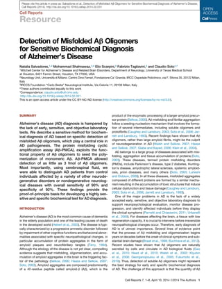
  • 1. Cell Reports Resource Detection of Misfolded Ab Oligomers for Sensitive Biochemical Diagnosis of Alzheimer’s Disease Natalia Salvadores,1,4 Mohammad Shahnawaz,1,4 Elio Scarpini,2 Fabrizio Tagliavini,3 and Claudio Soto1,* 1Mitchell Center for Alzheimer’s Disease and Related Brain Disorders, Department of Neurology, University of Texas Medical School at Houston, 6431 Fannin Street, Houston, TX 77030, USA 2Neurology Unit, Universita` di Milano, Centro Dino Ferrari, Fondazione Ca’ Granda, IRCC Ospedale Policlinico, via F. Sforza 35, 20122 Milan, Italy 3IRCCS Foundation ‘‘Carlo Besta’’ Neurological Institute, Via Celoria 11, 20133 Milan, Italy 4These authors contributed equally to this work *Correspondence: claudio.soto@uth.tmc.edu http://dx.doi.org/10.1016/j.celrep.2014.02.031 This is an open access article under the CC BY-NC-ND license (http://creativecommons.org/licenses/by-nc-nd/3.0/). SUMMARY Alzheimer’s disease (AD) diagnosis is hampered by the lack of early, sensitive, and objective laboratory tests. We describe a sensitive method for biochem- ical diagnosis of AD based on specific detection of misfolded Ab oligomers, which play a central role in AD pathogenesis. The protein misfolding cyclic amplification assay (Ab-PMCA), exploits the func- tional property of Ab oligomers to seed the poly- merization of monomeric Ab. Ab-PMCA allowed detection of as little as 3 fmol of Ab oligomers. Most importantly, using cerebrospinal fluid, we were able to distinguish AD patients from control individuals affected by a variety of other neurode- generative disorders or nondegenerative neurolog- ical diseases with overall sensitivity of 90% and specificity of 92%. These findings provide the proof-of-principle basis for developing a highly sen- sitive and specific biochemical test for AD diagnosis. INTRODUCTION Alzheimer’s disease (AD) is the most common cause of dementia in the elderly population and one of the leading causes of death in the developed world (Hebert et al., 2003). The disease is typi- cally characterized by a progressive amnestic disorder followed by impairment of other cognitive functions and behavioral abnor- malities associated with specific neuropathological changes, in particular accumulation of protein aggregates in the form of amyloid plaques and neurofibrillary tangles (Terry, 1994). Although the etiology of the disease is not yet clear, compelling evidence suggests that misfolding, oligomerization, and accu- mulation of amyloid aggregates in the brain is the triggering fac- tor of the pathology (Selkoe, 2000; Haass and Selkoe, 2007; Soto, 2003). Amyloid aggregates are composed predominantly of a 42-residue peptide called amyloid-b (Ab), which is the product of the enzymatic processing of a larger amyloid precur- sor protein (Selkoe, 2000). Ab misfolding and fibrillar aggregation follow a seeding-nucleation mechanism that involves the forma- tion of several intermediates, including soluble oligomers and protofibrils (Caughey and Lansbury, 2003; Soto et al., 2006; Jar- rett and Lansbury, 1993). Recent findings have shown that Ab oligomers, rather than large amyloid fibrils, might be the culprit of neurodegeneration in AD (Walsh and Selkoe, 2007; Haass and Selkoe, 2007; Glabe and Kayed, 2006; Klein et al., 2004). AD belongs to a large group of diseases associated with mis- folding, aggregation and tissue accumulation of proteins (Soto, 2003). These diseases, termed protein misfolding disorders (PMDs), include Parkinson’s disease, type 2 diabetes, Hunting- ton’s disease, amyotrophic lateral sclerosis, systemic amyloid- osis, prion diseases, and many others (Soto, 2003; Luheshi and Dobson, 2009). In all these diseases, misfolded aggregates composed of different proteins are formed by a similar mecha- nism resulting in the accumulation of toxic structures that induce cellular dysfunction and tissue damage (Caughey and Lansbury, 2003; Soto et al., 2006; Jarrett and Lansbury, 1993). One of the major problems in AD is the lack of a widely accepted early, sensitive, and objective laboratory diagnosis to support neuropsychological evaluation, monitor disease pro- gression, and identify affected individuals before they display the clinical symptoms (Parnetti and Chiasserini, 2011; Urbanelli et al., 2009). For diseases affecting the brain, a tissue with low regeneration capacity, it is crucial to intervene before irreversible neuropathological changes occur. Therefore, early diagnosis of AD is of utmost importance. Several lines of evidence point that the process of Ab misfolding and oligomerization begins years or decades before the onset of clinical symptoms and sub- stantial brain damage (Braak et al., 1999; Buchhave et al., 2012). Recent studies have shown that Ab oligomers are naturally secreted by cells and circulate in AD biological fluids (Gao et al., 2010; Head et al., 2010; Walsh et al., 2002; Klyubin et al., 2008; Georganopoulou et al., 2005; Fukumoto et al., 2010). Thus, detection of soluble Ab oligomers might represent the best strategy for early and specific biochemical diagnosis of AD. The challenge of this approach is that the quantity of Ab Cell Reports 7, 1–8, April 10, 2014 ª2014 The Authors 1 Please cite this article in press as: Salvadores et al., Detection of Misfolded Ab Oligomers for Sensitive Biochemical Diagnosis of Alzheimer’s Disease, Cell Reports (2014), http://dx.doi.org/10.1016/j.celrep.2014.02.031
  • 2. oligomers is likely very small in tissues other than the brain. An additional difficulty for specific detection of Ab oligomers is that their sequence and chemical structure is the same as the native Ab protein. Our strategy to detect misfolded oligomers is to use their functional property of being capable of catalyzing the polymeri- zation of the monomeric protein. For this purpose, we invented the protein misfolding cyclic amplification (PMCA) technology in order to achieve the ultrasensitive detection of misfolded aggre- gates through amplification of the misfolding and aggregation process in vitro (Saborio et al., 2001). So far, PMCA has been applied to detect minute quantities of oligomeric misfolded prion protein (PrPSc ) implicated in prion diseases (Morales et al., 2012). Using PMCA, we were able to detect the equivalent of a single particle of misfolded PrP oligomer (Saa´ et al., 2006b) and strikingly to identify PrPSc in the blood and urine of infected animals at symptomatic and presymptomatic stages of the dis- ease (Castilla et al., 2005; Saa´ et al., 2006a; Gonzalez-Romero et al., 2008). The basis for the PMCA technology is the fact that the process of misfolding and aggregation of Ab, PrP, and the other proteins implicated in PMDs follow a seeding-nucle- ation mechanism (Soto et al., 2002, 2006). In a seeded-nucle- ated polymerization, the limiting step is the formation of stable oligomeric seeds that, depending on the conditions, may take a very long time to form or not occur at all. Once formed, oligo- mers grow exponentially by recruiting and incorporating protein monomers into the growing polymer. Addition of preformed seeds into a solution containing the monomeric protein acceler- ates protein misfolding and aggregation (Soto et al., 2006; Jar- rett and Lansbury, 1993). Thus, measuring seeding activity could be used to estimate the presence and quantity of oligomers in a given sample. To increase the sensitivity of detection, PMCA combines steps of growing polymers with multiplication of olig- omeric seeds to reach an exponential increase of misfolding and aggregation (Soto et al., 2002). In this study, we describe the im- plementation and optimization of PMCA for highly sensitive detection of misfolded Ab oligomers and show its application to detect these structures in the cerebrospinal fluid (CSF) of AD patients. RESULTS Cyclic Amplification of Amyloid-b Misfolding To implement the experimental conditions for Ab-PMCA, we per- formed studies using in-vitro-produced oligomeric seeds. Because it is still unknown which of the different species of Ab oligomers is most relevant for AD pathology, we decided to work with a mixture of oligomers of different sizes generated dur- ing the process of fibril formation. Ab oligomers were prepared by incubation of monomeric (seed-free) synthetic Ab1-42 (10 mM) at 25 C with stirring. After 5 hr of incubation, we observed an abundance of globular oligomers by electron micro- scopy with only a small amount of protofibrils and fibrils (Figure 1A). These aggregates were positive with the A11 olig- omer-specific antibody (Kayed et al., 2003; data not shown). After longer incubation, protofibrillar and fibrillar structures were observed. The size of the aggregates was determined by filtration through filters of defined pore size and western blotting after SDS-PAGE separation. Oligomers formed by incubation for 5 hr migrated as $170 kDa SDS-resistant aggregates, with a mi- nor band at 17 kDa (Figure 1B). Low concentrations of seed-free Ab1-42 (2 mM) were incu- bated at 22 C with constant shaking (100 rpm) for different times alone or in the presence of distinct concentrations of synthetic Ab oligomers, prepared by incubation during 5 hr as described above. Ab aggregation was studied by the fluorescence emis- sion of the amyloid-binding dye Thioflavin T (ThT) (LeVine, 1993; Soto et al., 1995). The peptide concentration, tempera- ture, and pH of the buffer are critical to control the extent of the lag phase and reproducibility among experiments. Under these conditions, no spontaneous Ab aggregation was detect- able during the time in which the experiment was performed (125 hr). However, Ab aggregation was observed in the pres- ence of 0.3–8.4 fmol of Ab oligomers (Figure 1C). To increase the efficiency of seeding, and thus the limit of detection of Ab oligomers, we introduced cycles of amplification, combining phases of polymer growing with multiplication of seeds as in the PMCA assay. For this purpose, we subjected the samples to intermittent shaking, which has been previously shown to dramatically accelerate the seeded conversion of recombinant prion protein (Atarashi et al., 2008). Under these conditions, the kinetic of Ab aggregation induced by 8,400, 300, 80, and 3 fmol of Ab oligomers was clearly faster and easily distinguish- able from that observed in the absence of Ab seeds (Figure 1D). This result indicates that, using the Ab-PMCA assay, we should be able to detect as little as 3 fmol of Ab oligomers in a given sample. Detection of Ab Oligomers in the Cerebrospinal Fluid of AD Patients To study the usefulness of the Ab-PMCA assay to detect seed- ing-competent Ab oligomers in biological fluids, we analyzed aliquots of CSF from 50 AD patients, 39 cognitively normal indi- viduals affected by nondegenerative neurological diseases (NNDs), and 37 patients affected by non-AD neurodegenerative diseases (NANDs) including other forms of dementia. The exper- iments as well as the initial part of the analysis were done blindly, because the investigator was unaware of which samples were coming from AD or controls. Figure 2A shows the average kinetics of aggregation of five representative samples from the AD, NND, and NAND groups. The result indicates that CSF from AD patients accelerates significantly Ab aggregation as compared to control CSF (p 0.001). To determine the effect of individual samples on Ab aggregation, we estimated the lag phase (Figure 2B), defined as the time required to get a ThT fluo- rescence larger than 40 arbitrary units (after subtraction of the blank). This value was selected considering that it corresponds to approximately five times the reading of the buffer alone. We also estimated the P90, which corresponds to the extent of Ab aggregation at 90 hr (Figure 2C). By comparing both parameters among the experimental groups, a highly significant difference was observed between AD and control samples from individuals with nondegenerative neurological diseases or with non-AD neurodegenerative diseases. No correlation was detected between the aggregation parameters and the age of the AD pa- tients, which indicates that the levels of the marker are not simply 2 Cell Reports 7, 1–8, April 10, 2014 ª2014 The Authors Please cite this article in press as: Salvadores et al., Detection of Misfolded Ab Oligomers for Sensitive Biochemical Diagnosis of Alzheimer’s Disease, Cell Reports (2014), http://dx.doi.org/10.1016/j.celrep.2014.02.031
  • 3. a reflection of age but rather whether or not the patients contain seeding-competent Ab aggregates in their CSF. Using the values for lag phase, we calculated the sensitivity, specificity, and predictive value of the Ab-PMCA test (Table 1). To determine the appropriate cutoff points and the performance of the test, we carried out a detailed statistical analysis of the receiver oper- ating characteristics (ROC) (Figure S1). In relation to the control group consisting of age-matched individuals with nondegenera- tive neurological diseases, we estimated a 90.0% sensitivity and 84.2% specificity, whereas for the clinically more relevant differ- entiation of AD from other neurodegenerative diseases including other forms of dementia, we obtained a staggering 100% sensi- tivity and 94.6% specificity (Table 1). If confirmed with a larger number of patients, the ability of Ab-PMCA to distinguish AD from other forms of neurodegenerative diseases might be very useful in clinic. The overall sensitivity and specificity consid- ering all control individuals was 90% and 92%, respectively (Table 1). To confirm that Ab-PMCA detects a seeding activity associ- ated to Ab oligomers present in CSF, we performed immuno- depletion experiments. The methodology for efficient immuno- depletion of Ab oligomers was first optimized by using synthetically prepared Ab oligomers. Incubation with Dyna- beads conjugated with a mixture of sequence (4G8) and confor- mational (A11) antibodies led to the complete removal of these structures (Figure 3A). Next, application of immunodepletion to three AD CSF samples showed that the kinetic of Ab aggrega- tion in the Ab-PMCA reaction was comparable to that observed in control CSF samples, and both were significantly different from the aggregation observed with AD CSF prior to immunode- pletion (Figure 3B). A similar result was observed when immuno- depletion was done only using the A11 conformational antibody (Figure 3C), which recognizes specifically oligomers (Kayed et al., 2003). These results indicate that the seeding activity observed in AD CSF samples was indeed associated to Ab oligomers. Figure 1. Detection of Synthetic Ab Oligomers by Ab-PMCA (A) To prepare Ab oligomers, solutions of Ab1-42 at 10 mM were incubated for different times at 25 C with shaking. At various time points, samples were visualized by electron microcopy after negative staining. At 5 hr of incubation, aggregates consist mainly of globular oligomers, whereas at 10 hr, there are mostly pro- tofibrils, and at 24 hr, we can observe a large amount of long fibrils. (B) Preparations of oligomers were characterized by SDS-PAGE followed by western blot with 4G8 antibody. The mixture at 5 hr of incubation was also characterized by strong reactivity with A11 antibody and by a native size ranging from 30 to 1,000 kDa, as evaluated by filtration using filters of defined pore size. (C) Seeding of Ab aggregation was studied incubating a solution of 2 mM seed-free Ab1-42 in 100 mM Tris-HCl (pH 7.4) in the presence of 5 mM Thioflavin T, with or without different quantities of synthetic Ab oligomers (prepared by incubation during 5 hr, as indicated in A) with constant, but low, agitation (100 rpm) at 22 C. (D) The same samples as in (C) were incubated with cyclic agitation (1 min stirring at 500 rpm followed by 29 min without shaking). Aggregation was measured over time by the Thioflavin T (ThT) binding to amyloid fibrils using a plate spectrofluorometer (excitation: 435; emission: 485 nm). Graphs show the mean and SE of three replicates. The concentration of Ab oligomers was estimated assuming an average molecular weight of 170 kDa. Cell Reports 7, 1–8, April 10, 2014 ª2014 The Authors 3 Please cite this article in press as: Salvadores et al., Detection of Misfolded Ab Oligomers for Sensitive Biochemical Diagnosis of Alzheimer’s Disease, Cell Reports (2014), http://dx.doi.org/10.1016/j.celrep.2014.02.031
  • 4. DISCUSSION AD is one of the most important public health problems in the developed world. Among the ten leading causes of death, AD is the only one that lacks effective ways to prevent, cure, or even slow its progression. Part of the difficulties in developing an efficient treatment for this devastating disease is the absence of an early and objective biochemical diagnosis that will enable to recognize patients before substantial brain damage has occurred (Parnetti and Chiasserini, 2011; Blennow et al., 2010; Urbanelli et al., 2009). Indeed, many of the failures in recent clin- ical trials with compounds aimed to modify the disease progres- sion have been attributed to the lack of early diagnosis that prevents beginning treatment before it is too late (Galimberti and Scarpini, 2012). Currently, diagnosis is achieved by clinical assessment, neuroimaging, and CSF measurements of the levels of Ab42, tau, and phospho-tau, with definitive confirmation relying on postmortem identification of amyloid plaques and neurofibrillary tangles in the brain (Parnetti and Chiasserini, 2011; Urbanelli et al., 2009). Recent advances in neuroimaging using amyloid tracers offer a promising alternative to increase accuracy of diagnosis (Klunk et al., 2004; Klunk and Mathis, 2008), but it is unclear whether these methods will identify pa- tients before irreversible brain damage has occurred. Identifica- tion of biomarkers that could be used for noninvasive, sensitive, and objective detection of the disease process years or even decades before the onset of brain abnormalities and clinical symptoms are of utmost importance (Parnetti and Chiasserini, 2011; Blennow et al., 2010; Urbanelli et al., 2009). Although it is widely accepted that combining the measurements of Ab1-42, total tau, and phosphorylated tau in CSF can provide a relatively high sensitivity and specificity for AD diagnosis (Blennow et al., Figure 2. Detection of Seeding Activity in Human CSF Samples from Controls and AD Patients by Ab-PMCA (A) Representative aggregation curves of seed- free Ab1-42 in the presence of CSF samples from AD patients, people affected by non- neurodegenerative neurological diseases (NND), and patients suffering from neurodegenerative diseases other than AD (NAND). The values represent the average and SE of five different patients, representative of the average results in each group. For this purpose, we selected as representative for each group, the patients in which the kinetic parameters (lag phase and P90) were closer to the average obtained in each group. (B) The lag phase of Ab aggregation in Ab-PMCA was compared for the three groups of patients. Lag phase was defined as the time (in hours) required to reach a ThT signal more than 40 arbi- trary units. One sample from the NND group did not reach a ThT signal of 40 during the duration of the aggregation experiment. Thus, the lag phase for this sample is 300 hr. This sample was not included in the graph and was not utilized to calculate sensitivity and specificity in Table 1. (C) The extent of amyloid formation obtained after 180 Ab-PMCA cycles, i.e., 90 hr of incubation (P90), was measured in each patient. For the graphs in (B) and (C), we show the values obtained in individual samples, which correspond to the average of three independent experiments. Data were analyzed by one-way ANOVA, followed by the Tukey’s multiple comparison post hoc test. The differences between AD and samples from the other two groups were highly significant with p 0.001 (***). Table 1. Estimation of Sensitivity, Specificity, and Predictive Value for Ab-PMCA Using CSF Samples Groups Sensitivity (%)a Specificity (%)b Positive Predictive Value (%)c Negative Predictive Value (%)d AD versus NAND 100.0 94.6 96.2 100.0 AD versus NND 90.0 84.2 88.2 86.5 AD versus controlse 90.0 92.0 88.2 93.2 For estimation of sensitivity, specificity, and predictive value, we used the results of the lag phase as shown in Figure 2B. Cutoffs were estimated by receiver operating characteristics (ROC) curve analysis using the MedCalc software (see Figure S1). a Sensitivity was estimated by the formula (true positives/[true positives + false negatives] ) 3 100. b Specificity was estimated by the formula (true negatives/[false positives + true negatives]) 3 100. c Positive predictive value was estimated by the formula (true positives/[true positives + false positives]) 3 100. d Negative predictive value was estimated by the formula (true negatives/[true negatives + false negatives]) 3 100. e Controls refer to the samples from NND plus NAND. 4 Cell Reports 7, 1–8, April 10, 2014 ª2014 The Authors Please cite this article in press as: Salvadores et al., Detection of Misfolded Ab Oligomers for Sensitive Biochemical Diagnosis of Alzheimer’s Disease, Cell Reports (2014), http://dx.doi.org/10.1016/j.celrep.2014.02.031
  • 5. 2010), other CSF markers are needed that can more specifically differentiate AD from other dementias. It is also necessary to identify markers that correlate with the severity of dementia in patients with AD and, more importantly, can identify the patho- logical process earlier during the presymptomatic period of the disease. Recent compelling evidence indicates that the formation of misfolded Ab oligomers might be a critical event in AD pathogen- esis (Walsh and Selkoe, 2007; Haass and Selkoe, 2007; Glabe and Kayed, 2006; Klein et al., 2004). Indeed, both synthetic and natural Ab oligomers have been shown to induce apoptosis in cell cultures at very low concentrations (Dahlgren et al., 2002; El-Agnaf et al., 2000; Shankar et al., 2007), block long-term potentiation in brain slice cultures (Wang et al., 2002; Walsh et al., 2002), and impair synaptic plasticity and memory in ani- mals (Cleary et al., 2005; Shankar et al., 2007). These findings, in addition to the fact that Ab oligomers are small and soluble structures, suggest that they could be circulating in biological fluids and offer a specific marker for AD. Considering the well- established observation that the levels of total Ab1-42 are lower in AD than in controls, it might be surprising that, in our study, we found that Ab oligomers are elevated in AD patients. However, our assay measures the levels of seeding-competent Ab oligo- mers, which are likely a very small proportion of total Ab in CSF. A rough estimation based on the comparison with the amplification results obtained with synthetic oligomers is that the material we are measuring is below 100 pg/ml (expressed as monomer). Our assay does not distinguish oligomers pro- duced by Ab40 or Ab42; thus, in our estimation, oligomers repre- sent a very small fraction (less than 1%) of total Ab. Therefore, it is perfectly possible that a decline in total Ab42 coincides with an overall increase of oligomers in AD patients. Indeed, recent studies have shown that Ab oligomers might be present in low quantities in CSF and that their levels are elevated in the CSF of people affected by AD (Pitschke et al., 1998; Klyubin et al., 2008; Georganopoulou et al., 2005; Gao et al., 2010; Santos et al., 2012). In this study, we adapted the PMCA technology to cyclically amplify the process of Ab misfolding and aggregation, leading to the highly sensitive and specific detection of soluble Ab oligo- mers. PMCA was originally developed to replicate the misfolding and aggregation of the prion protein implicated in prion diseases and is now considered a major technological breakthrough that has been instrumental to understand the prion biology and to detect misfolded prions in various biological fluids (Morales et al., 2012). PMCA is a cyclical process that enables exponential increase of the detection signal in a manner analogous to the amplification of DNA by PCR (Soto et al., 2002). PMCA takes advantage of the fact that the process of protein misfolding and aggregation associated with Ab, PrP, and other proteins implicated in PMDs follows a seeding-nucleation mechanism and is dependent on the formation and number of oligomeric seeds present in the reaction (Soto et al., 2002, 2006). To achieve exponential amplification, PMCA consists of cycles of incubation for the elongation of the polymers, followed by breaking the aggregates to multiply the number of seeds. For amplification of prions, PMCA traditionally uses sonication as a mechanical force to break big polymers into smaller species (Morales et al., 2012). However, as shown by Caughey and colleagues, sonication can be replaced by strong shaking to achieve Figure 3. Seeding Activity in AD CSF Is Removed by Ab Oligomers Immunodepletion To test whether the seeding activity observed in human AD CSF was dependent on Ab aggre- gates, we removed these structures from CSF samples by immunodepletion using Dynabeads coated with a mixture of antibodies recognizing specifically the sequence of Ab (4G8) and the conformation of Ab oligomers (A11) (Kayed et al., 2003). (A) The procedure for immunodepletion was opti- mized using synthetically prepared Ab oligomers spiked into human CSF at quantities detectable by western blot. Three consecutive rounds of incuba- tion with antibody-coated Dynabeads were sufficient to efficiently remove the Ab oligomers observed at around 170 kDa in our western blots. (B) Samples of AD CSF before or after immu- nodepletion with 4G8 and A11 antibodies were used to seed Ab aggregation in the Ab-PMCA assay. (C) Samples of AD CSF were also depleted only with the A11 conformational antibody and aggregation monitored by Ab-PMCA assay. As in Figures 1 and 2, Ab aggregation was measured by ThT fluores- cence emission at different times of incubation. Values represent the average and SE of three different replicates. Cell Reports 7, 1–8, April 10, 2014 ª2014 The Authors 5 Please cite this article in press as: Salvadores et al., Detection of Misfolded Ab Oligomers for Sensitive Biochemical Diagnosis of Alzheimer’s Disease, Cell Reports (2014), http://dx.doi.org/10.1016/j.celrep.2014.02.031
  • 6. similar results using a procedure called shaking-based PMCA or quaking-induced conversion (Atarashi et al., 2008). In adapting PMCA for AD, we found that shaking is more compatible with the fragile nature of Ab aggregates. In our current setting, Ab-PMCA enables detection of as little as 3 fmol of Ab oligomers. This level of sensitivity can be further increased by additional development of the technology, as was also the case in prion diseases. Indeed, in our original proof- of-concept study using PMCA for prion detection, the increase of sensitivity was only around 60-fold (Saborio et al., 2001), which contrast with the striking 3 billion folds obtained after further development (Saa´ et al., 2006b). Nevertheless, even with the current status of the Ab-PMCA technology, we were able to distinguish with a high degree of sensitivity and specificity CSF samples coming from individuals diagnosed with AD, patients affected by other neurodegenerative disorders, and people suffering from nondegenerative neurological diseases (Table 1). Sensitivity, specificity, and predictive value of the Ab-PMCA assay are higher than the currently widely used ELISA tests measuring the levels of Ab42, tau, and phospho-tau in CSF. The sensitivity and specificity of these methods range between 80% and 90%, depending on the study (Mulder et al., 2010; Sun- derland et al., 2003; Maddalena et al., 2003). A crucial feature for an assay aimed for routine use in AD diagnosis is its reproduc- ibility and repeatability. Several variables need to be carefully controlled to maintain a good reproducibility, including Ab con- centration, temperature, buffer composition, pH, shaking rate, etc. It is also possible that, with different batches of Ab, the kinetic of aggregation and the parameters most useful to compare different group of samples may be different, but still AD samples induce accelerated Ab aggregation compared to controls (Figure S2). Experiments displayed in this article were carried out blindly and independently by two different investiga- tors at distinct times and using samples obtained from three different locations. However, during the course of the study, we found that an entire set of CSF samples coming from a fourth location was not amenable to the assay, because using these samples Ab did not aggregate at all, not even after spiking with large concentrations of synthetic oligomers. We suspect that preanalytical factors related to sample collection influenced the assay. We are actively investigating what this interference could be. Nevertheless, more work needs to be done to assess reproducibility in a large number of samples, with distinct reagents, equipment, and samples coming from different locations. Future studies should include larger populations of AD patients and controls and direct comparisons with the diag- nostic accuracy obtained by measuring Ab42, total tau, and phospho-tau in CSF and by positron emission tomography amyloid imaging with specific ligands. Also, it will be important to investigate the presence of Ab oligomers in patients affected by mild cognitive impairments, which is considered a precursor for AD (Petersen et al., 2009), as well as in asymptomatic car- riers of familial AD to analyze the utility of Ab-PMCA for pre- symptomatic detection of AD. Longitudinal studies from indi- vidual patients would also be needed to evaluate the relationship between the detection of Ab oligomers and the progression of the disease. Finally, adaptation of Ab-PMCA to detect Ab oligomers, which may potentially be circulating in the blood of AD patients, may offer a great opportunity for more-routine testing. EXPERIMENTAL PROCEDURES Biological Samples We used CSF samples from 50 patients with the diagnosis of probable AD as defined by the DSM-IV and the National Institute of Neurological and Com- municative Disorders and Stroke-Alzheimer’s Disease and Related Disorders Association guidelines (McKhann et al., 1984) and determined using a variety of tests, including routine medical examination; neurological evaluation; neuropsychological assessment; MRI; and measurements of CSF levels of Ab1-42, total tau, and phospho-tau. The mean age of AD patients at the time of sample collection was 71.0 ± 8.1 years (range 49–84). As controls, we used CSF from 39 patients affected by NND, including 12 cases of normal pressure hydrocephalus, seven patients with peripheral neuropathy, seven with diverse forms of brain tumor, two with ICTUS, one with severe cephalgia, three with encephalitis, one with hypertension, and six with unclear diagnosis. The mean age at which CSF samples were taken from this group of patients was 64.6 ± 14.7 years (range 31–83). We also used as controls CSF samples from 37 individuals affected by NAND, including ten cases of fronto-temporal dementia (five behavioral and five language variants); six patients with Parkinson’s disease (including four associated with dementia and one with motor neuron disease); six with progressive supranuclear palsy; six with spino- cerebellar ataxia (one associated with dementia); four with amyotrophic lateral sclerosis; two with Huntington’s disease; one with mitochondria encephalo- myopathy, lactic acidosis, and stroke-like episodes; one with Lewy body dementia; and one with vascular dementia. The mean age at sample collection for this group was 63.8 ± 11.1 years (range 41–80). CSF samples were collected in polypropylene tubes following lumbar puncture at the L4/L5 or L3/L4 interspace with atraumatic needles after one night fasting. The samples were centrifuged at 3,000 g for 3 min at 4 C, aliquoted, and stored at À80 C until analysis. CSF cell counts, glucose, and protein concentration were deter- mined. Albumin was measured by rate nephelometry. To evaluate the integrity of the blood brain barrier and the intrathecal immunoglobulin G (IgG) produc- tion, the albumin quotient (CSF albumin/serum albumin) 3 103 and the IgG index (CSF albumin/serum albumin)/(CSF IgG/serum IgG) were calculated (Sellebjerg and Christiansen, 1996). The study was conducted according to the provisions of the Helsinki Declaration and was approved by the Ethics Committee. Production of Synthetic Ab and Preparation of Ab Aggregates Ab1-42 was synthesized using solid-phase N-tert-butyloxycarbonyl chemistry at the W. Keck Facility at Yale University and purified by reverse-phase high- performance liquid chromatography. The final product was lyophilized and characterized by amino acid analysis and mass spectrometry. To prepare stock solutions free of Ab aggregated seeds, we used our previously described protocol (Morales et al., 2010), which involves dissolution of aggregates in high pH and filtration through 30 kDa cutoff filters to remove remaining aggregates. To prepare different types of aggregates, solutions of seed-free Ab1-42 (10 mM) were incubated for different times at 25 C in 0.1 M Tris-HCl (pH 7.4) with agitation. This preparation contains a mixture of Ab monomers as well as fibrils, protofibrils, and soluble oligomers in distinct proportions depending on the incubation time. The degree of aggregation was characterized by ThT fluorescence emission, electron microscopy after negative staining, dot blot studies with the A11 conformational antibody (Kayed et al., 2003), and western blot after gel electrophoresis using the 4G8 Ab antibody. Cyclic Amplification of Ab Misfolding and Aggregation Solutions of 2 mM aggregate-free Ab1-42 in 0.1 M Tris-HCl (pH 7.4; 200 ml total volume) were placed in opaque 96-well plates and incubated alone or in the presence of synthetic Ab aggregates or 40 ml of CSF aliquots. Samples were incubated in the presence of 5 mM ThT and subjected to cyclic agitation (1 min at 500 rpm followed by 29 min without shaking) using an Eppendorf thermomixer, at a constant temperature of 22 C. At various time points, ThT 6 Cell Reports 7, 1–8, April 10, 2014 ª2014 The Authors Please cite this article in press as: Salvadores et al., Detection of Misfolded Ab Oligomers for Sensitive Biochemical Diagnosis of Alzheimer’s Disease, Cell Reports (2014), http://dx.doi.org/10.1016/j.celrep.2014.02.031
  • 7. fluorescence was measured in the plates at 485 nm after excitation at 435 nm using a plate spectrofluorometer. Determination of Sensitivity, Specificity, and Predictive Value The differences in the kinetic of aggregation between different samples were evaluated by the estimation of two different kinetic parameters: the lag phase and P90. Lag phase is defined as the time required to reach a ThT fluorescence of 40 arbitrary units. This value was selected because it corresponds to approximately five times the value of the buffer alone. The P90 corresponds to the extent of aggregation (measured as ThT fluorescence) at 90 hr. Sensitivity, specificity, and predictive value were determined using the lag phase data, with cutoff thresholds determined by ROC curve analysis, using the MedCalc software (version 12.2.1.0). Statistical Analysis The significance of the differences in Ab aggregation kinetics in the presence of human CSF samples was analyzed by one-way ANOVA, followed by the Tukey’s multiple comparison post hoc test. The level of significance was set at p 0.05. Statistical tests were performed using the GraphPad Prism 5.0 software. SUPPLEMENTAL INFORMATION Supplemental Information includes two figures and can be found with this article online at http://dx.doi.org/10.1016/j.celrep.2014.02.031. AUTHOR CONTRIBUTIONS N.S. designed the experiments, optimized the Ab-PMCA technology, carried out many of the experiments with CSF samples, analyzed the results, and pre- pared the figures. M.S. performed various Ab-PMCA assays and analyzed the data. E.S. and F.T. provided some of the CSF samples and characterized them. C.S. is the principal investigator on the project and was responsible for coordinating research activity, analyzing the data, funding, and producing the final version of the article. ACKNOWLEDGMENTS We are very grateful to Dr. Justo Garcia de Yebenes (Hospital Ramon y Cajal) for providing us some of the CSF samples used in this study and Dr. Silvia Suardi (‘‘Carlo Besta’’ Neurological Institute) and Dr. Daniela Galimberti (Uni- versity of Milan) for help in collecting and characterizing CSF samples. We would also like to thank Dr. Lisbell Estrada (Catholic University of Chile) and Dr. Ines Moreno-Gonzalez (UTH) for valuable help in diverse aspects of this work. This research was supported by grants from the Alzheimer’s Associa- tion, CART Foundation, and Mitchell Foundation (to C.S.) and the Italian Ministry of Health (JPND, project BiomarkAPD) and MIUR (Prot. RBAP11FRE9; CUP: J41J11000330001) (to F.T.). C.S. is an inventor on several patents related to the PMCA technology and is currently Founder, Chief Scientific Officer, and Vice-President of Amprion Inc., a biotech company focusing on the commer- cial exploitation of PMCA for diagnosis of neurodegenerative diseases. Received: October 1, 2013 Revised: January 19, 2014 Accepted: February 20, 2014 Published: March 20, 2014 REFERENCES Atarashi, R., Wilham, J.M., Christensen, L., Hughson, A.G., Moore, R.A., Johnson, L.M., Onwubiko, H.A., Priola, S.A., and Caughey, B. (2008). Simpli- fied ultrasensitive prion detection by recombinant PrP conversion with shaking. Nat. Methods 5, 211–212. Blennow, K., Hampel, H., Weiner, M., and Zetterberg, H. (2010). Cerebrospinal fluid and plasma biomarkers in Alzheimer disease. Nat. Rev. Neurol. 6, 131–144. Braak, E., Griffing, K., Arai, K., Bohl, J., Bratzke, H., and Braak, H. (1999). Neuropathology of Alzheimer’s disease: what is new since A. Alzheimer? Eur. Arch. Psychiatry Clin. Neurosci. 249 (Suppl 3), 14–22. Buchhave, P., Minthon, L., Zetterberg, H., Wallin, A.K., Blennow, K., and Hans- son, O. (2012). Cerebrospinal fluid levels of b-amyloid 1-42, but not of tau, are fully changed already 5 to 10 years before the onset of Alzheimer dementia. Arch. Gen. Psychiatry 69, 98–106. Castilla, J., Saa´ , P., and Soto, C. (2005). Detection of prions in blood. Nat. Med. 11, 982–985. Caughey, B., and Lansbury, P.T. (2003). Protofibrils, pores, fibrils, and neuro- degeneration: separating the responsible protein aggregates from the innocent bystanders. Annu. Rev. Neurosci. 26, 267–298. Cleary, J.P., Walsh, D.M., Hofmeister, J.J., Shankar, G.M., Kuskowski, M.A., Selkoe, D.J., and Ashe, K.H. (2005). Natural oligomers of the amyloid-beta protein specifically disrupt cognitive function. Nat. Neurosci. 8, 79–84. Dahlgren, K.N., Manelli, A.M., Stine, W.B., Jr., Baker, L.K., Krafft, G.A., and LaDu, M.J. (2002). Oligomeric and fibrillar species of amyloid-beta peptides differentially affect neuronal viability. J. Biol. Chem. 277, 32046–32053. El-Agnaf, O.M., Mahil, D.S., Patel, B.P., and Austen, B.M. (2000). Oligomeriza- tion and toxicity of beta-amyloid-42 implicated in Alzheimer’s disease. Bio- chem. Biophys. Res. Commun. 273, 1003–1007. Fukumoto, H., Tokuda, T., Kasai, T., Ishigami, N., Hidaka, H., Kondo, M., Allsop, D., and Nakagawa, M. (2010). High-molecular-weight beta-amyloid oligomers are elevated in cerebrospinal fluid of Alzheimer patients. FASEB J. 24, 2716–2726. Galimberti, D., and Scarpini, E. (2012). Progress in Alzheimer’s disease. J. Neurol. 259, 201–211. Gao, C.M., Yam, A.Y., Wang, X., Magdangal, E., Salisbury, C., Peretz, D., Zuckermann, R.N., Connolly, M.D., Hansson, O., Minthon, L., et al. (2010). Ab40 oligomers identified as a potential biomarker for the diagnosis of Alzheimer’s disease. PLoS ONE 5, e15725. Georganopoulou, D.G., Chang, L., Nam, J.M., Thaxton, C.S., Mufson, E.J., Klein, W.L., and Mirkin, C.A. (2005). Nanoparticle-based detection in cerebral spinal fluid of a soluble pathogenic biomarker for Alzheimer’s disease. Proc. Natl. Acad. Sci. USA 102, 2273–2276. Glabe, C.G., and Kayed, R. (2006). Common structure and toxic function of amyloid oligomers implies a common mechanism of pathogenesis. Neurology 66 (2, Suppl 1), S74–S78. Gonzalez-Romero, D., Barria, M.A., Leon, P., Morales, R., and Soto, C. (2008). Detection of infectious prions in urine. FEBS Lett. 582, 3161–3166. Haass, C., and Selkoe, D.J. (2007). Soluble protein oligomers in neurodegen- eration: lessons from the Alzheimer’s amyloid beta-peptide. Nat. Rev. Mol. Cell Biol. 8, 101–112. Head, E., Pop, V., Sarsoza, F., Kayed, R., Beckett, T.L., Studzinski, C.M., Tomic, J.L., Glabe, C.G., and Murphy, M.P. (2010). Amyloid-beta peptide and oligomers in the brain and cerebrospinal fluid of aged canines. J. Alzheimers Dis. 20, 637–646. Hebert, L.E., Scherr, P.A., Bienias, J.L., Bennett, D.A., and Evans, D.A. (2003). Alzheimer disease in the US population: prevalence estimates using the 2000 census. Arch. Neurol. 60, 1119–1122. Jarrett, J.T., and Lansbury, P.T., Jr. (1993). Seeding ‘‘one-dimensional crystal- lization’’ of amyloid: a pathogenic mechanism in Alzheimer’s disease and scrapie? Cell 73, 1055–1058. Kayed, R., Head, E., Thompson, J.L., McIntire, T.M., Milton, S.C., Cotman, C.W., and Glabe, C.G. (2003). Common structure of soluble amyloid oligomers implies common mechanism of pathogenesis. Science 300, 486–489. Klein, W.L., Stine, W.B., Jr., and Teplow, D.B. (2004). Small assemblies of unmodified amyloid beta-protein are the proximate neurotoxin in Alzheimer’s disease. Neurobiol. Aging 25, 569–580. Klunk, W.E., and Mathis, C.A. (2008). The future of amyloid-beta imaging: a tale of radionuclides and tracer proliferation. Curr. Opin. Neurol. 21, 683–687. Cell Reports 7, 1–8, April 10, 2014 ª2014 The Authors 7 Please cite this article in press as: Salvadores et al., Detection of Misfolded Ab Oligomers for Sensitive Biochemical Diagnosis of Alzheimer’s Disease, Cell Reports (2014), http://dx.doi.org/10.1016/j.celrep.2014.02.031
  • 8. Klunk, W.E., Engler, H., Nordberg, A., Wang, Y., Blomqvist, G., Holt, D.P., Bergstro¨ m, M., Savitcheva, I., Huang, G.F., Estrada, S., et al. (2004). Imaging brain amyloid in Alzheimer’s disease with Pittsburgh Compound-B. Ann. Neurol. 55, 306–319. Klyubin, I., Betts, V., Welzel, A.T., Blennow, K., Zetterberg, H., Wallin, A., Lemere, C.A., Cullen, W.K., Peng, Y., Wisniewski, T., et al. (2008). Amyloid beta protein dimer-containing human CSF disrupts synaptic plasticity: preven- tion by systemic passive immunization. J. Neurosci. 28, 4231–4237. LeVine, H., 3rd. (1993). Thioflavine T interaction with synthetic Alzheimer’s disease beta-amyloid peptides: detection of amyloid aggregation in solution. Protein Sci. 2, 404–410. Luheshi, L.M., and Dobson, C.M. (2009). Bridging the gap: from protein mis- folding to protein misfolding diseases. FEBS Lett. 583, 2581–2586. Maddalena, A., Papassotiropoulos, A., Mu¨ ller-Tillmanns, B., Jung, H.H., Hegi, T., Nitsch, R.M., and Hock, C. (2003). Biochemical diagnosis of Alzheimer dis- ease by measuring the cerebrospinal fluid ratio of phosphorylated tau protein to beta-amyloid peptide42. Arch. Neurol. 60, 1202–1206. McKhann, G., Drachman, D., Folstein, M., Katzman, R., Price, D., and Stadlan, E.M. (1984). Clinical diagnosis of Alzheimer’s disease: report of the NINCDS- ADRDA Work Group under the auspices of Department of Health and Human Services Task Force on Alzheimer’s Disease. Neurology 34, 939–944. Morales, R., Estrada, L.D., Diaz-Espinoza, R., Morales-Scheihing, D., Jara, M.C., Castilla, J., and Soto, C. (2010). Molecular cross talk between misfolded proteins in animal models of Alzheimer’s and prion diseases. J. Neurosci. 30, 4528–4535. Morales, R., Duran-Aniotz, C., Diaz-Espinoza, R., Camacho, M.V., and Soto, C. (2012). Protein misfolding cyclic amplification of infectious prions. Nat. Pro- toc. 7, 1397–1409. Mulder, C., Verwey, N.A., van der Flier, W.M., Bouwman, F.H., Kok, A., van Elk, E.J., Scheltens, P., and Blankenstein, M.A. (2010). Amyloid-beta(1-42), total tau, and phosphorylated tau as cerebrospinal fluid biomarkers for the diag- nosis of Alzheimer disease. Clin. Chem. 56, 248–253. Parnetti, L., and Chiasserini, D. (2011). Role of CSF biomarkers in the diagnosis of prodromal Alzheimer’s disease. Biomarkers Med. 5, 479–484. Petersen, R.C., Roberts, R.O., Knopman, D.S., Boeve, B.F., Geda, Y.E., Ivnik, R.J., Smith, G.E., and Jack, C.R., Jr. (2009). Mild cognitive impairment: ten years later. Arch. Neurol. 66, 1447–1455. Pitschke, M., Prior, R., Haupt, M., and Riesner, D. (1998). Detection of single amyloid beta-protein aggregates in the cerebrospinal fluid of Alzheimer’s patients by fluorescence correlation spectroscopy. Nat. Med. 4, 832–834. Saa´ , P., Castilla, J., and Soto, C. (2006a). Presymptomatic detection of prions in blood. Science 313, 92–94. Saa´ , P., Castilla, J., and Soto, C. (2006b). Ultra-efficient replication of infec- tious prions by automated protein misfolding cyclic amplification. J. Biol. Chem. 281, 35245–35252. Saborio, G.P., Permanne, B., and Soto, C. (2001). Sensitive detection of path- ological prion protein by cyclic amplification of protein misfolding. Nature 411, 810–813. Santos, A.N., Ewers, M., Minthon, L., Simm, A., Silber, R.E., Blennow, K., Prvulovic, D., Hansson, O., and Hampel, H. (2012). Amyloid-b oligomers in cerebrospinal fluid are associated with cognitive decline in patients with Alzheimer’s disease. J. Alzheimers Dis. 29, 171–176. Selkoe, D.J. (2000). Toward a comprehensive theory for Alzheimer’s disease. Hypothesis: Alzheimer’s disease is caused by the cerebral accu- mulation and cytotoxicity of amyloid beta-protein. Ann. N Y Acad. Sci. 924, 17–25. Sellebjerg, F., and Christiansen, M. (1996). Qualitative assessment of intra- thecal IgG synthesis by isoelectric focusing and immunodetection: interlabor- atory reproducibility and interobserver agreement. Scand. J. Clin. Lab. Invest. 56, 135–143. Shankar, G.M., Bloodgood, B.L., Townsend, M., Walsh, D.M., Selkoe, D.J., and Sabatini, B.L. (2007). Natural oligomers of the Alzheimer amyloid-beta pro- tein induce reversible synapse loss by modulating an NMDA-type glutamate receptor-dependent signaling pathway. J. Neurosci. 27, 2866–2875. Soto, C. (2003). Unfolding the role of protein misfolding in neurodegenerative diseases. Nat. Rev. Neurosci. 4, 49–60. Soto, C., Castan˜ o, E.M., Frangione, B., and Inestrosa, N.C. (1995). The a-helical to beta-strand transition in the amino-terminal fragment of the amyloid beta-peptide modulates amyloid formation. J. Biol. Chem. 270, 3063–3067. Soto, C., Saborio, G.P., and Anderes, L. (2002). Cyclic amplification of protein misfolding: application to prion-related disorders and beyond. Trends Neurosci. 25, 390–394. Soto, C., Estrada, L., and Castilla, J. (2006). Amyloids, prions and the inherent infectious nature of misfolded protein aggregates. Trends Biochem. Sci. 31, 150–155. Sunderland, T., Linker, G., Mirza, N., Putnam, K.T., Friedman, D.L., Kimmel, L.H., Bergeson, J., Manetti, G.J., Zimmermann, M., Tang, B., et al. (2003). Decreased beta-amyloid1-42 and increased tau levels in cerebrospinal fluid of patients with Alzheimer disease. JAMA 289, 2094–2103. Terry, R.D. (1994). Neuropathological changes in Alzheimer disease. Prog. Brain Res. 101, 383–390. Urbanelli, L., Magini, A., Ciccarone, V., Trivelli, F., Polidoro, M., Tancini, B., and Emiliani, C. (2009). New perspectives for the diagnosis of Alzheimer’s disease. Recent Pat. CNS Drug Discov. 4, 160–181. Walsh, D.M., and Selkoe, D.J. (2007). A beta oligomers - a decade of dis- covery. J. Neurochem. 101, 1172–1184. Walsh, D.M., Klyubin, I., Fadeeva, J.V., Cullen, W.K., Anwyl, R., Wolfe, M.S., Rowan, M.J., and Selkoe, D.J. (2002). Naturally secreted oligomers of amyloid beta protein potently inhibit hippocampal long-term potentiation in vivo. Nature 416, 535–539. Wang, H.W., Pasternak, J.F., Kuo, H., Ristic, H., Lambert, M.P., Chromy, B., Viola, K.L., Klein, W.L., Stine, W.B., Krafft, G.A., and Trommer, B.L. (2002). Soluble oligomers of beta amyloid (1-42) inhibit long-term potentiation but not long-term depression in rat dentate gyrus. Brain Res. 924, 133–140. 8 Cell Reports 7, 1–8, April 10, 2014 ª2014 The Authors Please cite this article in press as: Salvadores et al., Detection of Misfolded Ab Oligomers for Sensitive Biochemical Diagnosis of Alzheimer’s Disease, Cell Reports (2014), http://dx.doi.org/10.1016/j.celrep.2014.02.031