Breaking tolerance to invoke an immune response to cancer
•Als DOC, PDF herunterladen•
1 gefällt mir•861 views


Empfohlen
Empfohlen
Weitere ähnliche Inhalte
Was ist angesagt?
Was ist angesagt? (20)
Andere mochten auch
Andere mochten auch (7)
Ähnlich wie Breaking tolerance to invoke an immune response to cancer
Ähnlich wie Breaking tolerance to invoke an immune response to cancer (20)
Breaking tolerance to invoke an immune response to cancer
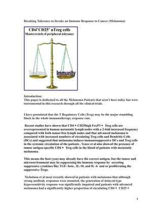
- 1. Breaking Tolerance to Invoke an Immune Response to Cancer (Melanoma) Introduction: This paper is dedicated to all the Melanoma Patients that aren’t here today but were instrumental in this research through all the clinical trials. I have postulated that the T Regulatory Cells (Treg) may be the major stumbling block in the whole immunotherapy response rate. Recent studies have shown that CD4+CD25high FoxP3+ Treg cells are overrepresented in human metastatic lymph nodes with a 2-fold increased frequency compared with both tumor-free lymph nodes and that advanced melanoma is associated with increased numbers of circulating Treg cells and Dendritic Cells (DCs) and suggested that melanoma induces immunosuppressive DCs and Treg cells in the systemic circulation of the patients . Vence et al also showed the presence of tumor antigen-specific CD4+ Treg cells in the blood of patients with metastatic melanoma. This means the host (you) may already have the correct antigen, but the tumor and microenvironment may be suppressing the immune response by secreting suppressive cytokines like TGF- beta , IL-10, and IL -6 and or proliferating the suppressive Tregs. Nicholaou et al most recently showed in patients with melanoma that although strong antibody responses were mounted, the generation of delayed-type hypersensitivity response was significantly impaired and patients with advanced melanoma had a significantly higher proportion of circulating CD4+ CD25+ 1
- 2. FoxP3+ Treg cells compared with those with minimal residual disease. So we now can blame the tolerance of our immune system to cancer on the T Regulatory Cells (Tregs). It has been reported that the large number of different cell types that are claiming to be directly targeted by FoxP3+ Treg cells are CD4+, CD8+ T cell, dendritic cells, B cells, macrophages, osteoblasts, mast cells, NK cells, and NKT cells. If this is the case we must temporality deplete or suppress the T- Regulatory Cells (Tregs). This can be accomplished in a number of ways. Dr. Steven A Rosenberg uses Cyclophosamide. This is accomplished by administering the drug prior to ACT Therapy. Another way to suppress the Treg function is to block the CTLA-4 receptor with an antibody. That antibody, known as Anti-CTLA-4, Yervoy (Ipilimumab) that was FDA approved in March of 2011. 2
- 3. Patients treated with Anti-CTLA-4 showed a statistically significant decrease in the median percentage of Treg cells by Day 28 of therapy (5.8% vs.3.6%) “Biologic and Immunomodulatory Events after CTLA-4 Blockade with Ticilimumab in Patients with Advanced Malignant Melanoma” et al Luis H. Camacho 2006. Also, after treatment the treated patients had a significantly higher median percentage of resting CD4_CD25_ T cells that coexpressed PD-1 compared with control subjects (66.4% vs. 25.0%). This mean the Anti-CTLA-4 therapy unregulated the PD-1 on resting T-cells. This is an unwanted effect, the upregulation PD-1 is known to cause T-cell exhaustion. This is why the combination of Anti-PD-1 and Anti-CTLA-4 would be a more synergistic treatment for Melanoma. <A HREF="https://www.box.net/s/mzxb1b2o0tue44a3z3xo">Breaking Tolerance to Invoke an Immune Response to Cancer (Melanoma</A> <BR> 3
- 4. CTLA-4 blockade therapy can succeed in breaking peripheral immune tolerance in some patients, but not in others. So what are we missing? We removed the suppressive function of the Tregs. We believe we have tumor specific antigens. What is the one thing that happens with most immune responses? The “DANGER SIGNAL”. You get an inflammatory response. 4
- 5. Proflammatory Cytokines and chemoattractants must be induced to produce an immune response. If not, the immune response doesn’t take hold and it (the response) is terminated. Now with this information, we now know that we need Proflammatory Cytokines and chemoattractants to initiate an immune response. Recently, in a paper by Dr. Thomas F. Gajewski, “Identifying and Overcoming Immune Resistance Mechanisms in the Melanoma Tumor Microenvironment” 2006, he states: "Immunotherapeutic approaches for the treatment of melanoma, such as tumor antigen- based vaccines, can frequently boost immune responses. However, clinical responses as measured by tumor shrinkage are seen in only a minority of patients. This observation has prompted careful analysis of the tumor microenvironment for biologic correlates to clinical response and also to identify mechanisms of tumor resistance. Patients with advanced melanoma treated with antigen-specific vaccines had pre-treatment tumor biopsies analyzed by gene expression profiling. Supervised hierarchical clustering was performed based on clinical outcome. An expanded bank of tumors was analyzed to increase the sample size and better understand gene patterns. Two major categories of melanoma metastases have been observed. One subgroup of patient has an inflamed phenotype that includes expression of chemokines, T-cell markers, and other immunoregulatory factors. Clinical responders to melanoma vaccines appear to fall within this subset. This group also contains the highest expression of negative regulatory factors, including PD-L1, IDO, and FoxP3, suggesting that these immunosuppressive mechanisms may dominantly inhibit anti-tumor –cell function in those patients. In addition, absence of B7 expression supports classical T-cell anergy. Preclinical experiments have confirmed a critical role for these mechanisms in limiting anti-tumor T–cell efficacy in vivo, giving candidate treatment strategies for translation back into the clinic. A second subset of patients is represented by tumors which are non-inflamed and lack chemokines for T cell recruitment. Therefore, a major barrier in these cases appears to be failed T –cell migration into tumor sites. Experimental strategies to augment T-cell migration can have important anti-tumor effects in preclinical models. The presence of the "inflamed" gene signature was associated with a type I IFN transcriptional profile, and murine experimental models have confirmed a critical role for type I IFN signaling in promoting adaptive immunity." 5
- 6. So, in the first subset of Patients, tumors had a suppressive nature that may be over- ridden by Anti-CTLA-4 (Yervoy) and or Anti-PD-1 Therapy. The second subset was missing the "danger signal" inflammatory cytokines and chemoattractants most likely due to STAT3 signaling from the Tumor. 6
- 7. Stimulation of Toll-like receptor 4 (TLR-4) activates macrophages and results in the release of TNF-alpha. It is hypothesized that melanoma inhibits macrophage activation by suppressing TLR-4 signaling. 7
- 8. So, if Melanoma suppresses Macrophage Activation, then the tumor microenvironment is missing IL-6, IL-1b and other cytokines and chemoattractants secreted by the Tumor- derived Antigen presenting cells (APCs) like the Dendritic Cells and Macrophages. 8
- 9. Activated Macrophages secrete the following cytokines under different conditions: IL-1, IL-12, IL-6, IFN-gamma/alpha/beta and TNF-alpha IL-6 Interleukin 6 is a pro-inflammatory cytokine and is produced in response to infection and tissue injury. IL-6 exerts its effects on multiple cell types and can act systemically. IL-6 stimulates liver secretion of acute phase proteins IL-6 stimulates B-lymphocytes to produce antibodies IL-6 in concert with IL-1b causes T-cell activation IL-6 induces STAT 3 Signaling IL-6 Plus TGF-b induces the Th17 cell phenotype IL-1 beta Interleukin-1b is a pro-inflammatory cytokine which is secreted by macrophages activated by a number of stimuli including TNF-alpha, bacterial endotoxin and IL-1b 9
- 10. itself. IL-1b exerts its effects on many different cell types locally at the site of production and systemically (at a distance). IL-12 Interleukin-12 is a heterodimer consisting of a p35 and a p40 subunit. Both subunits are required for receptor binding and biological activity. IL-12 stimulates growth of activated Natural Killer (NK) cells, CD8+ and CD4+ T- cells. IL-12 increases NK and T-cell g-IFN production which shifts T-cell differentiation towards a Th1-type response. IL-12 increases NK production of TNF-alpha which can act synergistically with IFN- gamma. IL-12 suppresses IL-4 induced IgE production. TNF-alpha Tumor Necrosis Factor alpha is made by many other cells as well as macrophages, which are major sources, especially after priming by Interferon gamma. TNF-alpha initiates a cascade of cytokines which mediate an inflammatory response. TNF-alpha effects are mediated through two types of receptor, a 75kDa TNFR-a receptor and a 55kDa TNFR-b receptor. TNF-alpha regulates the expression of many genes in many cell types important for the host response to infection. IFN-gamma/beta Macrophages, and many other cells produce these Type I interferons which act as immunomodulatory, as well as antiviral cytokines. Distinct receptor from interferon gamma, mediates overlapping or competing effects on macrophages. Cellular signaling pathways involve Jak/Stats, and other pathways. So, if Melanoma suppresses Macrophage Activation, then the tumor’s microenvironment is missing IL-6, IL-1b and other cytokines. This change in the tumor’s microenvironment results in the suppression of the T- cell Activation. 10
- 11. If you look at the above micrographs, you will see that the two patients that had relapsed (10710 and 10737) had IL-1b and IL-6 missing. The macrophages were not activated!!!! The "Danger Signal" known as inflammation was missing! The non-responders most likely had their macrophages polarized to a M2-like phenotype. 11
- 12. 12
- 13. So what if you introduced a mixture of cytokines that resembles the secretion from the Tumor derived APCs. Would you induce an immune response? This is just what a small Biotech company; CEL-SCI Corporation is trying. It is now in Phase 3 clinical trials. It is used prior to therapy/radiation to enhance the immune system. 13
- 14. “As we look at data from Phase I and Phase II clinical trials, we see strong indication that Multikine simulates the activities of a healthy person’s immune system, enabling it to use the body’s own anti-tumor immune response.” What if there was another way to activate the immune system. Recent research shows that if you use CpG ODNs (TLR9 antogonists) on mature dendritc cells, immune activation occurs. “Deoxycytidyl-deoxyguanosine [(CpG)3] oligodeoxynucleotides (ODNs) signal through TLR9 to induce innate immune responses and adjuvant effects that promote T cell responses.” et al C. V. Harding and colleagues. CpG ODNs can be divided into three Classes. Class C, CpG-B&C ODNs stimulate B cells efficiently to produce IgM and IL-6. This is very important because we want IL-6 produced because IL-6 in concert with IL-1b causes T-cell activation. TLR9 signaling regulates multiple aspects of antigen processing and APC function. In mice, TLR9 is widely expressed by APCs, including myeloid DCs (mDCs), pDCs, macrophages, and B cells. In humans, TLR9 expression is restricted to (pDCs) plasmacytoid DCs and B cells. CpG ODNs promote maturation of mDCs, which results in enhanced expression of MHC-I, MHC-II, and costimulatory molecules (i.e., CD40, CD80, and CD86) B7-1, B7-2 and increased MHC-I and MHC-II antigen-presentation function. 14
- 15. Activation of a T-cell According to Dr. Slingluff in a paper recently, he broke down the immunohistologic Characteristics into three immunotypes. In the research paper “Immunotype and Immunohistologic Characteristics of Tumor Infiltrating Immune Cells are Associated with Clinical Outcome in Metastatic Melanoma”1 et al Slingluff 2012, it breaks down the Immunohistologic Characteristics into three distinct immunotypes: A) No infiltration of immune cells in the tumor’s microenvironment. B) Infiltrating Immune cells only in close proximity to the tumor’s vascular system C) Diffuse immune cell infiltrates throughout a metastatic tumor and its microenvironment. 15
- 16. Immunohistologic Characteristics of Tumor Infiltrating Immune Cells Immunotype A B C Immune cell None(very Low) Some Throughout infiltration Estimated median 15 23 130 survival (months) Metastases (%) 29 63 8 B-cells Low Med High Macrophages High Med Low likely M1 or M2 M2 Mix M1 CD8+ T-cells Low Med High Overall, the most predominant immune cells were T cells (53%), followed by the B cell lineage cells (33%), and then by macrophages (13%), with NK and mature dendritic cells only hardly present. With the setting of the tumor’s microenvironment evaluated, we will focus the low survival immunotype A patients. How can we improve the overall survival and the immune response to Melanoma? We need to push the differentiation of the macrophages towards the M1 phenotype. Macrophages are important tumor-infiltrating cells and play pivotal roles in tumor growth and metastasis. Macrophages participate in immune responses to tumors in a polarized manner: classic M1 macrophages produce interleukin (IL) 12 to promote tumoricidal responses, whereas M2 macrophages produce IL10 and help tumor progression. The mechanisms governing macrophage polarization are unclear But in 1990 it was discovered treatment of M2 macrophages with GM-CSF or IFN-gamma led to production of M1 phenotypic markers upon LPS stimulation. The research paper “IRF5 promotes inflammatory macrophage 1 polarization and TH1-TH17 response” Et al Irina A. Udalova explains it in great detail. One of the hallmarks of M1 macrophage polarisation is acquired antigen presenting features leading to efficient Th1 response. 16
- 17. The M1 macrophage phenotype is induced by Interferon gamma (IFN-γ) followed by stimulation with bacterial products like lipopolysaccharides (LPS) or by treatment of monocytes with granulocyte-macrophage colony-stimulating factor (GM-CSF). Based on Immunohistologic Characteristics of Tumor Infiltrating Immune Cells presented by Dr. Slingluff, the macrophages in the A type (Non-responder) must be M2 phenotype. Since the Macrophage are known to have plasticity, one could in theory change their microenvironment to shift the macrophages from M2 to M1 phenotype. We need to somehow combine these immunotherapies with a cancer vaccine to invoke an immune response using the macrophages as the starter of immunity with memories T-cells so we can eliminate any reoccurrence. Oncologists have been working on this for decades. I believe they may have discovered the right combination to just that. Dr. Kingston Mills and colleagues from the Immune Regulation Research Group, School of Biochemistry and Immunology, Trinity Biomedical Sciences Institute, Trinity College Dublin Immunotherapy with PI3K Inhibitor and Toll-Like Receptor Agonist Induces IFN-γ IL-17þ Polyfunctional T Cells That Mediate Rejection of Murine Tumors Dr. Mills and colleagues discovered that if you combine a PI3k inhibitor with TRL agonists (TRL9 and or 5) with DC vaccine/tumor antigen, you invoke a tremendous immune response along with memory cells. This is because this combination (PI3k inhibitor with TRL 17
- 18. agonists) induces IFN gamma which differentiates M2 macrophages into the M1 phenotype which in turn activates the Th1 effector T-cells. TLR agonists, including CpG ODNs, have been shown to break tolerance to self-antigens by inhibiting the function of Treg cells via the production of IL-6 by DC. However, CpG ODNs have also been shown to stimulate the production of IL-10 by DC, and promote the induction of Treg cells. Therefore, CpG ODNs can generate effector as well as suppressive immune responses well as suppressive immune responses. Immune activation by CpG ODN initiates with specific binding to the TLR9 receptor in B cells and plasmacytoid DCs. TLR9 ligation in DCs results in secondary activation of lymphocytes, macrophage, monocyte, NK-cell, and T-cell populations through the elaboration of cytokines generating a TH1 cytokine milieu. This results in increased NK activity as well as improved antigen presentation and T cell help that can augment humoral and cell-mediated immune responses. In addition, TLR ligation results in the production of IL-6 by DCs, which helps overcome the suppressive effect of CD4 CD25 Treg cells. 18
- 19. TLR9 signaling regulates multiple aspects of antigen processing and APC function. In mice, TLR9 is widely expressed by APCs, including myeloid DCs (mDCs), pDCs, macrophages, and B cells. In humans, TLR9 expression is restricted to pDCs and B cellset al H. Wagner. CpG ODNs promote maturation of mDCs, which results in enhanced expression of MHC-I, MHC-II, and costimulatory molecules (i.e., CD40, CD80, and CD86) and increased MHC-I and MHC-II antigen-presentation function et al C.V. Harding. With the addition of a PI3K Inhibitor, one can tilt the T-cell Differentiation toward the Th1 Phenotype and limiting the propagation of Tregs. This is a winning combination. It not only awakens the innate immune system, but also awakens the Adaptive Immune System which can mount an immune response with memory. A VACINATION in vivo. This approach uses the full arsenal of the immune system. 19
- 20. One also thinks if you add Yervoy and anti-PD-1 with TRL9 agonist you might also produce the right immune response. This assumption needs to be tested with Clinical trials. 20
- 21. 21
- 22. 22
- 23. 22
- 24. 22