Applying dosing schedules to the clinical protocols of combinatorial therapy
•Als DOC, PDF herunterladen•
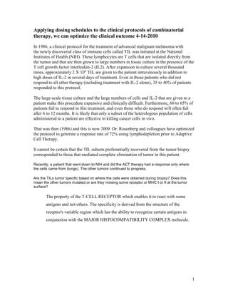
1 gefällt mir•215 views


Empfohlen
Empfohlen
Weitere ähnliche Inhalte
Was ist angesagt?
Was ist angesagt? (20)
Andere mochten auch
Andere mochten auch (20)
Ähnlich wie Applying dosing schedules to the clinical protocols of combinatorial therapy
Ähnlich wie Applying dosing schedules to the clinical protocols of combinatorial therapy (20)
Kürzlich hochgeladen
Kürzlich hochgeladen (20)
Applying dosing schedules to the clinical protocols of combinatorial therapy
- 1. Applying dosing schedules to the clinical protocols of combinatorial therapy, we can optimize the clinical outcome 4-14-2010 In 1986, a clinical protocol for the treatment of advanced malignant melanoma with the newly discovered class of immune cells called TIL was initiated at the National Institutes of Health (NIH). These lymphocytes are T cells that are isolated directly from the tumor and that are then grown to large numbers in tissue culture in the presence of the T cell growth factor interleukin-2 (IL2). After expansion in culture several thousand times, approximately 2 X 10" TIL are given to the patient intravenously in addition to high doses of IL-2 in several days of treatment. Even in those patients who did not respond to all other therapy (including treatment with IL-2 alone), 35 to 40% of patients responded to this protocol. The large-scale tissue culture and the large numbers of cells and IL-2 that are given to a patient make this procedure expensive and clinically difficult. Furthermore, 60 to 65% of patients fail to respond to this treatment, and even those who do respond will often fail after 6 to 12 months. It is likely that only a subset of the heterologous population of cells administered to a patient are effective in killing cancer cells in vivo. That was then (1986) and this is now 2009. Dr. Rosenberg and colleagues have optimized the protocol to generate a response rate of 72% using lymphodepletion prior to Adaptive Cell Therapy. It cannot be certain that the TIL subsets preferentially recovered from the tumor biopsy corresponded to those that mediated complete elimination of tumor in this patient. Recently, a patient that went down to NIH and did the ACT therapy had a response only where the cells came from (lungs). The other tumors continued to progress. Are the TILs tumor specific based on where the cells were obtained during biopsy? Does this mean the other tumors mutated or are they missing some receptor or MHC I or II at the tumor surface? The property of the T-CELL RECEPTOR which enables it to react with some antigens and not others. The specificity is derived from the structure of the receptor's variable region which has the ability to recognize certain antigens in conjunction with the MAJOR HISTOCOMPATIBILITY COMPLEX molecule. 1
- 2. Adoptive Cell Transfer.. 57 days after transfer. CD8+ T-cell (CTLs) at the Maximum Propagation A tumor lesion excised from patient 9 before nonmyeloablating chemotherapy ("pretreatment") exhibited scant CD8+ cells (left), strong stromal cell staining but weak staining of tumor cells with antibody to MHC class I (center), and sporadic cell staining with an antibody to MHC class II (probably tumor macrophages) but minimal staining of tumor cells (right). In contrast, a sample resected 57 days after cell transfer ("post treatment") exhibited a dense, diffuse CD8+ infiltrate and ubiquitous expression of both MHC class I and class II molecules in tumor cells. Source: Cancer Regression and Autoimmunity in Patients After Clonal Repopulation with Antitumor Lymphocytes Originally published in Science Express on 19 September 2002 Science 25 October 2002: Vol. 298. no. 5594, pp. 850 - 854 DOI: 10.1126/science.1076514 2
- 3. Patients undergoing Anti-CTLA-4 Blockadge. Week7 = Day 49 maximum ALC.. CD8+ T-cell (CTLs) Source: Dr. Jedd Wolchok 3
- 4. What I am trying to show is the growth curves for the various T-cells subsets with data from in vivo to back up the above graphic. With that in mind, we now have a better understanding on the growth patterns of these cells. By applying dosing schedules to the clinical protocols of combinatorial therapy, we can optimize the clinical outcome. 4