SlideShare a Scribd company logo
1 of 47
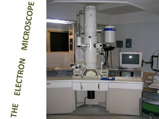
THE  ELECTRON  MICROSCOPE
Introduction
Introduction
Introduction
Introduction
Introduction
Introduction
Introduction
Introduction
Introduction
Introduction
[object Object],[object Object]
Electron Microscope ,[object Object],[object Object],The two types are:  Scanning and Transmission
Which is the most powerful kind of microscope?
THE LIGHT MICROSCOPE v THE ELECTRON MICROSCOPE   fluorescent (TV) screen, photographic film Human eye (retina), photographic film Focussing screen Vacuum Air-filled Interior Magnets Glass Lenses High voltage (50kV) tungsten lamp Tungsten or quartz halogen lamp Radiation source x500 000 x1000 – x1500 Maximum magnification 0.2nm Fine detail app. 200nm Maximum resolving power Electrons app. 4nm Monochrome Visible light 760nm (red) – 390nm  Colours visible Electromagnetic spectrum used ELECTRON MICROSCOPE LIGHT MICROSCOPE FEATURE
THE LIGHT MICROSCOPE v  THE ELECTRON MICROSCOPE Copper grid Glass slide Support Heavy metals Water soluble dyes Stains Microtome only. Slices    50nm Parts of cells visible Hand or microtome slices    20 000nm Whole cells visible Sectioning Resin Wax Embedding OsO 4  or KMnO 4 Alcohol Fixation Tissues must be dehydrated =  dead Temporary mounts  living or dead Preparation of specimens ELECTRON MICROSCOPE LIGHT MICROSCOPE FEATURE
Properties of electron ,[object Object],[object Object],[object Object],[object Object]
[object Object],[object Object],[object Object],[object Object]
Electron beam (A) Transmitted electron (B) Inelastically scattered electrons (C) Elastically scattered electrons (D) Back-scattered electrons (E) Secondary electrons X-rays Visible light
[object Object],[object Object],[object Object]
[object Object],[object Object],[object Object]
Working   &  Image formation ,[object Object],[object Object],[object Object],[object Object],[object Object],[object Object]
[object Object],[object Object],[object Object],[object Object]
[object Object],[object Object],[object Object],[object Object]
[object Object],[object Object],[object Object],[object Object]
Magnification ,[object Object],[object Object],[object Object],[object Object],[object Object]
Resolution ,[object Object],[object Object],[object Object],[object Object]
Transmission Electron Microscope (TEM) ,[object Object],[object Object],[object Object]
[object Object],[object Object],[object Object]
Basic Systems Making Up a Transmission Electron Microscope ,[object Object],[object Object],[object Object]
Basic Systems Making Up a Transmission Electron Microscope ,[object Object],[object Object]
Page 167 TABLE 6.3 Major Column Components of the TEM* Component Synonyms Function of Components Illumination System Electron Gun Gun, Source Generates electrons and provides first coherent crossover of electron beam Condenser Lens 1 C1, Spot Size Determines smallest illumination spot size on specimen (see Spot Size in Table 6.4) Condenser Lens 2 C2, Brightness Varies amount of illumination on specimen — in combination with C1 (see Brightness in Table 6.4) Condenser Aperture C2 Aperture Reduces spherical aberration, helps control amount of illumination striking specimen
Specimen Manipulation System Specimen Exchanger Specimen Air Lock Chamber and mechanism for inserting specimen holder Specimen Stage Stage Mechanism for moving specimen inside column of microscope Imaging System Objective Lens — Forms, magnifies, and focuses first image (see Focus in Table 6.4) Objective Aperture — Controls contrast and spherical aberration Intermediate Lens Diffraction Lens Normally used to help magnify image from objective lens and to focus diffraction pattern Intermediate Aperture Diffraction Aperture, Field Limiting Aperture Selects area to be diffracted Projector Lens 1 P1 Helps magnify image, possibly used in some diffraction work Projector Lens 2 P2 Same as P1
Observation and Camera Systems Viewing Chamber — Contains viewing screen for final image Binocular Microscope Focusing Scope Magnifies image on viewing screen for accurate focusing Camera — Contains film for recording
Scanning Electron Microscopy (SEM) Visualizes  Surface  Features
[object Object],[object Object],[object Object],[object Object]
[object Object],[object Object],[object Object],[object Object],[object Object]
[object Object],[object Object],[object Object]
Layout and performance of SEM 1-3 Electron gun 4, 10 Aperture 5-6 Condenser lenses 7 Scanning coils 8 Stigmator 9 Objective lens 11 X-ray detector 12 Pre-amplifier 13 Scanning circuits 14 Specimen 15 Secondary electron detector 16-18 Display/Control circuits
Specimen Preparation Specimens are coated with metals to deflect electrons from a beam scanned across the sample.
SEM  of Stereocilia Projecting from a Cochlear (inner ear) Hair Cell
Copper grid slides © 2007 Paul Billiet  ODWS
Higher Resolution Is Achieved by Viewing Sections of Fixed, Stained, and Embedded Samples A microtome cutting sections of an embedded sample.
Microtome knife © 2007 Paul Billiet  ODWS
Fig. 3-22

More Related Content

What's hot

Scanning transmission electron microscope
Scanning transmission electron microscopeScanning transmission electron microscope
Scanning transmission electron microscope
Gulfam Hussain
 
Electron Microscope Shanthakumar
Electron Microscope ShanthakumarElectron Microscope Shanthakumar
Electron Microscope Shanthakumar
guestcd4662
 

What's hot (20)

Transmission Electron Microscope
Transmission Electron MicroscopeTransmission Electron Microscope
Transmission Electron Microscope
 
SCANNING ELECTRON MICROSCOPY (SEM)
SCANNING ELECTRON MICROSCOPY (SEM)SCANNING ELECTRON MICROSCOPY (SEM)
SCANNING ELECTRON MICROSCOPY (SEM)
 
Scanning transmission electron microscope
Scanning transmission electron microscopeScanning transmission electron microscope
Scanning transmission electron microscope
 
SEM- scanning electron microscope
SEM- scanning electron microscope SEM- scanning electron microscope
SEM- scanning electron microscope
 
Transmission electron
Transmission        electronTransmission        electron
Transmission electron
 
Transmission electron microscope
Transmission electron microscopeTransmission electron microscope
Transmission electron microscope
 
Scanning Electron microscopy
Scanning Electron microscopy Scanning Electron microscopy
Scanning Electron microscopy
 
Scanning electron microscopy
Scanning electron microscopyScanning electron microscopy
Scanning electron microscopy
 
Scanning Electron Microscopy (SEM) lecture
Scanning Electron Microscopy (SEM) lectureScanning Electron Microscopy (SEM) lecture
Scanning Electron Microscopy (SEM) lecture
 
SEM and TEM
SEM and  TEMSEM and  TEM
SEM and TEM
 
Transmission Electron Microscope
Transmission Electron MicroscopeTransmission Electron Microscope
Transmission Electron Microscope
 
Scanning Electron Microscope (SEM)
Scanning Electron Microscope (SEM)Scanning Electron Microscope (SEM)
Scanning Electron Microscope (SEM)
 
Transmission electron microscopy (1) (1)
Transmission electron microscopy (1) (1)Transmission electron microscopy (1) (1)
Transmission electron microscopy (1) (1)
 
Scanning electron microscope
Scanning electron microscopeScanning electron microscope
Scanning electron microscope
 
Scanning Electron Microscopy - SEM
Scanning Electron Microscopy - SEM Scanning Electron Microscopy - SEM
Scanning Electron Microscopy - SEM
 
SCANNING PROBE MICROSCOPY .
SCANNING PROBE MICROSCOPY .SCANNING PROBE MICROSCOPY .
SCANNING PROBE MICROSCOPY .
 
Scanning electron microscopy-SEM
Scanning electron microscopy-SEMScanning electron microscopy-SEM
Scanning electron microscopy-SEM
 
ATOMIC FORCE MICROSCOPE MITHILESH CHOUDHARY
ATOMIC FORCE MICROSCOPE MITHILESH CHOUDHARYATOMIC FORCE MICROSCOPE MITHILESH CHOUDHARY
ATOMIC FORCE MICROSCOPE MITHILESH CHOUDHARY
 
Transmission electron microscope
Transmission electron microscopeTransmission electron microscope
Transmission electron microscope
 
Electron Microscope Shanthakumar
Electron Microscope ShanthakumarElectron Microscope Shanthakumar
Electron Microscope Shanthakumar
 

Viewers also liked (7)

Electron microscope
Electron microscopeElectron microscope
Electron microscope
 
Electron microscopy
Electron microscopyElectron microscopy
Electron microscopy
 
transmission electron microscopy
transmission electron microscopytransmission electron microscopy
transmission electron microscopy
 
Sem and tem
Sem and temSem and tem
Sem and tem
 
INTRODUCTION TO SPECTROSCOPY
INTRODUCTION TO SPECTROSCOPYINTRODUCTION TO SPECTROSCOPY
INTRODUCTION TO SPECTROSCOPY
 
Scanning electron microscopy
Scanning electron microscopyScanning electron microscopy
Scanning electron microscopy
 
UV visible spectroscopy
UV visible spectroscopyUV visible spectroscopy
UV visible spectroscopy
 

Similar to Electron microscope

Transmission electron microscope
Transmission electron microscopeTransmission electron microscope
Transmission electron microscope
SuganyaPaulraj
 
Principle & Applications of Transmission Electron Microscopy (TEM) & High Res...
Principle & Applications of Transmission Electron Microscopy (TEM) & High Res...Principle & Applications of Transmission Electron Microscopy (TEM) & High Res...
Principle & Applications of Transmission Electron Microscopy (TEM) & High Res...
Gulfam Raza
 
Unit – 2 Microscopy
Unit – 2 MicroscopyUnit – 2 Microscopy
Unit – 2 Microscopy
sanchu yadav
 

Similar to Electron microscope (20)

ELECTRON MICROSCOPY-Scanning Electron MicroscopySEM.pptx
ELECTRON MICROSCOPY-Scanning Electron MicroscopySEM.pptxELECTRON MICROSCOPY-Scanning Electron MicroscopySEM.pptx
ELECTRON MICROSCOPY-Scanning Electron MicroscopySEM.pptx
 
Transmission electron microscope
Transmission electron microscopeTransmission electron microscope
Transmission electron microscope
 
Microscope
MicroscopeMicroscope
Microscope
 
Transmission Electron Microscopy
Transmission Electron MicroscopyTransmission Electron Microscopy
Transmission Electron Microscopy
 
Tem
TemTem
Tem
 
E mintro
E mintroE mintro
E mintro
 
Transmission Electron Microscope (TEM) for research (Full version)
Transmission Electron Microscope (TEM) for research (Full version)Transmission Electron Microscope (TEM) for research (Full version)
Transmission Electron Microscope (TEM) for research (Full version)
 
Electron microscopy
Electron microscopyElectron microscopy
Electron microscopy
 
2018 HM-Transmission electron microscope
2018 HM-Transmission electron microscope2018 HM-Transmission electron microscope
2018 HM-Transmission electron microscope
 
Electron microscope
Electron microscopeElectron microscope
Electron microscope
 
Electron microscopy
Electron microscopyElectron microscopy
Electron microscopy
 
SEM,TEM & AFM
SEM,TEM & AFMSEM,TEM & AFM
SEM,TEM & AFM
 
Principle & Applications of Transmission Electron Microscopy (TEM) & High Res...
Principle & Applications of Transmission Electron Microscopy (TEM) & High Res...Principle & Applications of Transmission Electron Microscopy (TEM) & High Res...
Principle & Applications of Transmission Electron Microscopy (TEM) & High Res...
 
Chapter-3. Electron Microscopy.pptx
Chapter-3. Electron Microscopy.pptxChapter-3. Electron Microscopy.pptx
Chapter-3. Electron Microscopy.pptx
 
Electron microscopy
Electron microscopyElectron microscopy
Electron microscopy
 
TEM versatile tool (small version)
TEM versatile tool (small version)TEM versatile tool (small version)
TEM versatile tool (small version)
 
Lec 3; Microscopy ALI.pptx
Lec 3; Microscopy ALI.pptxLec 3; Microscopy ALI.pptx
Lec 3; Microscopy ALI.pptx
 
Transmission electron microscope (TEM)
Transmission electron microscope (TEM)Transmission electron microscope (TEM)
Transmission electron microscope (TEM)
 
Electron microscopy, M. Sc. Zoology, University of Mumbai
Electron microscopy, M. Sc. Zoology, University of MumbaiElectron microscopy, M. Sc. Zoology, University of Mumbai
Electron microscopy, M. Sc. Zoology, University of Mumbai
 
Unit – 2 Microscopy
Unit – 2 MicroscopyUnit – 2 Microscopy
Unit – 2 Microscopy
 

Recently uploaded

Seal of Good Local Governance (SGLG) 2024Final.pptx
Seal of Good Local Governance (SGLG) 2024Final.pptxSeal of Good Local Governance (SGLG) 2024Final.pptx
Seal of Good Local Governance (SGLG) 2024Final.pptx
negromaestrong
 
Activity 01 - Artificial Culture (1).pdf
Activity 01 - Artificial Culture (1).pdfActivity 01 - Artificial Culture (1).pdf
Activity 01 - Artificial Culture (1).pdf
ciinovamais
 

Recently uploaded (20)

How to Give a Domain for a Field in Odoo 17
How to Give a Domain for a Field in Odoo 17How to Give a Domain for a Field in Odoo 17
How to Give a Domain for a Field in Odoo 17
 
Food Chain and Food Web (Ecosystem) EVS, B. Pharmacy 1st Year, Sem-II
Food Chain and Food Web (Ecosystem) EVS, B. Pharmacy 1st Year, Sem-IIFood Chain and Food Web (Ecosystem) EVS, B. Pharmacy 1st Year, Sem-II
Food Chain and Food Web (Ecosystem) EVS, B. Pharmacy 1st Year, Sem-II
 
ICT Role in 21st Century Education & its Challenges.pptx
ICT Role in 21st Century Education & its Challenges.pptxICT Role in 21st Century Education & its Challenges.pptx
ICT Role in 21st Century Education & its Challenges.pptx
 
Holdier Curriculum Vitae (April 2024).pdf
Holdier Curriculum Vitae (April 2024).pdfHoldier Curriculum Vitae (April 2024).pdf
Holdier Curriculum Vitae (April 2024).pdf
 
Web & Social Media Analytics Previous Year Question Paper.pdf
Web & Social Media Analytics Previous Year Question Paper.pdfWeb & Social Media Analytics Previous Year Question Paper.pdf
Web & Social Media Analytics Previous Year Question Paper.pdf
 
psychiatric nursing HISTORY COLLECTION .docx
psychiatric  nursing HISTORY  COLLECTION  .docxpsychiatric  nursing HISTORY  COLLECTION  .docx
psychiatric nursing HISTORY COLLECTION .docx
 
TỔNG ÔN TẬP THI VÀO LỚP 10 MÔN TIẾNG ANH NĂM HỌC 2023 - 2024 CÓ ĐÁP ÁN (NGỮ Â...
TỔNG ÔN TẬP THI VÀO LỚP 10 MÔN TIẾNG ANH NĂM HỌC 2023 - 2024 CÓ ĐÁP ÁN (NGỮ Â...TỔNG ÔN TẬP THI VÀO LỚP 10 MÔN TIẾNG ANH NĂM HỌC 2023 - 2024 CÓ ĐÁP ÁN (NGỮ Â...
TỔNG ÔN TẬP THI VÀO LỚP 10 MÔN TIẾNG ANH NĂM HỌC 2023 - 2024 CÓ ĐÁP ÁN (NGỮ Â...
 
Seal of Good Local Governance (SGLG) 2024Final.pptx
Seal of Good Local Governance (SGLG) 2024Final.pptxSeal of Good Local Governance (SGLG) 2024Final.pptx
Seal of Good Local Governance (SGLG) 2024Final.pptx
 
Explore beautiful and ugly buildings. Mathematics helps us create beautiful d...
Explore beautiful and ugly buildings. Mathematics helps us create beautiful d...Explore beautiful and ugly buildings. Mathematics helps us create beautiful d...
Explore beautiful and ugly buildings. Mathematics helps us create beautiful d...
 
Mixin Classes in Odoo 17 How to Extend Models Using Mixin Classes
Mixin Classes in Odoo 17  How to Extend Models Using Mixin ClassesMixin Classes in Odoo 17  How to Extend Models Using Mixin Classes
Mixin Classes in Odoo 17 How to Extend Models Using Mixin Classes
 
General Principles of Intellectual Property: Concepts of Intellectual Proper...
General Principles of Intellectual Property: Concepts of Intellectual  Proper...General Principles of Intellectual Property: Concepts of Intellectual  Proper...
General Principles of Intellectual Property: Concepts of Intellectual Proper...
 
Measures of Dispersion and Variability: Range, QD, AD and SD
Measures of Dispersion and Variability: Range, QD, AD and SDMeasures of Dispersion and Variability: Range, QD, AD and SD
Measures of Dispersion and Variability: Range, QD, AD and SD
 
This PowerPoint helps students to consider the concept of infinity.
This PowerPoint helps students to consider the concept of infinity.This PowerPoint helps students to consider the concept of infinity.
This PowerPoint helps students to consider the concept of infinity.
 
Sociology 101 Demonstration of Learning Exhibit
Sociology 101 Demonstration of Learning ExhibitSociology 101 Demonstration of Learning Exhibit
Sociology 101 Demonstration of Learning Exhibit
 
Activity 01 - Artificial Culture (1).pdf
Activity 01 - Artificial Culture (1).pdfActivity 01 - Artificial Culture (1).pdf
Activity 01 - Artificial Culture (1).pdf
 
Advanced Views - Calendar View in Odoo 17
Advanced Views - Calendar View in Odoo 17Advanced Views - Calendar View in Odoo 17
Advanced Views - Calendar View in Odoo 17
 
INDIA QUIZ 2024 RLAC DELHI UNIVERSITY.pptx
INDIA QUIZ 2024 RLAC DELHI UNIVERSITY.pptxINDIA QUIZ 2024 RLAC DELHI UNIVERSITY.pptx
INDIA QUIZ 2024 RLAC DELHI UNIVERSITY.pptx
 
Basic Civil Engineering first year Notes- Chapter 4 Building.pptx
Basic Civil Engineering first year Notes- Chapter 4 Building.pptxBasic Civil Engineering first year Notes- Chapter 4 Building.pptx
Basic Civil Engineering first year Notes- Chapter 4 Building.pptx
 
microwave assisted reaction. General introduction
microwave assisted reaction. General introductionmicrowave assisted reaction. General introduction
microwave assisted reaction. General introduction
 
PROCESS RECORDING FORMAT.docx
PROCESS      RECORDING        FORMAT.docxPROCESS      RECORDING        FORMAT.docx
PROCESS RECORDING FORMAT.docx
 

Electron microscope

  • 1. THE ELECTRON MICROSCOPE
  • 12.
  • 13.
  • 14. Which is the most powerful kind of microscope?
  • 15. THE LIGHT MICROSCOPE v THE ELECTRON MICROSCOPE fluorescent (TV) screen, photographic film Human eye (retina), photographic film Focussing screen Vacuum Air-filled Interior Magnets Glass Lenses High voltage (50kV) tungsten lamp Tungsten or quartz halogen lamp Radiation source x500 000 x1000 – x1500 Maximum magnification 0.2nm Fine detail app. 200nm Maximum resolving power Electrons app. 4nm Monochrome Visible light 760nm (red) – 390nm Colours visible Electromagnetic spectrum used ELECTRON MICROSCOPE LIGHT MICROSCOPE FEATURE
  • 16. THE LIGHT MICROSCOPE v THE ELECTRON MICROSCOPE Copper grid Glass slide Support Heavy metals Water soluble dyes Stains Microtome only. Slices  50nm Parts of cells visible Hand or microtome slices  20 000nm Whole cells visible Sectioning Resin Wax Embedding OsO 4 or KMnO 4 Alcohol Fixation Tissues must be dehydrated = dead Temporary mounts living or dead Preparation of specimens ELECTRON MICROSCOPE LIGHT MICROSCOPE FEATURE
  • 17.
  • 18.
  • 19. Electron beam (A) Transmitted electron (B) Inelastically scattered electrons (C) Elastically scattered electrons (D) Back-scattered electrons (E) Secondary electrons X-rays Visible light
  • 20.
  • 21.
  • 22.
  • 23.
  • 24.
  • 25.
  • 26.
  • 27.
  • 28.
  • 29.
  • 30.
  • 31.
  • 32.
  • 33.
  • 34. Page 167 TABLE 6.3 Major Column Components of the TEM* Component Synonyms Function of Components Illumination System Electron Gun Gun, Source Generates electrons and provides first coherent crossover of electron beam Condenser Lens 1 C1, Spot Size Determines smallest illumination spot size on specimen (see Spot Size in Table 6.4) Condenser Lens 2 C2, Brightness Varies amount of illumination on specimen — in combination with C1 (see Brightness in Table 6.4) Condenser Aperture C2 Aperture Reduces spherical aberration, helps control amount of illumination striking specimen
  • 35. Specimen Manipulation System Specimen Exchanger Specimen Air Lock Chamber and mechanism for inserting specimen holder Specimen Stage Stage Mechanism for moving specimen inside column of microscope Imaging System Objective Lens — Forms, magnifies, and focuses first image (see Focus in Table 6.4) Objective Aperture — Controls contrast and spherical aberration Intermediate Lens Diffraction Lens Normally used to help magnify image from objective lens and to focus diffraction pattern Intermediate Aperture Diffraction Aperture, Field Limiting Aperture Selects area to be diffracted Projector Lens 1 P1 Helps magnify image, possibly used in some diffraction work Projector Lens 2 P2 Same as P1
  • 36. Observation and Camera Systems Viewing Chamber — Contains viewing screen for final image Binocular Microscope Focusing Scope Magnifies image on viewing screen for accurate focusing Camera — Contains film for recording
  • 37. Scanning Electron Microscopy (SEM) Visualizes Surface Features
  • 38.
  • 39.
  • 40.
  • 41. Layout and performance of SEM 1-3 Electron gun 4, 10 Aperture 5-6 Condenser lenses 7 Scanning coils 8 Stigmator 9 Objective lens 11 X-ray detector 12 Pre-amplifier 13 Scanning circuits 14 Specimen 15 Secondary electron detector 16-18 Display/Control circuits
  • 42. Specimen Preparation Specimens are coated with metals to deflect electrons from a beam scanned across the sample.
  • 43. SEM of Stereocilia Projecting from a Cochlear (inner ear) Hair Cell
  • 44. Copper grid slides © 2007 Paul Billiet ODWS
  • 45. Higher Resolution Is Achieved by Viewing Sections of Fixed, Stained, and Embedded Samples A microtome cutting sections of an embedded sample.
  • 46. Microtome knife © 2007 Paul Billiet ODWS

Editor's Notes

  1. © Ryan Barrow 2008