1. Clinical How I do it?
MR Mammography: How I Do It
Kai-Boris Reiter
Institute for Diagnostic and Interventional Radiology and Neuroradiology,
Essen University Hospital, Essen, Germany
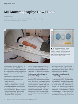
1A 1B
1 (A) Positioning the patient on the
breast coil (phased array breast coil;
Siemens, Erlangen) with the integrated
compression device, lateromedial.
(B) Compression wheels for lateromedial
immobilization of the breast.
In recent years, magnetic resonance to- In terms of planning and performance, Every patient receives a questionnaire
mography of the breast (MR Mammogra- as well as dealing with the patient, MR for recording prior breast diseases, genetic
phy) has become increasingly established mammography represents a particular predisposition to breast cancer, and cur-
as a diagnostic procedure to supplement challenge to the radiologic technologist. rent problems with the breast.
conventional mammography and sono-
graphy. Examination planning and Patient preparation and
The indications for MR mammography patient briefing positioning
include: When setting the appointment for MR For the examination, the patient receives
■ Clarification of recurrence of prior mammography, premenopausal patients a surgical gown (can be closed from the
breast carcinoma should be informed that the examination front) and pants to prevent possible MRT
■ Lymph node metastasis in the axilla takes place in the 2nd or 3rd week of her artifacts due to metal parts in their cloth-
when the primary tumor is unclear cycle. Examinations in the 1st or 4th week ing, and to enable more comfortable po-
■ Preoperative staging for histologically of the menstrual cycle could result in in- sitioning. Prior to beginning the examina-
certain breast carcinoma creased and therefore distorted contrast tion, an intravenous port is inserted with
■ Clinical monitoring of breast carcinoma agent enhancement due to hormones, an extension tube for mechanical contrast
during neoadjuvant chemotherapy making it much more difficult to deter- agent application. The patient should be
■ Unclear diagnostic findings from mam- mine the MR mammography diagnostic provided with hearing protection such
mography and/or sonography findings. as headphones or ear plugs to keep noise
104 MAGNETOM Flash · 3/2007 · www.siemens.com/magnetom-world
2. 2A 2B
2 Planning transverse slices using coronal and sagittal scouts.
3A 3B
3C 3D
3 In the T2_tse_tra(a) and T1_fl_3d_tra_native (B) measurements, no indication of suspected findings. Only in the T1_fl_3d_KM (C),
and in subtraction (D), is it possible to make out a tumor manifestation in the outer, lower quadrant.
exposure to a minimum. In addition, the in the breast coil (phased array breast coil; is performed in native technique. One
patient should be instructed to remain Siemens, Erlangen, Germany). A foam minute after the start of the second dy-
still during the examination to keep mo- cushion placed under the ankle enables namic T1-weighted GRE sequence (and
tion artifacts to a minimum. This includes comfortable positioning. 3 minutes after the start of the dynamic
gentle, shallow breathing during the ex- sequences), cm is applied.
amination. Examination sequence
For MR mammography, the patient is First, planning of the transverse slices Evaluation
placed in prone position on the breast coil is performed using coronal and sagittal After the examination, the dynamic
(Fig. 1). A cushion is placed under the scouts (Fig. 2). The examination begins sequences are subtracted: the post-cm
head, the arms are positioned at the sides with a T2-weighted Turbo-Spin-Echo sequences from the pre-cm sequences.
of the body. In exceptional cases for ex- (TSE) sequence at a 2 mm slice thickness, As a result we obtain 5 subtraction
tremely obese patients, the arms can be for the display of structures containing sequences. In MR mammography, sub-
folded at the head (disadvantage: the water, such as cysts and edema. Then, traction enables better visualization
breast is not fully covered by the breast 6 dynamic T1-weighted GRE (Flash 3D) of breast lesions that have taken up cm.
coil). If necessary, an additional OR cloth sequences are performed. The slice thick-
*WIP – Works in Progress. This information about this
can be placed on the patient cushions. ness of the dynamic sequences is 2 mm,
product is preliminary. The product is under develop-
Using the integrated compression device without gaps, lasting 2 minutes each. The ment and not commercially available in the U.S.,
(Fig. 1B), the breast is fixed lateromedially first dynamic T1-weighted GRE sequence and its future availability cannot be ensured.
MAGNETOM Flash · 3/2007 · www.siemens.com/magnetom-world 105