Dr digital diagnost_with_wpd_at_dartmouth_hitchcock_medical_center-usa_customer_story
•
1 gefällt mir•405 views


Empfohlen
Weitere ähnliche Inhalte
Was ist angesagt?
Was ist angesagt? (19)
Andere mochten auch
Ähnlich wie Dr digital diagnost_with_wpd_at_dartmouth_hitchcock_medical_center-usa_customer_story
Ähnlich wie Dr digital diagnost_with_wpd_at_dartmouth_hitchcock_medical_center-usa_customer_story (20)
Mehr von Jhon Arriaga Cordova
Mehr von Jhon Arriaga Cordova (20)
Dr digital diagnost_with_wpd_at_dartmouth_hitchcock_medical_center-usa_customer_story
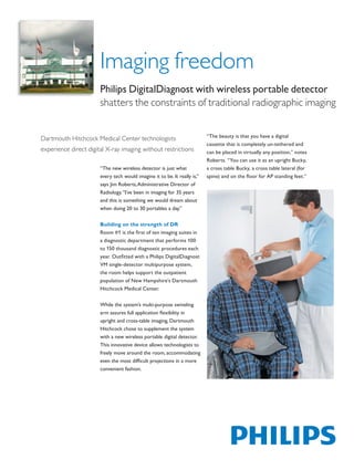
- 1. Imaging freedom Philips DigitalDiagnost with wireless portable detector shatters the constraints of traditional radiographic imaging “The beauty is that you have a digital Dartmouth Hitchcock Medical Center technologists cassette that is completely un-tethered and experience direct digital X-ray imaging without restrictions can be placed in virtually any position,” notes Roberts. “You can use it as an upright Bucky, “The new wireless detector is just what a cross table Bucky, a cross table lateral (for every tech would imagine it to be. It really is,” spine) and on the floor for AP standing feet.” says Jim Roberts, Administrative Director of Radiology. “I’ve been in imaging for 35 years and this is something we would dream about when doing 20 to 30 portables a day.” Building on the strength of DR Room #1 is the first of ten imaging suites in a diagnostic department that performs 100 to 150 thousand diagnostic procedures each year. Outfitted with a Philips DigitalDiagnost VM single-detector multipurpose system, the room helps support the outpatient population of New Hampshire’s Dartmouth Hitchcock Medical Center. While the system’s multi-purpose swiveling arm assures full application flexibility in upright and cross-table imaging, Dartmouth Hitchcock chose to supplement the system with a new wireless portable digital detector. This innovative device allows technologists to freely move around the room, accommodating even the most difficult projections in a more convenient fashion. The print quality of this copy is not an accurate representation of the original.
- 2. The wireless detector supports the special demands of “Once the wireless went in,” says Rider, “it was really trauma, orthopedic, bed and wheelchair examinations nice to leave the VM in the upright position for stitching with all benefits of direct digital radiography (DR): and knees and then use the wireless for lateral and • Images from the wireless digital detector are instantly supine views.” Nystrom concurs, “The detector has displayed on the Eleva workspot made the VM room much more versatile.” • Technologists can select pre-programmed settings for fast, reliable exposures “We do a lot of weight bearing feet exams,” points out • Philips advanced UNIQUE image processing technology Sleasman, “and we used to have to turn the VM detector assures excellent image quality into a horizontal position and then roll in this big stand for the patients to get up onto. With the new wireless Durable construction allows the detector to be used for detector, we just literally put it on the floor and have the bed thorax examinations and for weight-bearing exams. patient stand right on it.” Extraordinary versatility Patients and staff benefit Diagnostic Radiology Team Leaders Jessica Rider RT Patients undergoing X-ray exams often suffer from (R), Heidi Nystrom RT (R) (M) and Brenda Sleasman painful debilitation where mobility is limited. They RT (R) (M), feel the wireless detector opens up new may be confined to a wheelchair, unable to stand, possibilities. or bedridden and not able to sit up. These patients can challenge a technologist working with the rigid configuration of a typical X-ray system. Philips wireless detector breaks down those barriers. The print quality of this copy is not an accurate representation of the original.
- 3. “You get the flexibility of CR with the image quality of DR.” “Now, if a patient can’t assume a position, we can easily Off to the Emergency Department compensate,” says Roberts. After several months of outstanding work in outpatient room #1, the wireless detector is heading Recently an ICU patient with casts on both legs from for Dartmouth Hitchcock’s emergency department knees to feet was brought down to X-ray after a visit to (ED). Technologists in the ED will be using it for chest, the operating room. The request was for a bilateral feet, abdomen, cross table and extremity exams, much the ankles and tibia/fibula exam. In the past technologists same as in room #1, but in a more urgent context. would have had to use CR cassettes or transfer the patient to the table to use the built-in detector. “Everything we do is designed to make the lives of our patient’s easier and more comfortable.” insists Sleasman. “With our wireless detector, we did this exam in just “When we move the detector to the emergency room 10 minutes, which otherwise would have taken at least it’s going to be wonderful for the trauma patients – for 30 to 40,” states Nystrom. “The techs were all smiling those with a broken arm or leg. They won’t have to when they came out because of how quickly the images be moved onto the X-ray table. We can just use this were acquired. The patient actually snored through the detector.” exam. And we didn’t have to inflict any pain to get the best images.” Rider suggests workflow and throughput is positively impacted by this kind of speed. “What’s great is you don’t have to take the detector out to run it like you would a CR cassette. With this unit you can re-adjust quickly and the patient doesn’t have to move or wait. You get the flexibility of CR with the image quality of DR. We have found we actually use the wireless detector 51% of the time.” Improved image quality Not only does Dartmouth Hitchcock benefit from enhanced versatility and speed, but technologist and radiologist alike find the wireless detector’s DR image quality adds a sense of diagnostic confidence. Images are clear, clean and crisp, assisted by Philips advanced UNIQUE image processing and state-of-the-art Cesium iodide technology. “We’ve never received anything but compliments from our radiologists,” says Sleasman. But can a ‘wireless’ detector keep pace with a full schedule in a busy room? Concerns over the detector’s ability to maintain its charge have been quickly dispelled. “In between patients you clean it and set it down in the charger while you prep your room for the next patient,” explains Nystrom, “so it regularly gets a few minutes of charge. We’ve never had an issue with battery life.” The print quality of this copy is not an accurate representation of the original.
- 4. Sense and simplicity “In fact,” says Roberts, “one of our techs described the Advances in healthcare often come at the expense of technology as ‘gaining another room.’ That’s how much overworked professionals. Yet it is the Philips philosophy of an impact this has had – the freedom to image any to simplify, not complicate. Nystrom recalls initial patient, do any examination, with no limitations.” skepticism of the wireless detector. “Our technologists were reserving judgment to begin with, thinking this was Jim Roberts concludes, “I don’t think there is virtually just another technology they had to learn. It’s amazing any view that couldn’t be done using this wireless the rapid shift that occurred. In no time techs were detector.” really enjoying it and they continue to be excited about using it.” © 2010 Koninklijke Philips Electronics N.V. Philips Healthcare is part of Royal Philips Electronics All rights are reserved. www.philips.com/healthcare healthcare@philips.com Philips Healthcare reserves the right to make changes in specifications and/ fax: +31 40 27 64 887 or to discontinue any product at any time without notice or obligation and will not be liable for any consequences resulting from the use of this Printed in The Netherlands publication. 4522 962 62421 * MAY 2010 The print quality of this copy is not an accurate representation of the original.