

Empfohlen
Weitere ähnliche Inhalte
Was ist angesagt?
Was ist angesagt? (18)
Ähnlich wie Ct somatom sessions-33
Ähnlich wie Ct somatom sessions-33 (20)
Mehr von Jhon Arriaga Cordova
Mehr von Jhon Arriaga Cordova (12)
Ct somatom sessions-33
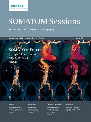
- 1. SOMATOM Sessions Answers for life in Computed Tomography December 2013 | International Edition Issue 33 SOMATOM Force: Bringing Personalized Medicine to CT Page 06 News syngo.via Frontier – Gateway to an Open Research Environment Page 31 Business Ready for the Next Revolution in Stroke Care? Page 40 Clinical Results Coronary CTA with Reduced Contrast and Radiation Dose of 0.19 mSv Page 50 Science Radiation Hygiene – Transparent and Easy Page 72
- 2. Editorial “In a general population with a very complex age and disease structure, the SOMATOM Force can solve the problems presented by every radiological situation for virtually every patient.” Professor Stefan Schönberg, MD, University Medical Center Mannheim, Germany Cover page: Dynamic CTA – 64 cm acquired with spiral 4D mode at 80 kV, 110 mAs, with 45 mL contrast. The vascular structures of the complete trunk are clearly demonstrated, and the suspected leaking from the aortic stent could be confidently ruled out. Courtesy of University Medical Center Mannheim, Germany 2 SOMATOM Sessions | December 2013 | www.siemens.com/SOMATOM-Sessions
- 3. Editorial SOMATOM Sessions | December 2013 | www.siemens.com/SOMATOM-Sessions 3 In today’s fast changing global health-care environment, Siemens’ aspiration is to contribute in two major directions. Together with our excellent network of academic partners, we continue to extend the frontiers of available diag-nostic and treatment capabilities. At the same time – and equally important – we innovate to make our technology accessible to more patients around the world. The cover article in this RSNA 2013 edition of SOMATOM Sessions intro-duces the latest frontier-shifting CT scanner from the Siemens innovation powerhouse. The new Dual Source CT SOMATOM Force* builds on the out-standing clinical success of Siemens’ unique Dual Source technology push-ing current capabilities and opening up new possibilities. SOMATOM Force features enhanced temporal, spatial and contrast resolution and intro-duces Turbo Flash scanning with up to 730 mm per second z-coverage for free-breathing CT imaging. Its out-standing tube power – already avail-able at 70 kV – makes low kV imaging accessible to virtually all patients and allows for unmatched iodine contrast enhancement. All this, together with a new level of spectral separation for high precision Dual Energy applica-tions, opens the door to CT examina-tions tailored to specific patient need. SOMATOM Force has the true poten-tial to deliver the right diagnostic pre-cision – at previously impossible low radiation and contrast dose levels. In the cover article, you will sense the excitement about the initial expe-rience of SOMATOM Force at the University Medical Centre Mannheim, Germany. The established SOMATOM Perspective, on the other hand, is an excellent example of how to leverage a leading technology position to develop a high-performance, affordable routine sys-tem with excellent economics. Origi-nally introduced as a 128- and 64-slice system, the SOMATOM Perspective family has now expanded into the 32- and 16-slice arena*. In the related article, you can see how affordability and full upgradeability within the product family together with high-tech features such as SAFIRE, iTRIM, and Single Source Dual Energy make these scanners a great choice – even for challenging economic environments. As a complement to our CT system portfolio, we are launching the syngo.via software VA30** with expanded functionality for existing applications as well as new applica-tions, such as syngo.CT Liver Analysis*. Finally, I would like to thank heartily all the participants in the International Right Dose Image Contest for so many truly wonderful contributions. Enjoy reading about these and a range of other interesting topics in this issue of SOMATOM Sessions. Dear Reader, Walter Maerzendorfer, CEO of the Computed Tomography & Radiation Oncology Business Unit, Imaging and Therapy Systems Division, Siemens Healthcare, Forchheim, Germany ** This product is 510(k) pending. Not available for sale in the U.S. ** The products/features (here mentioned) are not commercially available in all countries. Due to regulatory reasons their future availability cannot be guaranteed. Please contact your local Siemens organization for further details.
- 4. December 2013 Contents News 14 Getting Further in CT with New Imaging Possibilities 18 Improving Accuracy and Workflow Speed in Transcatheter Aortic Valve Implantation 22 Fighting Aortic Aneurysms with Modern CT Technology 26 Scientifically Validated: New Applications for CARE kV and Adaptive 4D Spiral 28 Back Among the Pioneers 31 syngo.via Frontier – Gateway to an Open Research Environment 32 Continuous Commitment to the Right Dose 34 Charting New Paths with True Dual Energy 36 Open Up New Opportunities with New Configurations 38 Getting to Grips with Stress Myocardial Perfusion Imaging Business 40 Ready for the Next Revolution in Stroke Care? 44 All-in-one 47 When Space is at a Premium – Compact High Quality Scanning Clinical Results Cardiovascular 48 Myocardial Ischemia Assessment using Adenosine-Stress Dynamic Myocardial CT Perfusion 50 Coronary CTA with Reduced Contrast and Radiation Dose of 0.19 mSv 52 Bicuspid Aortic Valve with Anomalous Coronary Artery Fistula – A Rare Incidental Coincidence Neurology 54 Dynamic Volume Perfusion CT in a Case of Childhood Moyamoya Disease before and after Surgical Revascularization 56 Differentiating an Intracranial Hemorrhage from Iodine in Acute Stroke after Intra-arterial Recanalization Acute Care 58 Diagnosis of Splenic Rupture in an 11-year-old Girl using a Sliding Gantry CT 4 SOMATOM Sessions | December 2013 | www.siemens.com/SOMATOM-Sessions 06 Cover Story SOMATOM Force: Bringing Personalized Medicine to CT
- 5. 14 18 32 72 SOMATOM Sessions | December 2013 | www.siemens.com/SOMATOM-Sessions 5 Pulmonology 60 Automated Quantification of Pulmonary Perfused Blood Volume in Acute Pulmonary Embolism using Dual Energy CTPA Urology 62 Diagnosing Small Renal Calculi using Low Dose Dual Energy CT at 0.8 mSv 64 Differentiating Stent from Stone: A New Approach using Dual Energy CT Pediatrics 66 Diagnosing Tracheal Stenosis in a 10-week-old Baby without Sedation Science 68 Image Quality in Computed Tomography 72 Radiation Hygiene – Transparent and Easy 74 Radiation Protection Scientifically Proven for Routine Practice Customer Excellence 76 Tips & Tricks: Easy Bone and Vessel Isolation 77 Clinical Workshops 2014 78 Upcoming Events & Congresses 2013/2014 79 Free DVD of the 11th SOMATOM World Summit in Orlando 80 Twenty Years of STAR – A Successful Educational Program for Radiologists 81 From Print to App: SOMATOM Sessions for Everyone 81 2014 Multislice CT Symposium in Garmisch 82 Subscriptions 83 Imprint Contents
- 6. Cover Story SOMATOM Force: Bringing Personalized Medicine to CT A quantum leap in CT engineering: Siemens’ new scanner, the SOMATOM® Force, takes over the lead in the Dual Source CT portfolio. As such it will enable radiologists not only to perform even more individualized diagnostics, but also to contribute to personalized medicine and new therapy concepts. Interdisci-plinary imaging experts at the University Medical Center Mannheim, Germany, share their experience of the first SOMATOM Force installed worldwide. By Irène Dietschi Curtain up on Siemens’ latest accom-plishment in outstanding engineering: The new SOMATOM Force CT scanner – the lead Dual Source scanner now in the market, re-writes the way CT will be used in the future for diagno-sis and treatment decisions. The premiere takes place at the German University Medical Center Mann-heim, where the Institute of Clinical Radiology is proud to be the very first research institution worldwide to 6 SOMATOM Sessions | December 2013 | www.siemens.com/SOMATOM-Sessions install the new CT system. The inter-disciplinary Mannheim specialists were excited to start working and doing translational research with their new scanner, not only because the SOMATOM Force is almost twice as 1A High resolution stent imaging – coronary CTA images acquired with Turbo Flash mode in only 0.18 s, at 70 kV and pitch 3.2, with 0.43 mSv. The patient’s heart rate varied between 58 to 70 bmp during the examination. The VRT image (Fig. 1A) shows nicely two long stents in both LAD and Cx. The curved MPR image (Fig. 1B) shows the details in the LAD stent. 1 1B
- 7. Cover Story The Institute of Clinical Radiology at the University Medical Center in Mannheim, Germany, is the very first place worldwide to install the new SOMATOM Force (Associate Professor Thomas Henzler, MD, left, Professor Stefan Schönberg, MD, right, patient, middle). SOMATOM Sessions | December 2013 | www.siemens.com/SOMATOM-Sessions 7 fast, more precise, and even more patient friendly than any of its prede-cessors, but also because they believe that it is possibly a vehicle for a new medical paradigm. “From now on, imaging is no longer limited to classi-cal diagnostics,” says Professor Stefan Schönberg, MD, director of the hospi-tal, and he explains: “As radiologists, we now have the possibility to create value-based medicine by targeting the clinical endpoint of medical proce-dures: the recovery of the patient.” CT is the imaging technique that in comparison with MRI or PET, delivers the most robust data in the long run, adds Professor Lothar Schad, PhD, director of computer-assisted clinical medicine at the faculty. “The consis-tency of quantitative data that we are able to produce using the high-end CT device cannot be equalled using any other imaging system,” he says. Schad thinks that CT will become more and more accepted as an imaging bio-marker, which will set the benchmark for other imaging techniques. The Medical Faculty Mannheim, part of Heidelberg University, has been focusing on medical technology for over a decade, according to the facul-ty’s dean Professor Uwe Bicker, MD, PhD. The renowned University Medical Center is located near the center of the city, on a campus designed for translational clinical research. The immediate proximity between the hospital, patients, and research is regarded as a huge advantage by the dean: Mannheim was successful in the national competition for the so-called research campus, funded by the German Ministry of Education and Research, which in Mannheim involves a public private partnership with Siemens. In this context, dean Uwe Bicker also points out some of the limits of tech-nological progress: “Technology by itself is useless unless its application is affordable for healthcare providers,” he says. In his opinion, this equation is one of the most challenging for the future. So, how does the SOMATOM Force contribute to solving this challenge? It does so in the first place with a number of engineering milestones, which are believed to change behav-ior patterns in CT imaging. Schönberg is enthusiastic: “In a general popula-tion with a very complex age and disease structure, this new scanner can solve the problems presented by every radiological situation for virtu-ally every patient,” he says. Associate Professor Thomas Henzler, MD, head of cardio-thoracic imaging at the Insti-tute of Clinical Radiology, is equally excited. He is convinced that “With the SOMATOM Force we have elimi-nated almost all contraindications for
- 8. The immediate proximity between hospital, patients, and research is regarded as a huge advantage by dean Professor Uwe Bicker, MD, PhD: University Medical Center Mannheim was successful in the national competition for the so called research campus, funded by the German Ministery of Educa-tion and Research, which in Mannheim involves a public private partnership with Siemens. Bicker is very proud of the reputation and the amount of expertise that has been accumulated at the campus lately, especially in imaging. He is reassured by research student Sonja Sudarski who considers Mannheim to be “invaluable for young researchers with a vision,” especially as the medical faculty is equipped with the latest technology. CT. The scanner allows precise and individualized imaging of all patients and thus changes our thinking of CT completely.” In his and Schönberg’s view the new system is especially promising in individualized diagnos-tics: Every patient should have his or her best possible diagnostic proce-dure, meaning that “the CT scan of an 85-year-old woman, weighing 60 kilograms, has in terms of parameter settings little in common with that of a 40-year-old morbidly obese man with a BMI of 40 as far as required “With the SOMATOM Force we have elimi-nated almost all contraindications for CT. The scanner allows precise and individu-alized imaging of all patients and thus changes our thinking of CT completely.” 8 SOMATOM Sessions | December 2013 | www.siemens.com/SOMATOM-Sessions dose are concerned. Only this high-end CT system is capable of offering the variety of parameters for such an individualized approach.” Individualized diagnostics is related to precision medicine. In the future, imaging will contribute substantially to the response evaluation of certain therapies, for example for cancer patients. Large nations are revising their healthcare policies radically in this respect: Henceforth, it will increas-ingly depend on the response rate – the ‘endpoints’, as Schönberg puts it – whether medical treatment will be reimbursed or not. In such an environ-ment, novel high-end systems such as the SOMATOM Force are fundamental for precise and sound decision-making by provision of quantitative data. The SOMATOM Force is expected to lead to positive changes in a number of areas. First of all, it is two steps ahead in contrast-to-noise. Low-kV imaging for all patients The engineers have put huge effort into lowering the tube voltage, while maintaining very high photon flux at a very small focal spot. Low kV exams are no longer only possible for small children and slim adults, but will be possible for practically all adults and even obese patients from now on. This, as a matter of routine, results in a reduction in radiation dose, and more: With the SOMATOM Force, the contrast-to-noise ratio has been Associate Professor Thomas Henzler, MD, University Medical Center Mannheim, Germany Cover Story
- 9. Cover Story “As radiologists, we now have the possibility to create value-based medicine by targeting the clinical endpoint of medical procedures: the recovery of the patient.” Professor Stefan Schönberg, MD, University Medical Center Mannheim, Germany SOMATOM Sessions | December 2013 | www.siemens.com/SOMATOM-Sessions 9 2 Low dose for challenging patient – 63 cm acquired with Turbo Flash mode in only 1.2 s, at 80 kV and pitch 2.4, with 1.9 mSv. The image quality is excellent although the patient’s left arm had to be kept in the scan field of view. 2 3 Dual Energy lung PBV – 32 cm acquired in only 4 s, with 55 mL contrast, at 90 / Sn 150 kV. An wedge shaped perfusion defect area is depicted in the left upper lobe, although no pulmonary emboli is present. The image quality is excellent due to greater spectrum separation. 3 improved to such an extent that a scan can be done with much lower con-trast medium amounts than previously. Whereas the average CT scanner requires between 90 and 110 milliliters for a certain application, the SOMATOM Force will produce the same image quality with just a fraction of the con-trast medium dose. For a thoracic CT, for example, volumes as low as 25 to 35 milliliters are expected. This aspect is especially important with regard to kidney protection, as Henzler explains: “In radiology, we’ve been discussing CT doses for years, even though we’ve known that nephro-pathy induced by iodinated contrast is the greater problem with some people undergoing computed tomog-raphy.” Up to 20 percent of patients, especially if they are older and suffer-ing from chronic diseases such as diabetes, might have to undergo pro-longed pre- and after-care because the contrast agent may harm their kidneys. With the new scanner, this time and cost intensive procedures might no longer be necessary. In short: SOMATOM Force is a versatile scanner. “We are expecting to be able to examine all patients adequately, even those suffering from renal insufficiency,” says Henzler.
- 10. Cover Story Dynamic CTA – 64 cm acquired with spiral 4D mode at 80 kV, 110 mAs, with 45 mL contrast. The vascular structures of the complete trunk are clearly demonstrated, and the suspected leaking from the aortic stent could be confidently ruled out. 4 10 SOMATOM Sessions | December 2013 | www.siemens.com/SOMATOM-Sessions 4 Lowest dose for early detection of lung and colon diseases For lung and colon scans, the contrast-to- noise ratio is also expected to rise significantly. Why? The SOMATOM Force has further developed the pop-ular Flash Spiral mode into the new ‘Turbo Flash mode’: Besides being almost twice as fast, it now uses two selective photon shields instead of one. The two tin filters optimize the X-ray spectrum to boost contrast between soft tissue and air in patient scans. The resulting increase in the contrast-to-noise ratio can be ‘rein-vested’ in lowering the dose, allowing a reduction of approximately 30 per-cent compared with other high-end CT’s. What this means for clinical practice is explained by Schönberg: “Computed tomography could very well become an important tool for the early detec-tion of lung cancer. The radiation dose for the risk evaluation of bronchial car-cinoma has dropped to an extent that dose is no longer an issue compared with the added value which you create with this exam.” Moreover, the ‘Turbo Flash mode’ might not only reveal lung lesions, but could also be used for the exclusion or early detection of two other major diseases: coronary heart disease and susceptibility to stroke. As for the detection of colon diseases, studies have produced excellent evidence in support of colon CT. “The results have shown that colon CT is almost equal to classical coloscopy, indicating that it could at least be applied in cases where classical coloscopy is not possible,” Schönberg says. The SOMATOM Force is not only characterized by low doses, new con-trasts, and reduced need for contrast medium, but also by speed. Compared with its predecessors, it moves breath-takingly fast. Free breathing for all patients One problem frequently found in con-ventional scans is motion artifacts, often resulting in insufficient image quality. Studies show that in cases of pneumonia, for example, a significant number of scans carried out with a standard system are unsatisfactory due to blurring. This leads to readmissions that could otherwise have been pre-vented. If doctors ask their colleagues in the radiology department to redo a scan, in one of three cases the reason is impaired image quality. The new SOMATOM Force and its novel Turbo Flash mode can help to minimize this problem: Compared with the former Flash Spiral scan mode, Turbo Flash is almost twice as fast, scanning at 737 mm/s. This means that the Turbo Flash mode literally freezes respira-tion, or other motion induced by the
- 11. Cover Story 5 Whole liver perfusion – 22 cm acquired at 80 kV, 100 mAs, with 17.58 mSv only, for an obese patient (118 kg) with liver tumor. SOMATOM Sessions | December 2013 | www.siemens.com/SOMATOM-Sessions 11 5A 5C 5B 5D diaphragm or the bowel. So, free breathing no longer impedes image quality. This is a relief, for instance, for older or maybe overweight patients who have trouble holding their breath. It is also relevant for trauma patients who are in considerable pain and/or unconscious, and in pediatric CT where doctors can now perform a CT exam without the need for sedation or controlled breath-ing in children. Finally, speed is a crucial factor in cardiac CT. “So far we have been able to perform a cardiac CT far below 1 mSv in patients whose heart rate was below 65,” says Henzler. “With the new system we will move to a situation in which we can examine higher heart rates and still remain below 1 mSv, acquiring robust results we haven’t seen before.” Henzler believes that those facts will also generate ‘clinical value’: Cardiac CT will be more consistently integrated in the workflow of the emergency room as an algorithm for patients with intermediate cardiac risk. Larger field of view With the SOMATOM Force, Siemens engineers have extended the field of view of the Flash Spiral mode to up to 50 centimeters. They accom-plished this major improvement by introducing the new powerful VECTRON tube which evolved from technology initially introduced with the renowned STRATON tube, and with the StellarInfinity detector. Based on the innovative Stellar technology, the new detector now additionally enhances resolution by 25%, and more-over extends the former z-coverage by 50%. The combination of two VECTRON tubes and two StellarInfinity detectors in a Dual Source CT enable the realizing of the unique Turbo Flash mode. Henzler is intrigued by this masterpiece of engineering and innovation. “The geometry of the detector has been changed in an ingenious way that we haven’t seen so far in computed tomography,” he says. “We will be able to show even the smallest vessels such as the coro-nary arteries or calcified lesions in perfect resolution, without having to worry about the dose or motion artifacts.” Moreover, the extended field of view will enable radiologists to scan practically all patients in Turbo Flash scan mode, including obese adults as well as patients with kidney disease. With the SOMATOM Force it is expected that the Turbo
- 12. Cover Story Flash mode will become standard, establishing ultra-high pitch scanning as the true successor of conventional spiral modes. Finally, the SOMATOM Force is likely to become the diagnostic CT tool of choice for personalized medicine: It offers precision CT at its best and is therefore two steps ahead in func-tional analysis and decision making. Dynamic perfusion at half the dose Although MRI will probably remain the benchmark for functional imaging, CT is gaining ground very fast. “CT has unmatched advantages if you need imaging in large quantities and within time limits,” Schönberg says. The SOMATOM Force offers dynamic perfusion – which usually requires high radiation doses – at up to half the dose compared with conventional state-of-the-art CT’s, e.g. for the per-fusion of the liver. The engineering solution lies in the new StellarInfinity detector (with TrueSignal technology plus its 50 percent wider coverage) and the redesign of the Adaptive Dose Shield, already known from the SOMATOM Definition Flash scanner. Matching the scan speed of the SOMATOM Force, the collimator blades can be opened and closed at twice the speed. Clinical application is possible for various organs, such as pancreas, abdomen, kidneys or the liver. Dynamic perfusion of the liver, for 12 SOMATOM Sessions | December 2013 | www.siemens.com/SOMATOM-Sessions 6 example, which at present requires doses between 60 and 70 mSv, is expected to be possible at the dose of a conventional 4-phase liver protocol. Why does this make sense? “Think of Bevacizumab,” says Henzler: “On the one hand, Bevacizumab is an impres-sive drug which suppresses angio-genesis in various cancers, including colorectal, lung or kidney. However, it is a costly drug. If you want to know whether patients are responding to the treatment, one way is to monitor these patients with repetitive perfu-sion CT.” Short-term monitoring can reveal which patients respond to anti-angiogenesis treatment, and which patients do not. Long-term CT moni-toring with functional parameters may help to detect recurrence. 6 Dynamic Runoff – 61 cm acquired with spiral 4D mode at 70 kV, 130 mAs, with 1.39 mSv and 45 mL contrast. MIP images show nicely the dynamic flow of the vascular details, and additionally, the tendons as well.
- 13. Cover Story Irène Dietschi is an award-winning Swiss science and medical writer. She writes for the public media, such as the Neue Züricher Zeitung and has published several books. The product is pending 510(k) clearance, and is not yet commercially available in the United States. The statements by Siemens customers described herein are based on results that were achieved in the customer’s unique setting. Since there is no “typical” hospital and many variables exist (e.g., hospital size, case mix, level of IT adoption) there can be no guarantee that other customers will achieve the same results. Further Information www.siemens.com/ SOMATOM-Force At the University Medical Center in Mannheim, the medical faculty has defined three fundamental topics it wants to pursue with the SOMATOM Force in various clinical studies: treatment response, nephroproctection and motion artifact reduction. From left to right: Professor Stefan Schönberg, MD, director of Radiology and Nuclear Medicine, Professor Lothar Schad, PhD, director of computer-assisted clinical medicine, Florian Lietzmann, MD, team leader of CT physics research at the institute of computer-assisted clinical medicine, Thomas Henzler, MD, head of cardio-thoracic imaging. SOMATOM Sessions | December 2013 | www.siemens.com/SOMATOM-Sessions 13 In other tumors such as gastrointestinal stromal tumors, the most promising way to assess treatment response is Dual Energy. In various studies conducted in Mannheim, iodine-related attenua-tion has proven to be a very robust response parameter, as Thomas Henzler explains. Whereas the Dual Energy scanners of the first generation had certain limits in coverage, the new scanner increases energy separation by 30 percent. “We expect that the SOMATOM Force will produce a clear-cut improvement because of the spectral upgrade,” says Henzler. In his view, Dual Energy is clearly gaining ground: Many vascular questions can be answered spectrally in post process-ing, because the two energies have been separated so effectively. In Mannheim, the medical faculty has defined three major topics that it wants to pursue with the SOMATOM Force in various clinical studies: treatment response, nephroprotec-tion, and motion artifact reduction. Researchers believe that the new standing of computed tomography could affect the workflow of a clinic substantially: CT could evolve into an all-in-one triage for new diagnostics and therapy models. One field in which this progress is already begin-ning to emerge is cardiology and the treatment of acute coronary syn-drome: At the University Medical Center Mannheim, if a patient at risk shows no relevant stenosis of the coronary arteries in cardiac CT, he or she is automatically excluded from cardiac catheter examination. “With this we have achieved three goals,” Schönberg explains: “First, we have supported our colleagues in cardiol-ogy in their daily work by making sure that catheter exams are conducted with higher therapeutic yield; second, we are more cost-effective; third, we’ve enriched the interventional scope of cardiology by referring to our colleagues those patients who actu-ally need an intervention.” Analysis of this new workflow modality has shown that it is actually cost effec-tive. In the view of dean Uwe Bicker, this is the key factor for any techno-logical innovation: If it is cost effec-tive, it will prove itself on the market. CT for cardiovascular issues is a role model for interdisciplinary workflow and decision making. But the other important domain that he and his radiology group are aiming for is oncology. Schönberg believes that cancer is the future market for the high-end CT system SOMATOM Force. “My vision is that in five years from now, oncologists around the world will prescribe innovative molecular substances based on functional imag-ing. “If you have to attend to millions of people globally, you need an efficient imaging system in order to apply those substances cost-effec-tively. And this will most likely be CT.”
- 14. News Getting Further in CT with New Imaging Possibilities Siemens continues to improve its advanced visualization platform syngo.via for CT: Combined with continuous scanner innovations, Siemens’ syngo.via VA30* offers a range of additional options for diagnosis and pre-procedural planning. By Arjen Bogaards, PhD, Jochen Dormeier, MD, Susanne Hölzer, Dominik Panwinkler, Philip Stenner, PhD Computed Tomography, Siemens Healthcare, Forchheim, Germany Comprehensive evaluation of myocardial perfusion with syngo.CT Cardiac Function – Enhancement. 2 1 2 14 SOMATOM Sessions | December 2013 | www.siemens.com/SOMATOM-Sessions rendered more flexible, too, as phy-sicians can view images on mobile devices. Sharing findings with col-leagues is also easier for fast and reliable clinical decisions. syngo.via software guides users through the entire workflow, identifying human anatomy, and enabling radiologists to deliver reliable and meaningful quan-titative results. Therefore, syngo.via VA30 is designed to meet as many clinical challenges as possible. all modalities, managing both day-to-day and more challenging cases suc-cessfully. For this reason, the software must be based on concepts that are efficient, flexible, and intelligent. Auto-mated pre-fetching of prior examina-tions and pre-processing saves valuable time, allowing physicians, technicians and IT professionals to focus on their core patient-centric tasks. Modular licensing models offer flexibility so that the system can grow in line with needs and budget. Workflows are Every year, clinical routine is becom-ing more and more demanding. Phy-sicians and clinical staff need to make best use of diagnostic technology tools available at their particular medical institution. It is essential to their job to understand diseases more comprehen-sively and make the right treatment decisions faster. This requires technol-ogy providers to continuously innovate medical imaging equipment. Siemens’ syngo.via software is designed to fur-thermore accelerate workflow across Automatic completion of manufacturer-specific AAA graft order forms with syngo.CT Rapid Stent Planning**. 1
- 15. syngo.CT Bone Reading enriched by Spine CAD. 3 4 syngo.CT Liver Analysis**: In-depth analysis of liver vascularization combined 4 with surgery planning. SOMATOM Sessions | December 2013 | www.siemens.com/SOMATOM-Sessions 15 3 Automated AAA stent planning Pre-procedural planning for the treat-ment of an abdominal or thoracic aortic aneurysm requires a precise assessment of several anatomical para-meters. Numerous vendors offer stent grafts, each of which requires its own set of measurements. Anatomical assessment and completion of the graft order forms can be tedious and time-consuming. The new syngo.CT Rapid Stent Planning** introduces automatic completion of manufacturer-specific stent order forms. That holds out the prospect of skipping all the cumber-some steps and streamlining abdomi-nal aortic stent planning. It represents an ideal extension to the Rapid Results Technology: Dedicated protocols guide the user through all length and dia-meter measurements, which are then automatically stored in the correspond-ing order form. For delivery purposes, syngo.CT Rapid Stent Planning** pro-vides three order forms as PDFs: Gore Excluder, Zenith Flex, and Medtronic Endurant. Furthermore, new order form templates can be generated to match the specific requirements of other vendors.1 Comprehensive myocardial perfusion analysis Coronary CTA is a well-established method of ruling out coronary artery stenosis. Often, an intermediate ste-nosis is found whose hemodynamic relevance may be unclear. In such cases, a myocardial stress perfusion exami-nation can help to decide whether a patient should undergo PCI2 or not. As a “one-stop shop”, CT is becoming increasingly important in the assess-ment of myocardial perfusion. Differ-ent approaches are currently available, but Siemens is the only manufacturer to offer the full spectrum of myocardial perfusion analysis: Whether simple first-pass enhancement, Dual Energy perfusion scanning, or quantitative dynamic myocardial perfusion. With syngo.via VA30 and the new perfu-sion evaluation feature in syngo.CT Cardiac Function-Enhancement, it is now possible to evaluate comprehen-sively all types of myocardial perfusion. Rather than simply looking at a first-pass enhancement scan, the quantifi-cation of iodine concentration in the myocardium and inspection of quan-titative blood flow and volume data provide additional clinical benefits.3 The visualization in AHA-compliant 17-segment polar maps and the direct overlay in MPR segments help to pinpoint the perfusion defect. With syngo.via VA30, the evaluation of myocardial perfusion becomes faster, easier, and more reliable. Advanced oncological analysis Assessment of tumor perfusion in follow-up examinations allows iden-tification of tumor viability before changes in tumor sizes are visible. Identifying these changes at an early stage of oncological treatment adds supplementary clinical information especially when following up on state-of-the-art treatment with anti-angiogenic drugs. The “body perfu-sion” functionality is now available in syngo.via and provides quantification of blood flow, blood volume, and per-meability, combined with automated motion correction for improved ana-tomical alignment. In addition to its 1 Adobe Acrobat Professional required; 2 PCI: Percutaneous coronary intervention; 3 CT DE Heart PBV and/or syngo VPCT Body-Myocardium required
- 16. News 5 6 use in oncology, a further clinical application is to assess perfusion in cases of organ transplantation. The new syngo.CT Liver Analysis** delivers in-depth clinical insights based on comprehensive analysis of CT data-sets and tools for surgery planning. For the surgeon it is crucial to know the precise size and location of tumors before the operation. It is also essen-tial to assess the amount of liver tissue that is to be resected and the exact anatomical vascular supply to the affected liver segments. By dissecting the liver virtually using the software, the physician is able to compare the amount of resected and residual liver tissue – one of the key factors in the surgery outcome. syngo.CT Liver Analysis** supports these pre-opera-tive planning steps by combining tailored functions and tools with intu-itive workflow guidance. Extended bone reading support Building on the success of syngo.CT Bone Reading, the application has been enhanced with CAD* (Computer Assessment of diffuse tumor infiltrations with syngo.CT DE Bone Marrow**. Aided Detection) functions to identify suspicious spine lesions. Intended for use as a second reader tool after the initial read has been completed, this supplementary tool draws the radiol-ogists attention to regions of interest (ROI) that may have been initially overlooked. In addition to the revolu-tionary new visualization in bone read-ing – which adapts complex anatomies to reading needs – this new feature has demonstrated potential in detect-ing lytic and blastic metastasis as reported in a scientific publication from the Department of Radiology, University Hospital Erlangen.[1] With these new additions, syngo.via VA30 offers a comprehensive portfolio enabling holistic oncological reading. CT imaging – the cornerstone of stroke care Across the globe, 1 in 6 people will suffer a stroke at some point in their life. It is one of the world’s most threat-ening diseases. Almost two million brain cells could be lost every minute if a stroke patient is left untreated. Fast treatment is essential to improve 16 SOMATOM Sessions | December 2013 | www.siemens.com/SOMATOM-Sessions the chances of a good outcome. How-ever, the time it takes from the stroke patient arriving in the emergency department to receiving thrombolytic drugs (door-to-needle times) remains a major challenge in many hospitals. An important element in this cascade of events is the imaging software that is connected to the CT scanner. It is decisive to increase speed and confi-dence of the diagnosis and conse-quently for the implementation and monitoring of effective treatment. Generally, a non-contrast CT scan and single phase CT Angiography will be administered to exclude bleeding and confirm the presence of an occlu-sion in order to determine eligibility for thrombolytic drug administration. syngo.CT Neuro Perfusion can help to visualize the size of the core infarct and penumbra; the latter represents tissues that may be salvaged through further reperfusion therapy. Excitingly, 4D CT Angiography is used increasingly and several novel applications are beginning to emerge. syngo.CT Dynamic Angio can create View of the neurovasculature from 6 arch to vertex with syngo.CT Neuro DSA. 5
- 17. 7 Evaluation of multiple monoenergetic ROIs with syngo.CT DE Monoenergetic Plus**. News ** The products/features (here mentioned) are not commercially available in all countries. Due to regulatory reasons their future availability cannot be guaranteed. Please contact your local Siemens organization for further details. ** This product is 510(k) pending. Not available for sale in the U.S. SOMATOM Sessions | December 2013 | www.siemens.com/SOMATOM-Sessions 17 movies that visualize the flow of con-trast from arterial to venous phase and depict tMIPs. This can help the clini-cian better assess the collateral status and define the occlusion length in stroke.[3] As such, 4D CT Angiography has potential in helping to select the patient optimally suited for interven-tional clot retrieval. All indications at the present suggest that CT imaging will remain the cornerstone of stroke care. New boost for Dual Energy CT with syngo.via VA30 True Dual Energy offers extended diagnostic possibilities taking CT imag-ing beyond morphology by enabling exploration of functional and quanti-tative aspects. And progress still con-tinues. A highlight of the syngo.via VA30 is the new Dual Energy application syngo.CT DE Bone Marrow**. The bone marrow can be affected by various pathologies, such as bone bruises after trauma as well as by diffuse tumor infiltrations. Until today, the major modality for imaging these patholo-gies has been MRI. With the benefit of True Dual Energy, CT imaging can now also aid in the diagnosis. syngo.CT DE Bone Marrow** allows for the seg-mentation and the visualization (color-coding) of the bone marrow based on a material decomposition into bone marrow and calcium. This application can be used for both Dual Source and Single Source Dual Energy datasets. Furthermore the syngo.CT DE Virtual Unenhanced* application has been complemented in order to address a wider clinical spectrum. While the well-established Liver VNC algorithm enables quantification of the iodine uptake in the liver tissue, the new Vir-tual Unenhanced algorithm has been improved for optimized visualization of those organs that – in contrast to the liver – do not contain variable amounts of fat, such as the lung, kid-ney, and pancreas. The iodine uptake may give additional indications about the malignancy of a lesion. Moreover, the effectiveness of a therapy can be validated by evaluating the develop-ment of the iodine uptake in the treated lesion before and after treatment. Monoenergetic imaging has become a reliable application to improve image quality as well as for effectively reducing metal artifacts. syngo.via VA30 together with syngo.CT DE Mono-energetic Plus** offers a new, power-ful algorithm allowing for a better quantitative assessment of different tissues and lesions by displaying multiple monoenergetic ROIs and the associated absorption curves. A further benefit for research and diagnostic tasks is the ability to export the statis-tical information to the file system for more in-depth evaluation. syngo.via VA30 offers a broader range of tools to meet today’s grow-ing clinical requirements with the support of high quality CT imaging. References [1] Automatic detection of lytic and blastic thoracolumbar spine metastases on computed tomography. Hammon M. et al; Eur Radiol. 2013 July; 23(7): 1862–1870. [2] Meretoja A et al. Reducing in-hospital delay to 20 minutes in stroke throm-bolysis. Neurology. 2012, 79:306-13. [3] Frölich AM et al. 4D CT Angiography More Closely Defines Intracranial Thrombus Burden Than Single-Phase CT Angiography. AJNR Am J Neuroradiol. Published online before print April 25, 2013. Further Information www.siemens.com/ ct-clinical-engines 7a 7B Further steps will follow opening up to users the opportunity to fully exploit their diagnostic technology. syngo.via can be used as a standalone device or together with a variety of syngo.via-based software options, which are medical devices in their own right. Not for diagnostic use.
- 18. News Improving Accuracy and Workflow Speed in Transcatheter Aortic Valve Implantation Computed tomography provides valuable information for the planning of transcatheter aortic valve implantation, and the syngo.CT Cardiac Function – Valve Pilot application of syngo.via speeds up workflow while increasing accuracy and safety for patients. By Sameh Fahmy, MS 18 SOMATOM Sessions | December 2013 | www.siemens.com/SOMATOM-Sessions selected. Choosing a prosthesis that is too small can lead to a paravalvular leak, for example, while fitting one that is too large could cause a catastrophic rupture of the aortic root. In addition, the catheters used in the procedure are relatively large, so physicians must be able to reliably assess calcifications, ment.[1] However, careful planning of this advanced procedure which is necessary for optimal patient outcome can present a number of challenges for physicians. Exact measurements of the anatomy of the heart are necessary so that the appropriate sized prosthesis is Transcatheter aortic valve implanta-tion (TAVI, also known as transcatheter aortic valve replacement (TAVR) in the U.S.) has been shown to signifi-cantly prolong the lives of those severe aortic valvular stenosis patients, who – because of comorbidities – are not candidates for surgical valve replace- syngo.CT Cardiac Function – Valve Pilot: physicians are able to work with zero-delay for quantitative assessment of the aortic annulus. 1 1
- 19. Professor Stephan Achenbach, MD, Department of Cardiology, University of Erlangen- Nürnberg SOMATOM Sessions | December 2013 | www.siemens.com/SOMATOM-Sessions 19 stenoses, and the diameter of the peripheral arteries, in order to select a suitable access route and avoid potentially fatal complications. Despite these challenges, physicians such as Professor U. Joseph Schoepf, MD and Professor Stephan Achenbach, MD are able to plan the TAVI procedure efficiently, accurately, and with confi-dence. Joseph Schöpf is Professor of Radiology and Medicine and Director of CT Research and Development at the Medical University of South Carolina in the United States. Stephan Achenbach is Chairman of the Department of Car-diology at the University of Erlangen- Nürnberg in Germany. They both were among the first to test the application syngo.CT Cardiac Function – Valve Pilot. The software provides a dedicated workflow for CT TAVI planning; auto-matically measuring the dimensions of the aortic annulus providing single-click localization and quantification of the smallest iliac diameter, and auto-matically calculating the corresponding C-arm angulation for a given projection. “It enhances our workflow efficiency, which is an aspect that is becoming increasingly significant – especially in centers with extremely high vol-umes,” Schoepf says. “What is more important for me is that it enhances accuracy and safety for patients.” Assessing critical structures easily Worldwide, an estimated 40,000 patients have received TAVI.[2] The landmark, multicenter trial PARTNER (Placement of AoRTic TraNscathetER Valve) demonstrated that the TAVI procedure reduced all-cause mortal-ity by nearly 50% in patients who were ineligible for the open proce-dure.[ 1] Furthermore, key secondary end points, such as patient condition, had significantly improved by the time of the one-year follow up. In the group of patients who were defined as having a high surgical risk, TAVI was found to be non-inferior to surgi-cal aortic valve implantation. Mortal-ity rates after one year were 24.2% for TAVI, compared with 26.8% for the surgical procedure.[3] While the clinical trials that led to the introduction of the TAVI procedure used echocardiography and conven-tional angiography for pre-procedural planning, Achenbach stresses that CT provides the information that improves the safety and accuracy of the proce-dure. “The question of whether there are arteries of the body, especially in the legs, available to use for an access route can, by far, be best answered by CT,” Achenbach says. “And we now have data that clearly show that CT is the best tool for choosing the correct size of prosthesis.” The manual detection and measure-ment of the annulus – the structure demarcated by the hinges of the aortic valve leaflets – is a particularly cumbersome and time-consuming process, but one at which the soft-ware excels. As the case is opened, it displays the annular plane and calcu-lates critical measures, such as the area, and long and short axes of the annulus. The ostium views help to determine the distance between the coronary ostia and the annulus plane. A process that could otherwise take “CT adds tremendously to the TAVI procedure by making it safer.” News
- 20. News up to approximately 20 minutes now happens almost instantaneously, and with an unparalleled level of repro-ducibility. In a study presented at the 2012 annual meeting of the Radiological Society of North America, Schoepf and his colleagues found that the software was in excellent agreement with human observers.[4] He adds that even in cases where manual adjustments are necessary, the use of the software still saves time by giving radiologists a good starting point from which they can work. “These sorts of measurements are crucial going into the procedure, but they’re also where substantial human error can occur – with pretty dire consequences,” Schoepf says. “The beauty of having a computer algo-rithm to do it is that if you give it the same task twice, it comes up with the same measure.” Choosing the appropriate prosthesis is a balancing act for physicians. Patients who develop a paravalvular leak have a higher likelihood of death following TAVI;[5] however, a recent study demonstrated that using CT substantially reduces the incidence of paravalvular aortic regurgitation, when compared to transesophageal echocardiography based sizing – with rates of 7.5% and 21.9%, respec-tively.[ 6] A similar balancing act occurs in measuring the ostia. A measurement that is too short will result in the unnecessary exclusion of a patient, while one that is too large has the potential to result in the implantation of a prosthesis that occludes a coro-nary artery. 20 SOMATOM Sessions | December 2013 | www.siemens.com/SOMATOM-Sessions Schoepf says that helping to deter-mine a suitable access route for the relatively large catheters required by the procedure is another area where the software excels. It offers single-click localization and quantification of the smallest iliac diameter, as well as visualization and subtraction of aortic calcifications. Furthermore, it auto-matically calculates the area and dia-meter of vessels: “Even the most expe-rienced observers derive substantial value from features like these because they improve quantitative accuracy and workflow,” Schoepf says. Minimizing contrast dose to improve safety According to Achenbach, one feature of syngo.via that is of particular bene-fit to patients is the automatic calcula-tion of the corresponding C-arm angu-lation for a given CT projection. This “The fundamental advantage of the software is that it finds the aortic annulus automatically.” Professor U. Joseph Schoepf, MD, Department of Radiology, Medical University of South Carolina, Charleston, U.S.
- 21. News With the SOMATOM Definition Flash very little amounts of contrast are required to acquire the entire anatomy relevant for TAVI planning (only 40 mL in this case) Courtesy of University of Erlangen-Nürnberg, Erlangen, Germany SOMATOM Sessions | December 2013 | www.siemens.com/SOMATOM-Sessions 21 feature improves workflow, while also increasing the accuracy of the proce-dure as physicians in the cath lab use the best-possible viewing angle. Achenbach notes that a large percent-age of patients undergoing TAVI have renal insufficiency, which makes keep-ing contrast dose usage to a minimum an important consideration for patient safety. “There are several methods to find the optimum viewing angle in the cath lab, but they all require contrast dose,” Achenbach says. “If you know which angle to use to look at the aortic valve, you don’t have to do extra imag-ing in the cath lab to find this out.” Achenbach and Schoepf both use a SOMATOM® Definition Flash Dual Source CT scanner for TAVI planning to fur-ther minimize contrast dose. Planning the procedure requires a relatively large scan range, from the shoulder to the hip, but the speed with which the scanner acquires data allows them to keep contrast dose to a minimum. In a study of 42 patients, Achenbach and his colleagues were able to assess aortic root anatomy and vascular access in less than 2 seconds, using 40 mL of iodinated contrast agent.[7] “That we can do everything so quickly and with so little contrast is of great benefit to patients undergoing the TAVI proce-dure,” Achenbach says, “and you’re not sacrificing any image quality.” Improving outcomes, reducing costs Patients who undergo TAVI have sub-stantially shorter hospital stays than those undergoing surgical valve replace-ment.[ 3] Also, patients treated medi-cally have higher rates of rehospital-ization than those undergoing TAVI.[1] By improving patient outcomes, the accuracy and safety offered by syngo.via has the potential to decrease costs further. Achenbach notes that TAVI pro-cedures require a large clinical team; therefore, even saving 10 to 15 min-utes during the procedure by deter-mining the optimal viewing angle in advance can make a big difference. As physicians’ experience with the pro-cedure grows, Schoepf and Achenbach believe that there will be fewer compli-cations and better outcomes. Currently, 2 2A two major manufacturers produce the prostheses, but the physicians expect increased competition from other manufacturers to drive down costs further. TAVI is currently indicated for patients who are inoperable because of comor-bid conditions, as well as those who are considered a high surgical risk. However, the minimally invasive nature of the procedure makes it appealing to younger and healthier patients: “As the results of the procedure get better and better, there’s less incen-tive to do conventional surgery, even maybe in healthier patients,“ Achenbach says. “So the question of who receives this procedure and who undergoes conventional surgery will constantly need to be recalibrated.” References [1] Leon MB, et al. Transcatheter aortic-valve implantation for aortic stenosis in patients who cannot undergo surgery. N Engl J Med. 2010;363:1597-607. [2] Holmes DR, et al. 2012 ACCF/AATS/SCAI/ STS expert consensus document on transcatheter aortic valve replacement. J Thorac Cardiovasc Surg. 2012 Sep; 144(3):e29-84. [3] Smith CR et. al., Transcatheter versus Surgical Aortic-Valve Replacement in High-Risk Patients N Engl J Med 2011; 364:2187-2198). [4] Schoepf JU et. al., Automated annulus assessment accuracy in comparison to standard software and manual assess-ment. RSNA 2012 [5] Tamburino C et. al., Incidence and predictors of early and late mortality after transcatheter aortic valve implan-tation in 663 patients with severe aortic stenosis. Circulation, 123 (2011), pp. 299-308 [6] Jilaihawi H, et al. Cross-sectional computed tomographic assessment improves accuracy of aortic annular sizing for transcatheter aortic valve replacement and reduces the incidence of paravalvular aortic regurgitation. J Am Coll Cardiol. 2012;59:1275-1286 [7] Wuest W, et al. Dual source multide-tector CT-angiography before Trans-catheter Aortic Valve Implantation (TAVI) using a high-pitch spiral acquisition mode. Eur Radiol. 2012 Jan;22(1):51-8. Sameh Fahmy, MS, is an award-winning freelance medical and technology reporter based in Athens, Georgia, USA. The statements by Siemens’ customers described herein are based on results that were achieved in the customer’s unique setting. Since there is no “typical” hospital and many variables exist (e.g., hospital size, case mix, level of IT adoption) there can be no guarantee that other customers will achieve the same results. 2B Further Information www.siemens.com/CT-TAVI
- 22. Svenja Hennigs, MD, is Head of the hospital’s Department of Radiology and Nuclear Medicine at the “Knappschaftskrankenhaus” in Bottrop, Germany. Fighting Aortic Aneurysms with Modern CT Technology A hospital that formerly served coal workers and their families has reinvented itself as a modern center of medical care. A distinct focus lies on the catheter-based treatment of life-threatening aortic aneurysms, a method that requires regular CT follow-up. The SOMATOM® Definition Edge has helped the hospital “Knappschaftskrankenhaus” in Bottrop in Germany to speed up these examinations and to reduce radiation exposure considerably. By Philipp Grätzel von Grätz, MD 22 SOMATOM Sessions | December 2013 | www.siemens.com/SOMATOM-Sessions
- 23. Without modern radiology, and particularly modern CT examinations, stent treatments of aortic aneurysms are unthinkable – even in Knappschaftskrankenhaus in Bottrop. News SOMATOM Sessions | December 2013 | www.siemens.com/SOMATOM-Sessions 23 The abdominal aorta is the main sup-plier of blood to the abdomen and the lower extremities. Technically, it is a flexible tube with an average diameter of around two centimeters. Most peo-ple will never be aware of what is the largest artery in the human body. It is an organ that normally works silently for decades. But there are exceptions: Approximately one in thirty adults will develop an aneurysm in the abdominal aorta – defined as an increase in vessel diameter to more than three centimeters. Stent treatment as a new standard of care Aortic aneurysms with a diameter of more than four to five centimeters are considered critical from a medical point of view. The larger the diameter, the higher the risk of a rupture. And a rup-ture of this high-volume, high-pressure artery can easily result in death: Nine out of ten patients with this condition will die. The perfidious thing about these ruptures is that they happen with-out warning, which is why abdominal aortic aneurysms are sometimes called the “silent killers.” Ruptures of aortic aneurysms, in other words, need to be avoided at all costs, and they can be. Aortic aneurysms are a treatable condition. For decades, open surgery was the method of choice. Today, most aortic aneurysms are treated by catheter-based implantation of aortic stents – a quicker and far less invasive method of permanently stabilizing the artery. The Knappschafts-krankenhaus in Bottrop is one of sev-eral hospitals that have specialized in this new method. “Our vascular sur-geons perform more than 150 of these procedures per year. This means that our hospital is among the leading insti-tutions in Germany in this field,” says Svenja Hennigs, MD, Head of the hos-pital’s Department of Radiology and Nuclear Medicine. CT as a tool for planning and follow-up There is a good reason why Svenja Hennigs, as a radiologist, is such an advocate of aortic stenting: Without modern radiology, and particularly modern CT examinations, stent treat-ments of aortic aneurysms would be unthinkable. Every single patient needs numerous CT examinations before and after the stent implanta-tion. The radiologist is the indispens-able partner of the vascular surgeon who is confronted with an aneurysm patient. First of all, the CT is a planning tool: “We need a good reconstruction of the aorta and the origins of the renal and mesenteric arteries before the intervention to choose the ideal pros-thesis,” explains Hennigs. “This is why we use thin slices of one millimeter to get the necessary raw data and to be able to provide a proper 3D model for our surgeons.” After the stent implantation, the CT examination becomes the single most important tool for following up the patients. The vascular surgeons at the Knappschaftskrankenhaus examine the patients on the day after the implantation. There are further follow-up examinations after three, six and twelve months. Later on, the frequency of examinations depends on the indi-vidual situation. Most patients come at least once a year. “This means that we have far more CT examina-tions of aortic aneurysm patients per year than we have surgeries. At the moment, the department of radiology performs 15 such examinations per week. And this number will probably increase further in the years to come.” Watching out for endoleaks The most important reason for regular CT follow-up examinations is the search for endoleaks. These are defined as persistent blood flow within the aneurysm sac. There are five dif-ferent types of endoleak with different characteristics and different degrees of clinical relevance. As a rule, an endo-leak increases the risk of an expan-sion of the aneurysm and, ultimately, the risk of rupture. This is why endo-leaks need to be detected and closely monitored. In some cases, a second intervention may be necessary. The problem with repeated CT exami-nations is that they add up to fairly high radiation dosages over the years. “Together with tumor patients, aortic aneurysm patients are probably the patients with the highest radiation exposure,” says Hennigs. But there is good news for the aneurysm patients at the Knappschaftskrankenhaus. Thanks to the new SOMATOM Definition Edge CT system that was installed in Bottrop in March 2013, the average radiation dose per exam-ination has been reduced considerably. Cutting-edge technology slashes radiation dose Hennigs recalls that the hospital had been working with a 64-slice CT sys-tem for many years. “At some time,
- 24. News The Knappschaftskrankenhaus in Bottrop is among the leading institutions in Germany in treating aortic aneurysms by catheter-based implantation of aortic stents. we started thinking about upgrading to a new one. When I heard about the SOMATOM Definition Edge in 2012, I was immediately interested. We learned that the new Stellar detector and the iterative reconstruction algo-rithm SAFIRE can lead to a reduction in radiation dose of up to 60 percent* under optimal conditions. This really thrilled us, because it was exactly what we were looking for.” Siemens Stellar detector is the first fully-integrated detector. It reduces electronic noise, which helps to reduce radiation dose and to improve spatial resolution by generating ultra-thin slices. When the new CT was installed in Bottrop, the radiologists there were quickly convinced of the system’s benefits. “In a lean patient with an aortic aneu-rysm, we often need less than half the radiation dose than we did with the previous 64-slice system.” As expected, adipose patients are some-what more challenging. “But even in these situations, the dose is down by 20 to 30 percent in many patients.” Together with Siemens, Hennigs is currently evaluating the average dose reduction that was achieved with the SOMATOM Definition Edge in a series of 50 aortic aneurysm patients. Assistants allowed to think The reduction in radiation dose is not only good for aortic aneurysm patients: “It’s good for every patient who needs 24 SOMATOM Sessions | December 2013 | www.siemens.com/SOMATOM-Sessions a CT. And it is particularly good for tumor patients or certain patients with neurological conditions who need to be examined again and again,” explains Hennigs. Another impressive example that she cites is patients who need preventive CT examinations for lung tumors. Such examinations are being carried out on asbestos workers. But they are also increasingly recommended for heavy smokers. “In past days, a conventional lung CT would require a radiation dose of 8 to 10 millisievert (mSv). Modern low-dose CTs bring that down to 3 to 4 mSv. With the SOMATOM Definition Edge, we are able to do a low-dose CT of the lung at 1 to 1.5 mSv. And believe it or not, we had one patient who needed as little as 0.8 mSv.” In combination with the new Stellar detector, the iterative image recon-struction technology SAFIRE is the key to achieving the outstanding low radi-ation doses. SAFIRE features a set of pre-specified programs. It also allows for a certain degree of manual control, as Svenja Hennigs explains: “We turn SAFIRE on for practically every patient. The radiological assistant then decides individually whether he or she can risk going down a little further or not. The SOMATOM Definition Edge is, in fact, the first CT system for many years that allows the radiological assistant to think in new directions.” Quicker examinations, higher image quality Having worked with the SOMATOM Definition Edge for four months, Hennigs and her colleagues have dis-covered various additional benefits The Knappschaftskrankenhaus Bottrop opened in 1931 as a hospital for miners who worked in the numerous coal mines of the Ruhr Basin in Germany – at that time, the powerhouse of Central Europe. There is still a small sculpture in the entrance hall that reminds visitors and patients of these roots: St. Barbara, patron saint of miners. Today, the Knapp-schaftskrankenhaus is a modern hospital for acute and regular care with 346 beds in nine clinical depart-ments. More than 50,000 patients are treated per year, a large number of which are outpatients. The department of radiology keeps nine radiologists and 15 radiology assistants busy. Apart from the SOMATOM Definition Edge, they have a Siemens MRI, three angiography systems, a mammography unit, and two workplaces plus nuclear medicine and ultrasound. Coal in the genes
- 25. Image comparison for follow-up scan of same patient between previous 64-slice system (Fig. 1A) and new SOMATOM Definition Edge (Fig. 1B) with SAFIRE at half the dose with comparable diagnostic image quality. Courtesy of Knappschaftskrankenhaus Bottrop, Germany News SOMATOM Sessions | December 2013 | www.siemens.com/SOMATOM-Sessions 25 to the new system. First of all, speed. “We are now able to perform a thoracic CT within 3 seconds, and an abdomen takes around 12 seconds. It’s extremely quick. The limiting factor is not the examination, but getting the patient in and out again.” Hennigs is also very impressed by the image quality that the SOMATOM Definition Edge provides: “I would put it this way: The images are more bril-liant. This becomes particularly obvious with CT examinations of bone fractures. When I compare high-resolution images of fracture lines from the same patient recorded with the previous 64-slice CT against the new one, the overall impres-sion is totally different. It is far better now, much clearer and more detailed.” A quantum leap When looking at the modern CT sys-tems available on the market last year, Hennigs also considered other vendors instead of SOMATOM Definition Edge system. “But I thought that the more compact system in combination with high end detector technology fitted our needs better.” The fact that the SOMATOM Definition Edge also fea-tures Dual Energy (DE) technology made the decision even easier: “Our urologists and nephrologists, in par-ticular, asked us to provide DE tech-nology for visualizing urinary tract stones and uric acid crystals. So we decided to also acquire the DE appli-cations that come with the SOMATOM Definition Edge, and we are now using it regularly. It provides excellent DECT images.“ All in all, neither the radiologists nor radiological assistants in Bottrop miss the previous 64-slice system: “The SOMATOM Definition Edge really is a quantum leap forward. We are still discovering new possibilities with it. And once you have learned to work with all its features, the results are fantastic.” * In clinical practice, the use of SAFIRE may reduce CT patient dose depending on the clinical task, patient size, anatomical location, and clinical practice. A consultation with a radiologist and a physicist should be made to determine the appro-priate dose to obtain diagnostic image quality for the particular clinical task. The following test method was used to determine a 54 to 60% dose reduction when using the SAFIRE reconstruc-tion software. Noise, CT numbers, homogeneity, low contrast resolution and high contrast resolu-tion were assessed in a Gammex 438 phantom. Low dose data reconstructed with SAFIRE showed the same image quality compared to full dose data based on this test. Data on file. 1A Previous 64-slice system (Fig. 1A) SOMATOM Definition Edge (Fig. 1B) kV-Setting 120 kV, 95 mAs 100 kV, 92 mAs DLP 318 mGy cm 158 mGy cm CTDI 7.32 mGy 3.66 mGy 1 1B Philipp Grätzel von Grätz is a medical doctor turned freelance writer and book author based in Berlin, Germany. His focus is on biomedicine, medical technology, health IT, and health policy. The statements by Siemens’ customers described herein are based on results that were achieved in the customer’s unique setting. Since there is no “typical” hospital and many variables exist (e.g., hospital size, case mix, level of IT adoption) there can be no guarantee that other customers will achieve the same results.
- 26. Recently published scientific studies present evidence of the additional benefits of CARE kV and the Adaptive 4D Spiral – which until now had only been described in terms of potential. CARE kV Adjusting the tube voltage for every CT scan can help deliver the right dose to every patient; and varying kV values for different applications can help achieve optimal image quality. This potential was known but the adjustments were too complicated to do manually, as the tube current then needs to be adapted accordingly. CARE kV automatically selects the tube voltage and CARE Dose4D adapts the tube current. Many scientific studies have shown the benefits of CARE kV for different types of examination.[1,2] Yet, stud-ies focusing on pediatric CT imaging with CARE kV had been lacking until researchers from Mallinckrodt Institute of Radiology, St. Louis, US, published their latest results. In their study they first evaluated the potential of CARE kV for CT Angiography exami-nations using three different-sized pediatric phantoms.[3] In the second step, these findings were used in a study with 87 pediatric patients.[4] The tube voltage set as reference was 120 kV. With CARE kV, the tube voltage was lowered to 100 kV, 80 kV, or even 70 kV in 82 of these 87 patients (i.e. 94% of the cases). Image quality was assessed subjectively; 15 of these cases were also compared with a previous CT scan at 120 kV. Contrast-to-noise ratio (CNR) was evaluated in these cases. The authors outline the implications for patient care: “Use of automated kilovoltage selection technology appears to be an effective strategy for optimizing tube voltage selection and reducing radiation dose while maintaining image quality in contrast-enhanced pediatric CT and should be introduced into routine clinical practice.”[4] Adaptive 4D Spiral CT Perfusion imaging with Adaptive 4D Spiral delivers qualitative and quantitative information about perfu-sion patterns. In recent years, scien-tific studies have been published that focus on different organs and tumor entities.[5,6] Usually, the examina-tions had to be performed with a tube This examination of a baby was included in a study.[4] The VRT shows well enhanced mediastinal vessels and a persistent left superior vena cava (arrow). The effective dose for this scan was 0.36 mSv. Courtesy of Mallinckrodt Institute of Radiology, Saint Louis, USA 26 SOMATOM Sessions | December 2013 | www.siemens.com/SOMATOM-Sessions 1 voltage of 100 kV. In a phantom study, CT perfusion examinations with SOMATOM® Definition Flash – either with conventional detector technology or the Stellar detector – were com-pared at 80 kV and 100 kV. In view of the minimized electronic noise, the authors conclude: “The Stellar detector allows the routine use of 80 kV for abdominal perfusion imaging. For identical CNR this reduces the dose by 35% compared to 100 kV.”[7] New cancer treatment options – including anti-angiogenic drugs that influence blood supply to a tumor – have been introduced and are still under intense evaluation. Researchers from University of Lille, France have used Adaptive 4D Spiral technology to assess treatment out-comes in the case of non-small-cell lung cancer (NSCLC).[8] In group 1, 17 patients received conventional chemotherapy, 23 patients in group 2 were also given an anti-angiogenic drug (Bevacizumab). The perfusion information was derived before treat-ment begin and then at three later points in time. Perfusion was quanti-fied using two new parameters: total tumor vascular volume (TVV, in mL), which is based on blood volume; and total tumor extravascular flow (TEF, in mL/min), which is based on the volume transfer constant ktrans – also known as flow extraction product. In addition, RECIST (Response Evaluation Criteria in Solid Tumors) data was col-lected to assess tumor size. Given the changes in perfusion parameters and in RECIST, the authors summarized a key finding: “Specific therapeutic effects of anti-angiogenic drugs can be detected before tumour shrinkage.”[8] News By Heidrun Endt, MD Computed Tomography, Siemens Healthcare, Forchheim, Germany 1 New Applications for CARE kV and Adaptive 4D Spiral
- 27. Outlook Since the introduction of CARE kV and Adaptive 4D Spiral, several studies have been published indicating broad potential application. This portfolio has now been extended. In the case of CARE kV, initial studies have shown the benefits when scanning young patients. Further research is expected on low kV imaging in pediatric CT, in particular. For Adaptive 4D Spiral, the perfusion evaluation of tumors was scientifically validated for different clinical questions.[5,6] New develop-ments in other areas, such as with the Stellar detector, may lead to new options for existing technologies.[7] The possibility of perfusion imaging at 80 kV will be of great interest to the scientific community. The study from France shows that with Adap-tive 4D Spiral technology a prediction of a treatment response to anti-angio-genic drugs is possible for cases of NSCLC. In their conclusion, the authors indicate the potential: “If these prom-ising preliminary results can be con-firmed by larger studies, perfusion CT could represent a very useful non-invasive tool for thoracic oncologists to manage anti-angiogenic treat-ments in clinical practice with the objective of avoiding pointless thera-pies and their potential adverse events as well as cost savings.”[8] The examination of this 62-year-old patient suffering from an adenocarcinoma of the lung in the left lower lobe was included in the study.[8] Images on the left-hand side show the situation before treatment, images on the right-hand side were obtained after one cycle of therapy (including anti-angiogenic drugs). Conventional images (mediastinal window) are shown in Fig. 2A and 2B. Perfusion information can be derived from Fig. 2C and 2D (TVV) and 2E and 2F (TEV). The perfusion maps show a decrease in vascularity (TVV from 4.4 mL to 1.6 mL; TEF from 4.3 mL to 2.2 mL) whereas no change in tumor size could yet be seen in the mediastinal images. Courtesy of University Hospital of Lille, France SOMATOM Sessions | December 2013 | www.siemens.com/SOMATOM-Sessions 27 2 News 2A 2C 2E 2B 2D 2F References [1] Eller A, et al. Invest Radiol. 2012 Oct;47(10):559-65. [2] Park YJ, et al. J Cardiovasc Comput Tomogr. 2012 May-Jun;6(3):184-90. [3] Siegel MJ, et al. Invest Radiol. 2013 Aug;48(8):584-9. [4] Siegel MJ, et al. Radiology. 2013 Aug;268(2):538-47. [5] Goetti R, et al. Invest Radiol. 2012 Jan;47(1):18-24. [6] Reiner CS, et al. Invest Radiol. 2012 Jan;47(1):33-40. [7] Klotz E, et al. Performance evaluation of a new CT detector with minimal electronic noise for low dose abdominal perfusion imaging. Insights Imaging (2013) 4 (Suppl 1):200 [8] Tacelli N, et al. Eur Radiol. 2013 Aug;23(8):2127-36.
- 28. The initial positive assessment of the SOMATOM Perspective has continued at Sainte-Marie Medical Imaging Center in Osny, near Paris, France. Back Among the Pioneers One of the first ever installations of a SOMATOM® Perspective CT scanner was at Sainte-Marie Medical Imaging Center in Osny, near Paris, France in January 2012. One and a half years later, SOMATOM Sessions returned to the center to discover whether the initial enthusiasm and hopes were justified. The positive assessment made at that time was entirely confirmed. And – particularly attractive in this era of austerity – at an affordable price. By Christian Rayr 28 SOMATOM Sessions | December 2013 | www.siemens.com/SOMATOM-Sessions
- 29. “eMode reduces material wear, extends the machine’s lifetime, and eliminates downtime. We’ve had no breakdowns or annoying problems to report.” Alexandre Fuchs, MD, Sainte-Marie Medical Imaging Center, Osny, France News SOMATOM Sessions | December 2013 | www.siemens.com/SOMATOM-Sessions 29 Alexandre Fuchs, MD is a doctor, spe-cialising in diagnostic and co-director of Imagerie Medicale Sainte Marie. His initial, positive assessment of the SOMATOM Perspective has not changed. The center is guided by the principle of achieving the utmost excellence and, therefore always seeks the best diag-nostic equipment for its patients. And this means that Fuchs is in a position to make comparisons. “The SOMATOM Perspective delivers perfect diagnostic efficiency“ he notes. So far, almost 10,000 patients have benefited from its use. Franck Lamesa, general super-visor of the Sainte-Marie Medical Imaging Center, adds: “The number of scans conducted currently stands at 12,400. With the SOMATOM Perspective, we have performed approximately 5,500 abdominopelvic scans, as well as 2,300 thoracic scans, 1,200 lumbar scans, 800 brain scans, and 800 sinus scans.” Good results have been achieved in all pathological areas: cancer, pediatrics, rheumatology, cardiology, and neurol-ogy, to name just a few. “Work in oncol-ogy is ongoing here,” Fuchs points out, “because we collaborate closely with the Sainte-Marie Medical Imaging Center and its cancer treatment center next door. For us, the work involves standard scans. Image acquisition is perfect, and all the preparatory and analytical work is carried out with the help of syngo.via. We are one of the major users of this software, espe-cially its applications for oncology.” Post-treatment image data are vali-dated by the radiologist and are then stored automatically so that treat-ment process can be tracked. Significantly lower radiation doses Levels of radiation dose pose an acute problem both in oncology and pedi-atrics. There must be no question of radiation overdoses when examining a child’s abdomen, thorax, or head. Extreme caution is also essential with cancer pathology where multiple images are required for diagnosis, during treatment, and at the regular check-ups that follow. Thanks to iterative reconstruction with SAFIRE (Sinogram Affirmed Iterative Recon-struction), significantly lower radia-tion doses are possible. “In overall terms, we are satisfied with SAFIRE for pediatrics as well as oncology,” Fuchs comments. Based on experience, the technicians and radiologists at the Sainte-Marie Imaging Center stated that SAFIRE enables an average dose reduction of 30 to 40 percent, or even 50 percent compared to scans without SAFIRE. In most cases, reduc-ing the power – and therefore the radiation – does not affect the quality of the image. Surgery and treatment for overweight persons are among the fields in which the Sainte-Marie Medical Imaging Center excels. This year again, the clinic was placed among the top ten clinics in the Ile-de- France region according to the 2013 Ranking of Hospitals and Clinics” published by le Figaro Magazine.“ “We work in liaison with the obesity treatment center at the Sainte-Marie Medical Imaging Center,” Fuchs explains. “Radiography and echography are the first investi-gations requested prior to bariatric surgery.” When talking about CT-scans, Fuchs explains, “we mostly deploy the SOMATOM Perspective to detect pathologies – or, more often, multi-pathologies – related to overweight.” Improved temporal resolution for heart scans In cardiology, temporal resolution is the most important factor. To achieve the lowest possible value, the spiral must rotate as fast as possible. On the SOMATOM Perspective, especially with the help of iTRIM software, satisfactory results can be achieved.
- 30. With optimizing protocols to the right dose, a perfect balance between image quality and radiation exposure can be achieved. 30 SOMATOM Sessions | December 2013 | www.siemens.com/SOMATOM-Sessions Technicians praise the simple operation of the SOMATOM Perspective. Christian Rayr is a freelance journalist based in Paris, France. He writes for various medical publications and covers medical topics for the general media. Likewise for imaging of the inner ear and the petrous portion of the tem-poral bone, centralized collection and analysis of the raw data have made it possible to optimize the protocols. That delivers more than satisfactory results in terms of slice thickness and perfect balance between image qual-ity and radiation dose. An efficient, economic scanner Overall it is evident that the SOMATOM Perspective is the preferred choice for radiology centers. A large number of SOMATOM Perspective scanners have been sold in France and a lot of them are now in use in the Paris region. Ever since the SOMATOM Perspective was installed at the Sainte-Marie Imaging Center, it has attracted visits from numerous specialists from countries such as Belgium, Switzerland, the USA, Korea, Japan, and Australia. Although economic constraints exercise ever-greater pressure on budgets, reducing the quality of care is not an accept-able option at all. Everyone is aware of the good price position and low operating costs for the SOMATOM Perspective. It can be installed easily and quickly – in just one day. It is very lightweight and so does not require floor reinforcements, nor does it take up much space. Thanks to its air-cooling system, it does not require water-cooling, and use of the eMode software makes this scanner even more reliable and durable. eMode for a perfect scan eMode is a software that automati-cally sets the scan parameters to encourage economical use of the sys-tem, but without ever compromising image quality or dose. “This feature reduces material wear, extends the machine’s lifetime, and eliminates downtime. We’ve had no breakdowns or annoying problems to report,” Fuchs notes. Technicians praise the simple opera-tion of the SOMATOM Perspective. They use eMode on almost every scan, with an average usage of at least 99 percent. Only cases of massive obesity leads to non-eMode scans. They also appreciate the machine’s rapid image acquisition with eMode. If the slightest problem arises in the scan settings – for example, should a patient go beyond the standard protocols − a warning lamp lights up. To adjust the scan parameters, the technician simply has to press the ‘Fast Adjust’ button to automatically adjust the scan parameters and to scan on eMode again. “With this machine, a technician could easily carry out 12 scans per hour,” Fuchs comments. “We perform six per hour: One patient every ten minutes, including emergen-cies, which is a fairly good rate. What’s more, we investigate some patholo-gies that take longer such as cancers or vascular problems. As a matter of fact, it’s no longer the machine that sets the limit nowadays, it’s actually the radiologist. We need to be able to duplicate ourselves!” Further Information www.siemens.com/ SOMATOM-Perspective News The statements by Siemens’ customers described herein are based on results that were achieved in the customer’s unique setting. Since there is no “typical” hospital and many variables exist (e.g., hospital size, case mix, level of IT adoption) there can be no guarantee that other customers will achieve the same results.
- 31. Gateway to an Open Research Environment online community to share experi-ences with fellow researchers and Siemens experts. The global exchange of research ideas may help to trans-form individual research endeavors with limited resources into effective collaborative efforts and may even facilitate the set-up and management of international multicenter studies. New developments With the launch of syngo.via Frontier, Siemens is opening up access to a range of cardiovascular and Dual Energy CT research prototypes. In the future, new prototypes may also be made available in other fields and from other external partners, giving the user the chance of a head start on current research questions. For customers with strong programmer know-how, an optional package is available that allows design and implementation of new prototypes. That will help to leverage personal research endeavors. via Frontier – cated prototype store. The research prototypes are not medical devices and are therefore not intended for use in clinical routine. They are not tied to the regular product develop-ment cycle. Thus new prototypes are available for research much sooner than released applications. The idea behind opening access to research software with syngo.via Frontier is as follows: Traditional stand-alone research software is often installed on a computer away from the everyday reading location. This is a clear downside, because this lack of integration into the routine reading workflow results in tedious data and result transfers. The new syngo.via Frontier, however, provides a direct connection between the clinical syngo.via server and the dedicated syngo.via Frontier server. The research prototypes may thus be accessed from any syngo.via client in the institution and are directly integrated in the usual syngo.via user interface. This tight integration enables the researcher to send and retrieve data and result images easily for inclusion in an ongoing on site research study, for instance. With syngo.via Frontier, the user also obtains access to an international SOMATOM Sessions | December 2013 | www.siemens.com/SOMATOM-Sessions 31 At the RSNA 2013, Siemens introduced syngo.via Frontier*, a novel concept in the field of medical imaging. It opens up access to research prototypes, pro-vides the means for individual prototype development, and allows participation in a global network of fellow research-ers. Engaging in state-of-the-art research will therefore be possible for a larger group of interested CT users. Research in medical imaging is as important and rewarding as it is excit-ing – whether evaluating new scan protocols for clinical practice, monitor-ing treatment success, or testing and analyzing new advanced imaging software. Before entering the market, underlying algorithms within these applications have already been thor-oughly tested by Siemens together with collaborating customers. Active participation in an initial evaluation of a prototype has been reserved to insti-tutions with a collaboration agreement with Siemens – until now. Opening access to research syngo.via Frontier is a novel research tool offered to literally every clinical institution. Any interested syngo.via user can buy and install it. Prototypes currently under development can then easily be downloaded from the dedi-syngo. 1 The prototype Siemens DE Rho/Z maps helps to differentiate tissue based on electron density and effective atomic number.** 1 The prototype Siemens DE Scatter Plots visualizes energy dependencies for detailed analysis of material homogeneity.** 2 Further Information www.siemens.com/ syngo.via-frontier By Philip Stenner, PhD Computed Tomography, Siemens Healthcare, Forchheim, Germany News 2 IodineLine BoneLine TissueLine Low kV (100) Value [HU] High kV (Sn 140) Value [HU] * This product is 510(k) pending. Not available for sale in the U.S. ** Accessible with syngo.via Frontier. Not for clinical use.
- 32. News Continuous Commitment to the Right Dose tube or the fully integrated detector design from the Stellar and StellarInfinity detectors. By implementing new advanced models of these crucial scanner geometry com-ponents into the iterations cycles, ADMIRE can support new levels of image quality. Without compromising on the dose reduction capabilities, ADMIRE now enables improved sharpness or low-contrast detectability, minimized artifacts – even applied to thicker slices of 3 or 5 mm. This, combined with a new genera-tion of image-processing computers, will allow ADMIRE to transfer its potential into clinical practice. Introduced together with the SOMATOM Force at the RSNA 2013, ADMIRE will be made available for all systems with Stellar detectors later in 2014. International Right Dose Image Contest 2013 Once again, the International CT Image Contest has attracted excellent submissions from users of SOMATOM CT scanners from across the globe. Siemens Healthcare announced the winners of the competition in eight clinical categories. Over 320 cases were submitted from more than 135 institutes and hospitals in countries from all continents. Any users of a CT scanner from the SOMATOM® family had the chance to present their best clinical images to an international jury of recognized experts. The winning images were shown during the congress of the 99th RSNA 2013 in Chicago. New award for sustainable dose management In addition to the existing eight clinical categories (Cardiac, Dual Energy, Neuro, Oncology, Pediatrics, Routine, Trauma, and Vascular) a further award was included in this year’s competition for the institution with the best dose reduction strategy. Expert jury Leading radiologists from around the world formed the jury: Professor Harold Litt, MD, University of Pennsylvania, Philadelphia (USA), Professor Willi A. Kalender, MD, PhD, University of Erlangen-Nuremberg Germany), Professor Marilyn J. Siegel, MD, Mallinckrodt Institute of Radiology, St. Louis (USA), By Ivo Driesser and Jan Freund Computed Tomography, Siemens Healthcare, Forchheim, Germany At the 99th Radiological Society of North America (RSNA) 2013 in Chicago, Siemens underlined its commitment to delivering the right balance between image quality and radiation dose – or in short: the CARE Right philosophy. Showcasing innovations as well as impressive clinical results from the “Right Dose Image Contest”, Siemens highlighted clearly its role as trendsetter in delivering sustainable solu-tions to minimize radiation exposure. ADMIRE – Next generation iterative reconstruction Along with the SOMATOM Force, Siemens also introduced its latest milestone in right dose technology: Advanced Modeled Iterative Reconstruction – ADMIRE. In 2010, Siemens introduced its raw-data based iterative recon-struction SAFIRE (Sinogram Affirmed Iterative Reconstruc-tion). With proven dose reduction potential of up to 60%* together with performance values that make it truly suitable for clinical routine, SAFIRE is now used daily at hundreds of sites – often for every examination. Building on these proven outcomes, ADMIRE now addition-ally leverages Siemens’ superior scanner technologies such as the flying focal spot in the STRATON and VECTRON ADMIRE now addition-ally leverages Siemens superior scanner technologies like the flying focal spot in the STRATON and VECTRON tube or the fully-integrated detector design from the Stellar and StellarInfinity detectors. 32 SOMATOM Sessions | December 2013 | www.siemens.com/SOMATOM-Sessions
- 33. News Pick of the Month June* Submitter: Ronald Booij, Erasmus Medical Center Rotterdam, the Netherlands Patient History: A seven-month-old child with severe aortic coarctation was referred for CT imaging. The patient indicated absence of groin pulsations and hypertension in upper body part. Examination by ultrasound suggested presence of double aortic arch. Diagnosis: The investigation results showed indication of a normal relationship between the atria, ventricles, and large vessels. A severe aortic coarctation distal of the left subclavian artery and strong collaterals through the intercostal artery to the aorta descendens could be depicted. There was no evidence of double aortic arch. Dose management: We scanned the young patient with the CARE kV option. We use almost all of our adult and child protocols with this option to keep our image quality preferences constant. In this case, the system used 70 kV and 16 eff. mAs. With the help of SAFIRE, CARE kV (the dose optimization slider on position 11) and a strong dose modulation curve for CARE Dose4D the optimal image quality with the lowest dose was achieved. Comments: Due to the high pitch technology, even this free-breathing patient had no motion artifact. No anesthetics were used. Scanner: SOMATOM Definition Flash Effective dose: 0.28 mSv *Winners had not been decided at the time of the editorial deadline. Associate Professor Peter Schramm, MD, University Medicine Goettingen (Germany), Professor Elliot K. Fishman, MD, Johns Hopkins University, Baltimore (USA), Professor Hyun Woo Goo, MD, University of Ulsan (South Korea), Professor Hatem Alkadhi, MD, University Hospital Zurich, (Switzerland), Aaron Sodickson, MD, PhD, Harvard Medical School, Boston (USA), Kheng-Thye Ho, MD, PhD, Khoo Teck Puat Hospital (Singapore) and Professor Uwe Joseph Schoepf, MD, Medical University of South Carolina (USA). Facebook community This year, the Facebook fan page has been particularly successful inviting everyone to interesting discussions about the most impressive cases submitted. Over the five-month duration of the contest – from June to October 2013 – a fan community of over 17,200 users “liked”, viewed, and commented on the images. Image Contest fans could also vote for their favorite picture in a public vote. The Siemens Internet page devoted to the contest received over 84,700 hits. This level of interest suggests that the aim of the contest was achieved – to raise awareness of sustainable dose management and the importance of balancing low dose with diagnostic quality imaging. More information on the Image Contest including all clinical details and respective protocols is available at: Further Information www.siemens.com/care-right www.siemens.com/image-contest * In clinical practice, the use of SAFIRE may reduce CT patient dose depending on the clinical task, patient size, anatomical location, and clinical practice. A consultation with a radiologist and a physicist should be made to determine the appropriate dose to obtain diagnostic image quality for the particular clinical task. The following test method was used to determine a 54 to 60% dose reduction when using the SAFIRE reconstruction software. Noise, CT numbers, homogeneity, low-contrast resolution and high contrast resolution were assessed in a Gammex 438 phantom. Low dose data reconstructed with SAFIRE showed the same image quality compared to full dose data based on this test. Data on file. SOMATOM Sessions | December 2013 | www.siemens.com/SOMATOM-Sessions 33
- 34. News Charting New Paths with True Dual Energy Siemens True Dual Energy provides many applications available for daily clinical use. True Dual Energy not only enables faster and more reliable diagnoses, but also further extends the application spectrum of CT and turns complex examinations into easy routine. By Susanne Hölzer and Jürgen Merz, PhD Computed Tomography, Siemens Healthcare, Forchheim, Germany Thanks to pioneering application development, CT examination meth-ods such as Dual Energy (DE) scan-ning have expanded into many new clinical fields. Single Source DE to charac-terize tissue or calculi The introduction of Single Source DE imaging for the SOMATOM® Definition Edge and SOMATOM Definition AS made it possible to add tissue charac-terization to morphology. The routine-ready Single Source DE scan mode is available on every SOMATOM Definition AS – even on the 20-slice configuration – and has just recently also been introduced for the SOMATOM Perspective family. With Single Source DE, a range of applica-tions has emerged such as syngo.CT DE Calculi Characterization.* By visu-alizing uric acid crystals in joints, a diagnosis of gout can be confirmed with certainty. Monoenergetic imag-ing for routine-ready metal artifact reduction can overcome many diffi-culties in CT imaging. More confident diagnostic evaluation prior to surgical procedures – such as the removal of metal plates or screws – is also possible. 34 SOMATOM Sessions | December 2013 | www.siemens.com/SOMATOM-Sessions Introducing new applications For SOMATOM Definition AS+ and SOMATOM Definition Edge scanners, two more application classes are being introduced: syngo.CT DE Virtual Unenhanced* is designed to perform a material decomposition into iodine contrast agent, fat, and liver tissue. It also creates a virtual non-contrast image. Additionally, syngo.CT DE Brain Hemorrhage* is designed to identify bleedings and lesions by displaying the contrast agent concentration in the brain. Dose-optimized DE All of these DE applications are per-formed in a dose-optimized DE scan mode. In order to avoid doubling the dose, both scans are performed at approximately half the dose of a con-ventional 120 kV scan. Furthermore, Siemens Single Source DE scan mode utilizes all dose reduction functional-ities: e.g. CARE Dose4D for real-time tube current modulation, or SAFIRE** for the reduction of tube current through iterative reconstruction. Full flexibility for system configuration and future upgrades The new Single Source DE functionality is not only limited to new installations. Systems already installed can also benefit: SOMATOM Definition AS+ and SOMATOM Definition Edge scanners can easily be upgraded with the new Single Source DE applications. Single Source DE scan: Monoenergetic shows a metal artifact-reduced image for undisturbed view of the implants and the surrounding tissue. Courtesy of LMU Grosshadern, Munich, Germany 1 1
- 35. Single Source DE scan: syngo.CT DE Brain Hemorrhage* shows iodine concentration in the brain, to rule-out intra-cranial bleeding. Courtesy of CHU Carémeau, Nîmes, France 3 * This product is 510(k) pending. Not available for sale in the U.S. ** In clinical practice, the use of SAFIRE may reduce CT patient dose depending on the clinical task, patient size, ana-tomical location, and clinical practice. A consultation with a radiologist and a physicist should be made to determine the appropriate dose to obtain diagnostic image quality for the particular clinical task. 3 SOMATOM Sessions | December 2013 | www.siemens.com/SOMATOM-Sessions 35 2 2 Further Information www.siemens.com/ dual-energy Single Source DE scan: syngo.CT DE Virtual Unenhanced* shows enhanced lesion in the liver. Courtesy of LMU Grosshadern, Munich, Germany News
- 36. News Open Up New Opportunities with New Configurations Service providers in healthcare are facing growing economic challenges. At the same time, the demand for seamless healthcare has intensified. To address both these aspects, new SOMATOM® Perspective 16- and 32-slice configurations have been introduced to the SOMATOM Perspective family. By Florian Hein Computed Tomography, Siemens Healthcare, Forchheim, Germany The majority of countries in Europe have to deal with the consequences of the crisis in the global economy leading to shrinking purchasing power and reduced national budgets. Health-care service providers are greatly affected by this vicious circle, as health-care expenditure is one of the largest costs for these countries. In the U.S., too, where affordable healthcare is a major goal for the next few years, healthcare institutions have to do more with less, because of tremen-dous budget cuts. On the other hand, clinical demands worldwide are increasing rapidly – high-end clinical care, which a decade ago was avail-able only in selected regions and for some patients, has now become the standard level of care. This is why a well thought-out investment and the efficient use of medical devices are key today to success in clinical prac-tice worldwide. To meet these requirements, Siemens offers the SOMATOM Perspective – the most economical CT of its class. With a new 16- and 32-slice configu-ration*, the SOMATOM Perspective is entering into a new market segment. The two new configurations combine first-class clinical care and an opti-mized total-cost-of-ownership posi-tion for healthcare institutions. The features and technologies of the SOMATOM Perspective family are designed to accomplish these two objectives, especially in the 16- and 32-slice market segments. 36 SOMATOM Sessions | December 2013 | www.siemens.com/SOMATOM-Sessions A broad clinical portfolio is now available with the new SOMATOM Perspective family from routine scanning in oncology and neurology to complex cardiac imaging. Courtesy of Radiology Department of Israelitisches Krankenhaus, Hamburg, Germany and SAMS Hospital, Lisboa, Portugal 1 1