Call Girls Service Nandiambakkam | 7001305949 At Low Cost Cash Payment Booking
Image of the Day 2: mediastinal mass
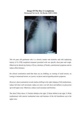
1. Image Of The Day 2: Lymphoma
Muhamad Na’im B. Ab Razak (MD USM)
This 46 years old gentleman who is a chronic smoker and alcoholic and with underlying
history of Ex PTB completed treatment presented with non specific chest pain and cough.
Otherwise he denied any history of fever, shortness of breath, constitutional symptoms and no
reduce effort tolerance.
On clinical examination noted that there are no clubbing, no wasting of small muscle, no
resting or intentional tremor, no cynosis, no ptosis and no hyperthyroidism symptoms.
However, chest examination reveals trachea shifting to the right, bulging of left mediastinum,
reduce left chest wall movement, reduce air entry over left chest and dullness on percussion
up to left upper zone. Otherwise, reduce vocal resonance and fremitus.
The chest X Ray shows 1) Trachea shifted to the right, 2) Heart shifted to the right, 3) Wide
mediastinum with anterior mediastinal mass and hazinnes of the left hemithorax up to the
upper zone.
2. In Image of the Day 1, [Link] i put a case of Dissecting aortic aneurysm with
haemothorax secondary to the leaking. Today’s image would broaden up our mind to think
about non vascular cause of enlarged mediastinal mass. The differential diagnosis would be
Hodgkin’s lymphoma, non hodgkin’s lymphoma, Thyroid mass, Teratoma and Tumors of the
thymus (cyst or thymoma).
Other rarer cases of widened mediastinum reported in case studies include venous aneurysm,
mediastinal lipomatosis, descending necrotizing mediastinitis, spontaneous rupture of
common carotid artery, malignant fibrous histiocytoma, traumatic extravasation of
cerebrospinal fluid and others.
Lateral chest film may help to differentiate the origin of the mass. However, with the advance
of radiological modalities, CT scan and MRI are found to be more superior than plain
radiograph. Radiological intervention would alse very helpful for diagnostic purpose
especially in doing biopsy.

![In Image of the Day 1, [Link] i put a case of Dissecting aortic aneurysm with
haemothorax secondary to the leaking. Today’s image would broaden up our mind to think
about non vascular cause of enlarged mediastinal mass. The differential diagnosis would be
Hodgkin’s lymphoma, non hodgkin’s lymphoma, Thyroid mass, Teratoma and Tumors of the
thymus (cyst or thymoma).
Other rarer cases of widened mediastinum reported in case studies include venous aneurysm,
mediastinal lipomatosis, descending necrotizing mediastinitis, spontaneous rupture of
common carotid artery, malignant fibrous histiocytoma, traumatic extravasation of
cerebrospinal fluid and others.
Lateral chest film may help to differentiate the origin of the mass. However, with the advance
of radiological modalities, CT scan and MRI are found to be more superior than plain
radiograph. Radiological intervention would alse very helpful for diagnostic purpose
especially in doing biopsy.](data:image/gif;base64,R0lGODlhAQABAIAAAAAAAP///yH5BAEAAAAALAAAAAABAAEAAAIBRAA7)