

Empfohlen
Weitere ähnliche Inhalte
Was ist angesagt?
Was ist angesagt? (20)
Ähnlich wie Acute intestinal obstruction, small bowel
Ähnlich wie Acute intestinal obstruction, small bowel (20)
Mehr von AR Muhamad Na'im
Mehr von AR Muhamad Na'im (20)
Kürzlich hochgeladen
Kürzlich hochgeladen (20)
Acute intestinal obstruction, small bowel
- 1. Image of the Day 10 : Acute Intestinal Obstruction, Small Bowel. Muhamad Na’im B. Ab Razak (MD USM) Disclaimer: The history was modified and no breach of patient’s confidentiality are made in the publication of this case study. A 70 years old gentleman with Benign Prostate Hypertrophy and history of laparotomy few years back presented with abdominal pain, nausea and altered bowel habit for three days duration. Patient was co-morbidly well but since for the past few days, he was having colicky abdominal pain centering at periumbilical region and radiates to the whole abdomen. The pain score was 5-6/10 and not relieve by medication taken at Klinik Kesihatan. He also complains of passing out small, hard and black stool once daily since three days ago and not passing flatus on the day of admission. His oral intake was also reduce in view of feeling nauseated each time taking meal and only took clear fluids. On examination, he look cachexic, afebrile, normal vital sign and pink conjunctiva. Per abdomen examination, there is a lower midline incision scar and right transverse lower incision scar. No discoloration of the abdomen noted. On palpation, the abdomen is soft and slightly distended with voluntary guarding around the umbilical area. No mass palpable per abdomen. Auscultation reveals hyperactive bowel sound and percussion note is resonance. Per rectal examination reveals empty rectum and moderately enlarged prostate which is firm, present of median sulcus and normal mucosa. Examination of other systems reveal no
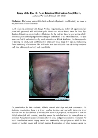
- 2. abnormality. Bedside ultrasound shows dilated small bowel with fluid and faecal material inside the bowel lumen. There is no free fluid collection and no abnormality of other organ noted. Labarotory investigation shows normal FBC, coagulation profile and no acidosis. The pottasium however is borderline high but other electrolytes are normal. The Abdominal X Rays shows dilated small bowel with intraluminal gas and minimal faecal materials. There was no sign of extra luminal gas. A well defined rounded opacity also noted inside the pelvic cavity, ? of bladder stone. A diagnosis of Acute Intestinal Obstruction of the Small Bowel secondary to Adhesion Colic was made and managed conservatively with 1) 4 hourly vital sign monitoring, 2) Ryle’s tube insertion with free flow and 4 hourly aspiration, 3) Keep nil by mouth, 4) CBD insertion, 5) Strict monitoring of Input/Output charting, 6) Analgesia PRN with IV Fentanyl 50 mcg, 7) IVD fluids with 3 pints of NS and 2 pints of D5% over 24 hours, 8) DXT monitoring, 9) IV Omeprazole 40 mg OD, 10) KIV for CVP insertion if patient become unwell 11) Replacement of loss fluid from RT, 12) Group Screen and Hold (GSH) and 13) KIV for laparotomy if non resolving of obstruction or signs of bowel perforation or gangrene present.
- 3. Discussion Intestinal obstruction is a very common surgical emergency that pose a high morbidity and mortality to the patient with inappropriate management. Being divided into large and small bowel obstruction, both are caused by mechanical obstruction and non mechanical. 80% of bowel obstructions involves the small bowel. The mechanical small bower obstruction is further divided into the luminal cause (foreign body, impacted fecal material, gallstone, bezoars, parasites and polypoidal tumors), intrinsic (atresia, tumors and inflammatory structures like TB and Crohn disease) and and extrinsic cause (adhesion, hernias, volvulus, intussuception, band, inflammation and neoplastic mass). Other type of obstruction in small bowel is coined as ‘Paralytic Ileus’ and most commonly occurs post operatively (up to 72 hours), pancreatitis and mesenteric infarct. Less common cause would be Pseudo obstruction (Ogilvie’s syndrome), Opiates, anticholinergics, retroperitoneal hemorrhage and metabolic cause (ketoacidosis, severe hypokalaemia). Although this entry is to discuss about small bowel obstruction, it is vital to differentiate between small vs large bowel obstruction as the definitive management is different. The table illustrated below will give a summary for it. My Version is modified from original table by Dr Chew Keng Sheng, Emergency Physician of University Science Malaysia. You can refer to his original table via this link. http://emergencymedic.blogspot.cm/2010/11/bowel- obstruction_15.html Acute Intestinal Obstruction Character Small bowel Large bowe Pain Colicky periumbilical pain. Not predominant, if present then located at lower abdomen Vomiting Early in proximal obstruction It is often a late sign due to and late in distal obstruction the incompetency of the ileo- caecal valve Abdominal Distension Little or no in proximal Significant abdominal obstruction and significant in distension. distal obstruction Bowel opening Late sign Altered bowel opening. Absolute obstruction when no bowel opening and not passing flatus.
- 4. Radiologic features Small bowel features: large bowel features: Valvulae conniventes – folds Haustrations: incomplete that cross the lumen crossing of folds across the completely lumen Normal features of small bowel: - No more than 3 mm wall thickness - Generally no more than 3 air fluid levels - No more than 3 cm diameter 3,6,9 rule Maximal normal diameter in small bowel 3 cm Maximal normal diameter in large bowel 6 cm Maximal normal diameter in cecum 9 cm Misscelenous Paralytic ileus usually In colorectal Carcinoma painless. Left sided tumors: generally presented with altered bowel habit, blood or mucus PR, mass PR Right sided tumors: generally presented with weight loss, anemia, less obstructive symptoms Other Physical sign to look - Dehydration for - Hyper peristaltic bowel sound. Diminish in paralytic ileus or perforation/ infarction. - Hypovolumic shock in late stage - Abdominal mass - Hernia orifices - Rectal examination – blood, palpable mass. Specific type of mechanical - Strangulated obstruction - Closed loop - Volvulus - Intussusception. Warning sign - Constant severe pain of sudden onset is omnious sign of bowel strangulation or infarct. - Presence of shock, leukocytosis, peritoneal irritation should rise of suspicion of strangulated obstruction.
- 5. Investigation that you should take includes 1) blood investigation (FBC, Coagulation profile, BUSE/Creat, LFT if indicated, Ca2+, Mg2+ and Po4- if indicated, blood culture if patient is septic), 2) Imaging (Plain radiograph, Ultrasound abdomen, water soluble contrast study if needed. As for management, the conservative management as i outlined in the case history is adequate and almost 80% of the cases will resolve with conservative management. Indication for surgical intervention includes 1) non resolving obstruction after 48 hours of conservative management, 2) present of primary underlying cause like hernia, obstructing tumor, 3) sign of peritoneal irritation. Reference Christian M & Gordon LC, “Acute Abdomen: Intestinal Obstruction”, Emergency Surgery, Surgery 26:3, Elsevier Ltd 2008