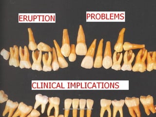
Dental education guide on tooth eruption problems
•
27 gefällt mir•5,989 views

This document discusses dental eruption and problems with eruption from the Indian Dental Academy website. It provides information on the mechanisms, etiology, diagnosis, and treatment of eruption problems. The key points are: - Tooth eruption is a complex process involving root development, periodontium establishment, and functional occlusion. - Problems can occur at any phase of eruption due to ectopic tooth position, obstacles in the eruption path, or failures in the eruption mechanisms. - Common causes of delayed eruption are local factors like scarring, supernumerary teeth, and systemic factors such as nutrition deficiencies, endocrine disorders, cerebral palsy, and genetic syndromes. - Accurate

Empfohlen
Weitere ähnliche Inhalte
Was ist angesagt?
Was ist angesagt? (20)
Ähnlich wie Dental education guide on tooth eruption problems
Ähnlich wie Dental education guide on tooth eruption problems (20)
Mehr von Indian dental academy
Mehr von Indian dental academy (20)
Kürzlich hochgeladen
Kürzlich hochgeladen (20)
Dental education guide on tooth eruption problems
- 2. INDIAN DENTAL ACADEMY Leader in continuing dental education www.indiandentalacademy.com www.indiandentalacademy.com
- 3. Contents Introduction Mechanisms Of Eruption Etiology Of Eruption Problems and Classification Delayed Tooth Eruption and Classification Local, Systemic, Genetic Diagnosis Of Eruption Problems Treatment Of Eruption Problems References www.indiandentalacademy.com
- 4. Eruption is the developmental process responsible for moving a tooth from its crypt position through the alveolar process into the oral cavity to its final position of occlusion with its antagonist. It is a dynamic process that encompasses completion of root development, establishment of the periodontium, and maintenance of a functional occlusion. Emergence, on the other hand, should be reserved for describing the moment of appearance of any part of the cusp or crown through the gingiva. Emergence is synonymous with moment of eruption, which is often used as a clinical marker for eruption. www.indiandentalacademy.com
- 5. (Tencate)The movements teeth make are complex and may be described in general as: Preeruptive tooth movement: Made by the deciduous and permanent tooth germ within tissues of the jaw before they begin to erupt. Eruptive tooth movement: Made by tooth to move from its position within the bone of the jaws to its functional position in occlusion. It is divided into intraosseous and extraosseous components. Posteruptive tooth movement: Maintaining the position of the erupted tooth in occlusion while the jaws continue to grow and compensate for occlusal and proximal tooth wear. www.indiandentalacademy.com
- 6. The four possible mechanisms for eruption are (Tencate,1998) 1. Root Formation 2.Hydrostatic Pressure 3.Bony Remodelling 4. Periodontal Ligament Root Formation: Root formation apears to be an obvious cause of tooth eruption because it undoubtedly causes an overall increase in the length of the tooth that must be accommodated by the growth of the root into the bone of the jaw, by an increase in jaw height, or by the occlusal movement of the crown. www.indiandentalacademy.com
- 7. This situation is however substantiated by the fact that: Some teeth erupt a greater distance than the total length of their roots. teeth still will erupt after the completion of root formation. teeth will still erupt when the Hertwig epithelial root sheath is removed surgically rootless teeth erupt. In conclusion root formation is accomodated during tooth eruption and is a consequence, not a cause of the eruption process. However it may accelerate tooth eruption. www.indiandentalacademy.com
- 8. Bone Remodelling: Marks and Cahill(1984) demonstrated that the dental follicle needs to be present for tooth eruption. Bony Remodelling occurs around the erupting follicle regardless of the presence of a tooth crown or tooth ,suggesting that remodelling process may be under the control of the dental follicle.However experiments do not prove that the dental follicle is involved in determination of final tooth position. Hydrostatic Pressure: Van Hassel and McMinn(1972) hypothesised that the tissue pressure apical to the erupting tooth was greater than occlusally,thus theoretically generating an occlusal force. However no association was demonstrated between the magnitude of force and the rate of eruption. www.indiandentalacademy.com
- 9. Periodontal Ligament: Tencate believed that the fibroblasts in the periodontal ligament have a contractile potential and are responsible for tooth eruption. Berkovitz1990 also favoured Tencate and agreed that no one hpothesis can fully explain the mechanism of tooth eruption as it is likely to be a multifactorial process. www.indiandentalacademy.com
- 10. ETIOLOGY OF ERUPTION PROBLEMS: Disturbances may occur in any of the phases of eruption. From an Etiologic point of view three main causes of eruption disturbances can be distinguished: 1.Ectopic position of the tooth germ, 2.Obstacles in the eruption path lack of space, follicular collision, presence of compact bone, supernumerary teeth, odontomas, scar tissue, non-attached mucosa, giant cell fibromatosis, odontogenic tumors or cysts. 3.Failures in the eruption mechanisms (i.e. follicle or PDL defects according to the stage of eruption) due to trauma, surgery, congenital diseases or other causes. www.indiandentalacademy.com
- 11. The first two conditions lead to impaction and the last according to the stage in which the eruption disturbance occurs as primary(before emergence) or secondary retention(after emergence) It is therefore necessary to develop a rational treatment approach in order to diagnose which of the prerequisites for eruption have been violated and to what extent. www.indiandentalacademy.com
- 12. Delayed tooth eruption is the most commonly encountered deviation from the normal eruption Conditions reported to be associated with Delayed tooth eruption are: Local: Mucosal Barriers-scar tissue: trauma/surgery Gingival Fibromatosis/ gingival hyperplasia Supernumerary teeth Odontogenic tumours Non Odontogenic Tumours Enamel Pearls Injuries to Primary teeth Ankylosis of deciduous teeth Premature loss of primary teeth Lack of resorption of deciduous tooth www.indiandentalacademy.com
- 13. Apical periodontitis of deciduous teeth Regional Odontodysplasia Impacted primary teeth Ectopic eruption Arch- Length deficiency and skeletal pattern Radiation damage Oral Clefts Segmented odontomaxillary dysplasia www.indiandentalacademy.com
- 14. Systemic: Nutrition Vitamin D resistant rickets Endocrine disturbances Hypothyroidism Hypoparathyroidism Hypopituitarism Pseudohypoparathyroidism Long term chemotherapy Hiv Infection Cerebral Palsy Dysosteosclerosis Drugs Phenytoin www.indiandentalacademy.com
- 15. Anemia Celiac disease Prematurity/ Low birth weight Ichthyosis Genetic disorders Familial/ Inherited Tobacco Smoke Idiopathic www.indiandentalacademy.com
- 16. Genetic disorders associated with DTE Amelogenesis Imperfecta Apert syndrome Carpenter syndrome Cherubism Chondro ectodermal dysplasia Cleidocranial dysplasia Congenital hypertrichosis lanuginosa Dentin dysplasia Mucopolysaccharidosis Down syndrome Dyskeratosis congenita Ectodermal dysplasia Ekman-Westborg-Julin syndrome www.indiandentalacademy.com
- 17. Epidermolysis bullosa GAPO syntlrome (growth retardation, alopecia, pseudoanodontia.and optic atrophy) Gaucher disease Gingival fibromatosis associated syndromes Gardners syndrome Gorlins syndrome Mc-Cune-Albright syndrome(Polyostotic fibrous dysplasia) Osteopetrosis (marble syndrome (polyostotic fibrous dysplasia) Neurofibromatoses Oculoauriculo vertebral spectrum (Goldenhar syndrome/hemifacial microsomia) Osteoglophonic dyspalsia www.indiandentalacademy.com
- 18. Osteogenesis imperfecta osteopetrosis Otodental dysplasia Parry-Romberg syndrome (progressive hemifacial atrophy) Progeria (Hutchinson-Gilford syndrome) Rothmund-Thompson syndrome Sclerosteosis Shokier syndrome (hereditary anodontia spuria) SHORT syndrome Singleton-Merten syndrome VonRecklinghausen neurofibromatosis 22q 11 deletion syndrome www.indiandentalacademy.com
- 19. Supernumerary teeth: About 28% to 60% of white people in 1984 have been reported with supernumerary teeth which can cause crowding, displacement, rotation, impaction,or delayed eruption of the associated teeth. The most common supernumerary tooth is the mesiodens followed by a fourth molar in the maxillary arch. Different forms of supernumerary teeth have been associated with different effects on the dentition: the tuberculate type is more common in patients with delayed tooth eruption the conical form has been associated with displacement. Supplemental Odontome(not accepted universally) www.indiandentalacademy.com
- 20. Arathi R, Ashwini R(MCODS, Mangalore )JIPDS,2005 Reported a case with both supplemental and tuberculate type of supernumerary teeth. The supernumerary teeth was found in the region of premolar predominantly and lower incisor region. The central incisor showed clear signs of tuberculate type with many cusps and dens invagination. www.indiandentalacademy.com
- 22. Regional odontodysplasia, also called "ghost teeth:' is an unusual dental anomaly that might result from a somatic mutation or could be due to a latent virus in the odontogenic epithelium. Affected teeth exhibit a delay or total failure in eruption. Their shapes are markedly altered, generally very irregular, often with evidence of defective mineralization. Central incisors, lateral incisors. and canines are the most frequently affected teeth, in either the maxillary or mandibular arch, and deciduous and permanent teeth can be affected. Abnormality in the tooth structure itself might be responsible for the eruptive disorders seen in this condition. www.indiandentalacademy.com
- 23. Odontomas: are also associated with delayed tooth eruption. Actually delayed tooth eruption is an alerting sign to diagnose odontomas. Mucosal Barrier: Any failure of the follicle of an erupting tooth to unite with the mucosa will entail in a delay in the breakdown of the mucosa and constitute a barrier to emergence. Gingival hyperplasia resulting from various causes (hormonal or hereditary causes, vitamin C defi-ciency, drugs such as phenytoin) might cause an abundance of dense connective tissue or acellular collagen that can be an impediment to tooth eruption. www.indiandentalacademy.com
- 24. Injuries to deciduous teeth have also been implicated as a cause of DTE of the permanent teeth. Traumatic injuries can lead to ectopic eruption or some disruption in normal odontogenesis in the form of dilacerations or physical displacement of the permanent germ. Cystic transformation of a nonvital deciduous incisor might also cause delay in the eruption of the permanent successor. The eruption of the succedaneous teeth is often delayed after the premature loss of deciduous teeth before the beginning of their root resorption. This can be explained by the abnormal changes that might occur in the connective tissue overlying the permanent tooth and the formation of thick, fibrous gingiva. www.indiandentalacademy.com
- 25. Ankylosis, resulting from the fusion of the cementum or dentin with the alveolar bone, is the most common local cause of delayed deciduous tooth exfoliation. Ankylosis occurs commonly in the deciduous dentition, usually affecting the molars, and has been reported in all 4 quadrants, although the mandible is more commonly affected than the maxilla. Ankylosed teeth will remain stationary while adjacent teeth continue to erupt through continued deposition of alveolar bone, giving the clinical impression of infraocclusion. www.indiandentalacademy.com
- 26. Arch-length deficiency is often mentioned as an etiologic factor for crowding and impactions. In a recent study of the relationship between formation and eruption of the maxillary teeth and the skeletal pattern of the maxilla, a shortened palatal length was found to delay the eruption of the maxillary second molar, although no delay in tooth formation was observed. Arch-length deficiency might lead to DTE, although more frequently the tooth erupts ectopicaliy X-radiation has also been shown to impair tooth eruption. Ankylosis of bone to tooth was the most relevant finding in irradiated animals. Root formation impairment, periodontal cell damage, and insufficient mandibular growth also seem to be linked to tooth eruption disturbances due to x-radiation. www.indiandentalacademy.com
- 27. Alveolar Clefts: Failure of union of the embryonic facial processes leads to discontinuity and disorientation of cells of the primodial dental lamina. Alveolar cleft occurs in the region of Maxillary lateral incisor. This leads to variation in number, morphology and enamel formation of teeth www.indiandentalacademy.com in the cleft area.
- 28. Canine Impaction was seen in 36% of the cases when lateral incisors where missing. Supernumerary teeth is also found associated with this condition and it decreases with the increase in clefting. www.indiandentalacademy.com
- 29. Systemic factors: Nutrition: The influence of nutrition on calcification and eruption is less significant compared with other factors.Nevertheless, delayed eruption is often reported in patients who are deficient in some essential nutrients. (1966,1988) Disturbance of the endocrine glands usually has a profound effect on the entire body, including the dentition. Hypothyroidism, hypopituitarism, hypoparathyroidism, and pseudohypoparathyroidism are the most common endocrine disorders associated with DTE. www.indiandentalacademy.com
- 30. In hypothyroidism, failure of thyrotropic function on the part of the pituitary gland or an atrophy or destruction of the thyroid gland per se leads to cretinism (congenital hypothyroidism) in a growing person. The dentofacial changes in cretinism are related to the degree of thyroid dcficiency. In hypopituitarism or pituitary dwarfism, the eruption and shedding of the teeth are delayed, as is the growth of the body in general. The dental arch has been reported to be smaller than normal; thus it cannot accommodate all the teeth, so a malocclusion develops. The roots of the teeth are shorter than normal in dwarfism, and the supporting structures are retarded in growth. www.indiandentalacademy.com
- 31. Weinberg and Berkowitz 2001 studied the dental manifestations in 70 children perinatally infected with HIV and indicated that delayed dental eruption was directly associated with clinical symptoms. DTE did not seem to correlate with CD4 positive T-lymphocyte depletion. The investigators concluded that HIV infection itself is not associated with DTE, but, rather, the onset of the clinical symptoms is. Pope and Curzon1991 found that unerupted deciduous and permanent teeth were more common in individuals with cerebral palsy compared with the controls. The first permanent molar erupted significantly later. However no etiology or implicated mechanisms were elaborated. www.indiandentalacademy.com
- 32. Giglio and Sanz 1987,1990,1994 found that other systemic conditions like anemia (hypoxic hypoxia, histotoxic hypoxia, and anemic hypoxia) and renal failure, have also been correlated with DTE and other abnormalities in dentofacial development. A Nagpal, Gsharma. A Sarkar and KM Pai 2005 reported a rare case of multiple unerupted permanent teeth without any known cause www.indiandentalacademy.com
- 33. Diagnosis of Eruption Disturbances: Accurate diagnosis of eruption problems is an important but complicated process. When teeth do not erupt at the expected age, a careful evaluation should be performed to establish the etiology and the treatment plan accordingly Diagnosis encompasses a history and clinical examination, as well as a detailed radiographic examination allowing a three-dimensional concept of the region involved. The radiographic technique differs according to the specific type of teeth involved. For localization of impacted canine ,3D C.T. utilizing shade surface display method has been found to be a very useful & accurate method compared to conventional techniques.( V.Ravinder,Nikhar Verma, Ashima Valiathan , JIOS 2002) www.indiandentalacademy.com
- 34. Use of 3D C.T. is recommended in cases with complex anatomical situations after a careful risk/benefit analysis www.indiandentalacademy.com
- 35. Familial information and information from affected patients about unusual variations in eruption patterns should be investigated. Clinical examination should be done methodically and must begin with the overall physical evaluation of the patient. Although the presence of syndromes is usually obvious in the mild forms. Only a careful examination will reveal the abnormalities Intraoral examination should include inspection. palpation, percussion, and radiographic examination. When the cause of the eruption disturbance has been identified, the problem of how to remedy it arises. Presently, there exists a series of treatment approaches aimed at assisting specific parts of the eruption process. Most of these treatments have the purpose of activating or establishing an eruption pathway. www.indiandentalacademy.com
- 36. Treatment of Eruption Disturbances: One established principle in the treatment of eruption disturbances is that an erupting tooth usually takes the path of least resistance, Likewise, an ectopically erupting permanent canine can be guided into a correct eruption path by removing either adjacent bone or a primary predecessor. Some of the treatment modalities are: 1. Ectopic Position of the tooth germ Observation: In some cases spontaneous uprighting of ectopically positioned permanent teeth may take place. However, this up righting is unpredictable. www.indiandentalacademy.com
- 37. Extraction of primary predecessors has been found to induce eruption and sometimes even up righting of ectopically positioned teeth such as canines and premolars. Removal of a primary tooth can both accelerate and retard eruption of the permanent successor Eruption spurt was found to be especially prominent in cases of periapical breakdown of bone due to pulpal complications in the primary molar. www.indiandentalacademy.com
- 38. However, if the extraction was performed very early, for instance at 4 years of age, the eruption spurt stopped and the permanent successor erupted later .This delayed effect has been suggested to be due to the formation of marginal dense bone or fibrous scar tissue. Surgical Exposure i)Surgical exposure usually involves removal of mucosa, bone and sometimes part of the follicle covering the permanent tooth so as to establish an eruption pathway. ii) a. In case of deep impaction, when the surgically created tract cannot be kept open postoperatively, the follicle should be left intact so that the eruption process can be completed.(without exposure of enamel) www.indiandentalacademy.com
- 39. b) If the follicle is damaged, or is in a superficial position, then an orthodontic bracket should be bonded to the crown so that an orthodontic extrusion force can take over the role of the follicle in the eruption process. (with exposure of enamel) The flap is apically repositioned in order to prevent labial retraction of the gingiva. The flap is then replaced and sutured. www.indiandentalacademy.com
- 40. FOLLOW-UP Surgical exposure usually induces partial eruption over 2-4 month (range 1 month to 2 years) and the speed is apparently not age dependent. Full eruption to the occlusal level usually takes about a year . The limitation of this procedure appears to be when ectopic teeth deviate more than 90° from the normal eruptive path Surgical Repositioning Autotransplantation of ectopic tooth germs has recently been shown to be an effective treatment, especially for displaced canines and premolars www.indiandentalacademy.com
- 41. Drawback: Injury to the periodontal ligament root resorption damage to the pulp may cause unsuccess,ful revascularization leading to pulp necrosis.Finally, injury to Hertwig's root sheath may result in partial or total arrest of further root development . This procedure should, therefore, only be used when other treatment alternatives are not indicated or are not available www.indiandentalacademy.com
- 42. 2.Obstruction of the Eruption Pathway This condition can be divided into the following three categories: 1. Lack of eruption space (crowding) 2. Follicular collision 3. Obstruction of the eruption pathway by compact bone, scar tissue, fibromatosis, unattached mucosa, giant cell fibroma-tosis, odontogenic keratocysts or odon-togenic tumors When the etiology has been established, the treatment principle is to determine the type of obstruction and then make it possible for the tooth to erupt by eliminating the cause www.indiandentalacademy.com
- 43. Lack of Eruption Space ( Crowding) Orthodontic evaluation should reveal whether expansion can increase the reduced eruption space or if extraction of the impacted tooth or adjacent teeth is the treatment of choice. Follicle collision a) seen in the maxillary anterior region, b) between the second and the third molar tooth germs. www.indiandentalacademy.com
- 44. The reason for the impaction effect of colliding follicles is probably neither the follicles can resorb the other, and therefore the eruption of both teeth becomes impossible. The treatment of choice in these cases is to remove one of the colliding tooth germs. In the case of supernumerary teeth, this decision is easy, whereas in the second and third molar region it is more complicated (depending on which one is in a clinically more favourable position) www.indiandentalacademy.com
- 45. Obstruction of Eruption Pathway by Hard or Soft tissue ERUPTION CYST The eruption cyst is a rare phenomenon, which appears as a bluish, translucent, elevated domeshaped lesion of the alveolar mucosa overlying the erupting tooth. It is not a true cyst, but rather results from hemorrhage into the dental follicle. It may occur during the eruption of both primary and permanent teeth. The most common treatment is removal of a portion of the gingiva overlying the crown of the tooth to facilitate eruption www.indiandentalacademy.com
- 46. ERUPTION SEQUESTRUM Tiny fragment of non vital hard tissue overlying an erupting permanent molar that consists of bone or cementum particles induced by the follicle. The condition is of little clinical significance because it generally does not seem to interfere with normal eruption.(No Treatment). Compact Bone a)In the case of premature extraction of primary molars,. b)After transplantation of an iliac graft to a cleft palate site, (it may present as an obstacle to an erupting canine) The treatment of choice in both cases is a surgical uncovering procedure, with or without enamel exposure depending on the depth of the tooth germ www.indiandentalacademy.com
- 47. ODONTOMAS Odontomas occur in the maxilla more often than in the mandible and in almost half of the cases are known are combined with impacted teeth.They also lead to impaction of the involved tooth. The treatment of choice is removal of the odontoma. Thus, three out of four impacted teeth can be expected to erupt subsequent to removal of odontomas. www.indiandentalacademy.com
- 48. The surgical procedure for removing an odontoma firstly consists of a flap procedure whereby full access to the odontoma is created. Bone covering the odontoma is excised and it is removed. Deep position - The follicle around the impacted tooth is left intact Superficial position - Exposure to the enamel is carried out www.indiandentalacademy.com
- 49. Scar Tissue: • Normally seen after cleft palate surgery and after trauma . • Apparently obstructs the collagenolytic capacity of the follicle. • There is also the possibility that a defect has occurred in the follicle. • Irrespective of etiology, the treatment of choice is an uncovering procedure, including enamel exposure. www.indiandentalacademy.com
- 50. ODONTOGENIC GIANT CELL FIBROMATOSIS It is a pathologic condition with development of a fibrous tissue layer dominated by giant cells. They prevent normal eruption. This condition has been recorded in the first and second molar region, with a strong preference for the mandible. The treatment of choice is surgical exposure, which routinely leads to successful eruption www.indiandentalacademy.com
- 51. ADENOMATOID ODONTOGENIC TUMOR Occurs in the second decade of life. Radiographically :usually has a pericoronal location and the radiographic appearance of a follicular cyst extends below the cervical region of the tooth. Histologically: very similar to ameloblasts . Treatment - Curettage has been found sufficient, without evidence of recurrence , and normal tooth eruption has been reported following conservative excision or even incomplete removal of www.indiandentalacademy.com the tumor
- 52. ODONTOGENIC KERATOCYST The lesions represent cystic changes in remnants of the dental lamina (Serres pearls). During ex pansion of these cysts, adjacent unerupted teeth may be secondarily involved, resulting in the radiographic appearance of a follicular cyst. Usually occurs in the second and third decade of life, The third molar and canine regions are most frequently involved. www.indiandentalacademy.com
- 53. Radiographically, small keratocysts have a unilocular appearance, whereas larger cysts have a multilocular appearance. Treatment: Cystectomy, and to prevent recurrence, a part of the alveolar mucosa covering the alveolar process is resected as it contains large number of epithelial remnants. www.indiandentalacademy.com
- 54. 3.Defects in the Follicle or PD L Found to occur subsequent to traumatic dental injuries and cleft-lip-palate surgery , in relation to congenital disorders such as amelogenesis imperfecta and cleidocranial dysplasia , and associated with hyperplastic follicles, central odontogenic fibromas, and follicular cysts . All of these events may lead to primary or secondary retention Diagnosis of a folliclerelated defect relies primarily on a radiographic examination. www.indiandentalacademy.com
- 55. Ankylosis in the Follicle or the PD L Recent studies have shown that 20% of eruption disturbances affecting the first and second molars are caused by an ankylosis of the tooth The etiology of this ankylosis is unknown at present, and treatment attempts such as breaking the ankylosis sites by tooth luxation have not proved successful. Thus, in most cases, the tooth has to be removed in order to prevent further complications. www.indiandentalacademy.com
- 56. Amelogenesis Imperfecta In this hereditary condition it is known that the tooth follicle is defective, and this may lead to tooth retention So far, no systematic treatment approaches have been published, and in all the reported cases the impacted teeth have been removed. However, considering that the defect is most likely situated in the enamel epithelium, surgical exposure of the tooth should be a treatment possibility www.indiandentalacademy.com
- 57. Cleidocranial Dysplasia In this hereditary condition, folliclemediated osteoclast activation is deficient, leading to severe disturbances in eruption . The treatment of choice for this condition is either surgical uncovering with enamel exposure or surgical repositioning. Follicular Cysts: A follicular cyst is one that encloses the crown of an unerupted tooth and is attached to its neck Seen mostly between10 40 years Cyst development can lead to displacement of the impacted tooth as well as neighboring teeth Treatment of choice for follicular cysts is usually a fenestration, which after 612 months leads to eruption of www.indiandentalacademy.com the displaced tooth germ
- 58. The treatment regimen depends entirely on the cause of impaction or retention. The following treatments have been found effective in activating eruption in the clinical situations. Ectopic Position of the Tooth Germ Extraction of primary predecessor Surgical uncovering with or without enamel exposure Surgical repositioning Obstruction of the Eruption Pathway Crowding Orthodontic expansion Extraction of the unerupted tooth or an adjacent tooth www.indiandentalacademy.com
- 59. Follicle collision Removal of one of the colliding tooth germs Compact bone Surgical uncovering with or without enamel exposure Scar tissue, giant cell fibromatosis, unattached oral mucosa, odontogenic tumors Surgical uncovering with enamel Exposure Supernumerary teeth or odontomas Removal of the hard tissue obstacle Follicle or PDL Defects Surgical uncovering exposure www.indiandentalacademy.com Surgical repositioning with enamel exposure
- 60. Summary: It is essentail to diagnose and treat eruption disturbances as early as possible because treatment at a later stage is usually more complicated due to the tendency of malocclusion to increase with time and reduced ability of remaining dentition to adjust. www.indiandentalacademy.com
- 61. References: 1.V. Ravinder, Nikhar Verma. Ashima Valiathan: 3 Dimensional Computed TomographyA new method for localization of Impacted Canines,JIOS 2002 page 7375 2.A Nagpal, Gsharma. A Sarkar and KM Pai: Eruption Disturbances: an etiologicalcummanagement perspective(Case Report),Dentomaxilofacial Radiology 2005 page 59 63 3.L. Suri, E.Gagari, H.Vastardis:Delayed tooth eruption: Pathogenesis,Diagnosis, and treatment. A literature Review, AJO DO October 2004,vol 126, number 4, page433445. 4.Arathi R, Ashwini R :Supernumerary teeth: A case report, JIPDS 2005 vol23 page 103105 www.indiandentalacademy.com
- 62. 5.Andreason, Petersen, Laskin DM. Normal and disturbed eruptions in humans. Textbook and colour Atlas of Tooth Impactions, 1997 Page 4964 6.H.L Craddock, C.C. Youngson: Eruptive tooth movement The current state of Knowledge.BDJ October 2004 vol 197 page385391 7.Tencate A.R. Oral Histology:Physiologic tooth movement: Eruption and shedding , 7th edition 2005,page 276 297 8.J. Kurol, Early treatment of tooth eruption disturbances, AJO DO June 2002 vol 121 number 6 page 588591. 9.Becktor et al: Association between ectopic eruption of maxillary canines and first molars, EJO 2005 vol27 page 186 189 www.indiandentalacademy.com
- 63. 10.William Proffit: Contemporary Orthodontics:3rd edition, 2000 page 430 432, 471 11.L.Mitchell, T.G Bennett: Supernumerary teeth causing delayed Eruption A retrospective Study, EJO February 1992 page 56 62 12. MG, Barry HJ, Blake M. Supernumerary Teeth An Overview of Classification, Diagnosis and Management, J Can Dent Assoc 1999;65:6126. 13.Regezi and Sciubb. Oral Pathology, W. B. Saunders Co. 2nd Ed. 1993. 14.Brook AH. Dental anomalies of number, form and size: Their prevalence in British schoolchildren. J Int Assoc Dent Child 1974;5:37 www.indiandentalacademy.com .
- 64. 15. Kinirons MJ. Unerupted premaxillary supernumerary teeth. A study of their occurrence in males and females. Br Dent J 1982;153:110. 16.RamosGomez FJ, Petru A, Hilton JF, Canchola AJ, Wara D,Greenspan JS. Oral manifestations and dental status in paediatric HIV infection. Int J Paediatr Dent 2000;10:311. 17. Pope JE, Curzon ME. The dental status of cerebral palsied, children. Pediatr Dent 1991;13:15662 18.B. Neville, D.Damm,C.Allen,J.Bouquot:Oral and Maxillofacial Pathology, 2nd Edition, 2004 page 40,41,598 www.indiandentalacademy.com
- 65. Thank you www.indiandentalacademy.com Leader in continuing dental education www.indiandentalacademy.com