
Empfohlen
Weitere ähnliche Inhalte
Was ist angesagt?
Was ist angesagt? (20)
Ähnlich wie Microscopy - an introduction
Ähnlich wie Microscopy - an introduction (20)
Mehr von ihmcbiology1213
Mehr von ihmcbiology1213 (20)
Kürzlich hochgeladen
Kürzlich hochgeladen (20)
Microscopy - an introduction
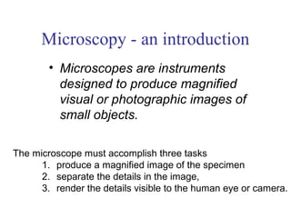
- 1. Microscopy - an introduction • Microscopes are instruments designed to produce magnified visual or photographic images of small objects. The microscope must accomplish three tasks 1. produce a magnified image of the specimen 2. separate the details in the image, 3. render the details visible to the human eye or camera.
- 2. Scale
- 3. Microscope One or more lenses that make an enlarged image of an object.
- 4. Simple Microscope • Light passes through only 1 lens. • Example: magnifying glass
- 5. Compound Microscope • Lets light pass through an object and then through two or more lenses.
- 6. Stereoscopic Microscope • Gives a three dimensional view of an object. (Examples: insects and leaves) • Used for dissections
- 7. Electron microscopes – use a beam of electrons instead of a beam of light to magnify the image
- 8. Electron Microscopes • can achieve 3D images using electrons
- 9. The Scanning Electron Microscope • produces a 3-dimensional image of specimen’s surface features spider head of a butterfly
- 10. Scanning electron microscopy (SEM) Types of specimens: -Whole organisms -Natural tissue surfaces -Exposed tissue structure A flea magnified 50 000 X What is this?
- 12. Transmission electron microscopy (TEM). • Allows the observation of molecules within cells • Allows the magnification of objects in the order of 100, 000’s.
- 13. Transmission electron microscope (TEM) – Provides for detailed study of the internal ultrastructure of cells – a beam of electrons Longitudinal Cross section section of of cilium 1 µm is transmitted through cilium the specimen for a 2D view Figure 6.4 (b) cilia on rabbit lungs
- 14. Transmission electron microscope Chloroplast from a tobacco leaf H1N1 virus
- 15. Confocal Laser Scanning Microscope (CLSM) • laser beam used to illuminate spots on specimen • computer compiles images created from each point to generate a 3-dimensional image • used on specimens that are too thick for a light microscope
- 16. A, B, C pollen grains: Scanning electron microscope D pollen grains: Confocal Laser Scanning Microscope E pollen grains: Transmission electron microscope F pollen grains: Light microscope G Mixed pollen grains (bright field light microscope, stained) H pollen grains confocal laser scanning microscope
- 17. Look at the following micrographs (a picture made by a microscope) and try to determine what the object is!
- 18. DENTIST’S DRILL
- 19. TOILET PAPER
- 21. VELCRO
- 23. BLACK WIDOW SPIDER CLAW
- 24. PORCUPINE QUILL
- 25. MASCARA BRUSH
- 26. ANT
- 27. BLACK FLY
- 28. MOSQUITO
- 29. CAT FLEA
- 30. MITE FEEDING
- 31. POLLEN GRAIN
- 32. ANT EYE
- 33. APHID ON A LEAF
- 34. EYELASHES
- 35. DOG FLEA
- 36. H1N1 VIRUS
- 37. What is the difference between a… VIRUS and CELL? E.coli bacterial cells
- 38. VIRUS BACTERIA - can’t live on its own- must - can exist on its own live inside another cell - much smaller (20 – 400nm) - larger (1000 nm = 1μm) - none are beneficial - some can be beneficial (bacteria in gut) - no cell wall, only a protein - outer cell wall coat - cannot be killed by antibiotics - are killed by antibiotics