
Recommended
Recommended
More Related Content
Featured
Featured (20)
Bsci202 practical 3
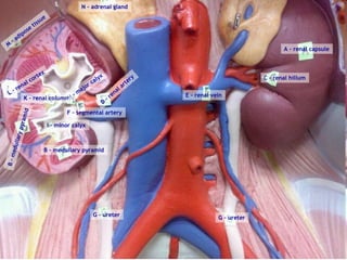
- 1. A – renal capsule C – renal hillum E – renal vein D – renal artery F – segmental artery H – major calyx B – medullary pyramid B – medullary pyramid I – minor calyx K – renal column L – renal cortex M – adipose tissue N – adrenal gland G – ureter G – ureter
- 2. K – minor calyx A – renal artery J – renal vein M – renal pelvis L – major calyx Q – cortex P – medullary pyramid P – medullary pyramid E arcuate artery C – interlobar vein H – arcuate vein N - ureter G – cortical radiate vein > F – cortical radiate artery O – renal column K – minor calyx
- 3. 7f – pyloric region |-- 7g – lesser curvature --| 7a – gastroesophageal sphincter 7b – pyloric sphincter 7c – cardiac region 7h – greater curvature 7e – body region 7d – fundus region
- 4. 9d – transverse colon 9i – haustra |---------| 9i – haustra |---------| 8 – small intestine 9i – haustra |---------| 9e – descending colon 9h – tenae coli |---------|
- 5. 9h – tenae coli |---------| 9e – descending colon 9e – descending colon 9h – tenae coli |---------| 9i – haustra |---------| 9i – haustra |---------| 9a - cecum 9i – haustra |---------| 9b – appendix 9f – sigmoid colon
- 6. 12 - pancreas 12a – pancreatic duct 13 – greater ommentum
- 7. 10a – hepatic duct 10b – bile duct > 11 - gallbladder 11a – cystic duct >
- 9. A - rectum B - anus C - pubis E – prostate gland F – ejaculatory duct > < G – prostatic urethra < H – membranous urethra J – bulb of penis L – corpus spongiosum K – corpus cavernosum I – spongy (penile) urethra > M – glans penis N - prepuce P - testis Q – epididymis O - scrotum
- 12. A - rectum B - anus C – symphysis pubis P – labium minus Q – labium majus R – clitoris > K – uterus L -cervix M – anterior fornix > F – urethra > O - vagina
- 13. P – hymen > C – symphysis pubis D – mons pubis R – labium majus Q – labium minus B - anus < O - vagina < F - urethra E – urinary bladder A - rectum M – anterior fornix > N – posterior fornix > K - uterus J – uterine tube G - ovary H - fimbriae I - infundibulum
- 16. OVARY + look for different stage follicles (ref Fig 43.4 p. 646) PENIS + look for a. corpus cavernosum (indicated by pointer) , and b. corpus spongiosum (ref Fig 42.1 p. 630) TESTIS + spermatozoid indicated by pointer (ref Fig 432.2 p. 632) EPIDIDYMIS + look for spermatozoids
- 17. ENDOMETRIUM DURING MENSTRUATION + functional layer, necrotic fragments, basal layer + myometrium (ref Fig 43.5 p. 647) CROSS SECTION OF URETER (x 100) + transitional epithelium + 2 layers of smooth muscle (ref p. 700 plate 43) TRANSITIONAL EPITHELIUM (X 400) + location + function (ref Fig 6A.3 h p. 73)
- 18. DETAILED STRUCTURE OF A GLOMERULUS (x 400) + glomerulus at pointer + cuboidal epithelium of the renal tubule (ref p. 700 plate 41) ACCESSORY ORGAN: TONGUE + foliate papillae + taste buds at pointer Ref. p. 695 plate 17 ACCESSORY ORGAN: TOOTH - MOLAR + to see full structure use 5x magnification + tooth oriented with crown to the right + enamel, dentin, pulp cavity, root canal, gingival, etc. (ref. Fig 38.12 p. 587)
- 19. ACCESSORY ORGAN: PANCREAS + acinar tissue (exocrine) and pancreatic islet (endocrine) + use 40x objective lens (ref. p. 699 plate 38) ACCESSORY ORGAN: SALIVARY GLAND + mucous cells and ducts + 40 x objective lens (ref. Fig 38.13 p. 588, & plate 37, p. 699) ACCESSORY ORGAN: LIVER + liver lobule, central vein, triad region (portal venous, portal arteriole, bile duct) ACCESSORY ORGAN: LIVER – CIRRHOSIS OF THE LIVE + compare to healthy liver slide + look for hexagonally shaped liver lobule + functionality
- 20. ALIMENTARY CANAL: SMALL INTESTINE - DUODENUM + lumen, villi (consists of simple columnar epithelial cells), plicae, circulares, goblet cells, duodenal glands (Brunner’s glands – simple cuboidal epithelium) (ref. p. 698 plate 33) ALIMENTARY CANAL: SMALL INTESTINE – JEJUNUM + lumen, plicae circulares, villi (individual columnar epithelial cells) (ref. plate 33, p. 698) ALIMENTARY CANAL: SMALL INTESTINE – ILEUM + lumen, villi (individual columbar epithelial cells), peyer’s patches at pointer (ref. plate 33, p. 698) ALIMENTARY CANAL: LARGE INTESTINE - COLON + lumen, goblet cells, tunics (mucosa, submucosa, mucularis externa, and serosa) (ref. p. 577, Fig 38.2)
- 21. ALIMENTARY CANAL: ESOPHAGUS/STOMACH JUNCTION + locate the gastroesophageal junction (which side belongs to the stomach/esophagus?) + id epithelial tissues of both organs (ref. Fig 38.6c p 581, and plate 30 p. 698) ALIMENTARY CANAL: STOMACH – PYLORIC REGION + gastric pit & gastric glands (10x) + mucosal tunic: epithelial tissue, muscularis mucosae + submucosa: note blood vessels +muscularis externa: oblique layer, circular layer, longitudinal layer