1. Annu. Rev. Immunol. 1997. 15:563–91
Copyright c 1997 by Annual Reviews Inc. All rights reserved
THE IFNγ RECEPTOR: A Paradigm
for Cytokine Receptor Signaling
Erika A. Bach, Michel Aguet∗ and Robert D. Schreiber
Center for Immunology and Department of Pathology, Washington University School
of Medicine, St. Louis, Missouri 63110; e-mail: schreiber@immunology.wustl.edu;
∗ Swiss Institute for Experimental Cancer Research (ISREC), Ch. des Boveresses,
CH-1066 Epalinges, Lausanne, Switzerland; e-mail michel.aguet@isrec.unil.ch
KEY WORDS: signal transduction, JAK-STAT pathway, transcription factors, tyrosine kinases,
Stat1
ABSTRACT
During the last several years, the mechanism of IFNγ -dependent signal trans-
duction has been the focus of intense investigation. This research has recently
culminated in the elucidation of a comprehensive molecular understanding of the
events that underlie IFNγ -induced cellular responses. The structure and function
of the IFNγ receptor have been defined. The mechanism of IFNγ signal trans-
duction has been largely elucidated, and the physiologic relevance of this process
validated. Most recently, the molecular events that link receptor ligation to sig-
nal transduction have been established. Together these insights have produced a
model of IFNγ signaling that is nearly complete and that serves as a paradigm
for signaling by other members of the cytokine receptor superfamily.
INTRODUCTION
The elucidation of the molecular understanding of the IFNγ receptor has been
an odyssey that has evolved over the past 15 years. Our understanding of this
receptor system has been the result of combined discoveries from several lab-
oratories working on different portions of the IFNγ signaling pathway. These
discoveries have led to the formulation of a cytokine receptor signaling model
that is currently one of the most complete. In addition, these studies have
provided information that has facilitated the understanding of many other cy-
tokine receptor systems. Specifically, the study of the IFNγ receptor has been
critical in revealing (a) tyrosine phosphorylation of the cytokine receptor intra-
cellular domain as a mechanism that couples the activated receptor to its signal
563
0732-0582/97/0410-0563$08.00
2. 564 BACH, AGUET & SCHREIBER
transduction system, (b) a novel pathway of signal transduction that mediates
biologic responses of many different cytokine receptors, and (c) the molecular
basis of specificity for the induction of many cytokine-dependent cellular re-
sponses. The purpose of this review is to summarize the major advances that
have produced the current comprehensive model of IFNγ receptor signaling.
HISTORICAL PERSPECTIVES
The current understanding of the IFNγ receptor system represents the synthesis
of two distinct experimental approaches aimed at analyzing IFNγ -dependent
gene induction. One focused on the cell surface with the intention of proceeding
into the cell nucleus, while the other focused on the nucleus of IFNγ -treated
cells and tracked the molecular trail back to the membrane. The meeting of
these two experimental approaches occurred at the inner leaflet of the plasma
membrane and resulted in the establishment of a clear molecular model of the
IFNγ signaling system.
The IFNγ receptor was initially characterized in the early 1980s in radio-
ligand binding studies conducted in several laboratories, including our own, on a
variety of different cell types (1). These experiments showed that most primary
and cultured cells expressed a moderate level of high affinity binding sites
for IFNγ . The interaction of IFNγ with its receptor was not inhibited by other
interferon classes, which explained the basis for the biologic specificity of IFNγ .
In addition, human and murine IFNγ bound to their respective receptors in a
strictly species-specific manner and thereby induced biologic responses only in
species-matched cells. The latter observation proved to be critical in defining
the subunits of the functionally active IFNγ receptor and in determining the
structure-function relationships operative within each subunit.
A major step forward in defining the subunit composition of IFNγ receptors
came from key genetic experiments conducted by Pestka and associates in 1987
(2). These studies employed a family of stable murine:human somatic cell hy-
brids that contained the full complement of murine chromosomes and a random
assortment of human chromosomes. All hybrids that contained human chro-
mosome 6 bound human IFNγ with high affinity, an observation later explained
by the presence of the human IFNγ receptor α chain gene on this chromosome
(3). However, biologic responsiveness to human IFNγ was found only in hy-
brids that contained both human chromosomes 6 and 21. These observations,
together with similar studies using hamster:murine somatic cell hybrids, led to
the hypothesis that functionally active human or murine IFNγ receptors consist
of two (or more) species-matched subunits (2, 4). The first is the receptor sub-
unit responsible for binding ligand in a species-specific manner. The second is
a species-matched subunit that is required for induction of biologic responses.
3. IFNγ RECEPTOR SIGNALING 565
This concept was further refined by independent reports in 1987–1988 of the
purification of the ligand-binding component of the human IFNγ receptor (5–7)
and the subsequent cloning of its gene initially by Aguet and colleagues (8).
This event was followed one year later by the isolation of the gene encoding the
murine homologue (9–13). When the ligand-binding chains of the human or
murine IFNγ receptor were expressed at high levels in murine or human cells,
respectively, they bound human or murine ligand in a manner that was identical
to endogenous receptors expressed on homologous cells. However, treatment
of the transfected cells with heterologous ligand failed to effect induction of
cellular responses. In contrast, when the human IFNγ -binding protein was
expressed in murine cells that also contained human chromosome 21, these
cells not only bound the human ligand but also responded to it (14–16). These
observations thus added significant support to the concept that functionally
active IFNγ receptors require a second, species-specific subunit. Definitive
proof of this concept came in 1994 when the second subunit of both the human
and murine IFNγ receptors were simultaneously identified by the Pestka and
Aguet laboratories using complementation cloning approaches (17, 18).
The nomenclature for the IFNγ receptor subunits has not been formally
established by the investigators in the field. Currently, the ligand-binding com-
ponent of the IFNγ receptor is referred to as either the IFNγ receptor α chain,
IFNγ R1, or CDw119. The second subunit has been designated the IFNγ re-
ceptor β chain, accessory factor-1 (AF-1) or IFNγ R2. For purposes of clarity
in this review and to maintain consistency with the nomenclature for other cy-
tokine receptors, we shall use only the designations IFNγ receptor α and β
chains to refer to the two receptor subunits.
At the same time that the IFNγ receptor was being identified on a molecu-
lar level, seminal biochemical and genetic experiments were being conducted
independently in the laboratories of James Darnell, Ian Kerr, George Stark,
and James Ihle that identified a novel signaling pathway activated following
treatment of cells with either IFNα or IFNγ (reviewed in 19–21). This work
resulted in the identification of two classes of signaling proteins that partici-
pated in this pathway. One was a family of latent cytosolic transcription factors
that eventually became known as STAT proteins (for signal transducers and
activators of transcription). The other was a family of structurally distinct
protein tyrosine kinases known as Janus family kinases or JAKs. The unique
feature of this signaling pathway, now known as the JAK-STAT pathway, was
that receptor ligation resulted in the activation of specific cytosolic STAT pro-
teins that dimerized and translocated directly from the membrane to the nucleus
and effected transcriptional activation of specific target genes. However, the
events linking receptor ligation with signal transduction remained ill-defined.
This missing step was filled in 1994 when the Schreiber laboratory showed that
4. 566 BACH, AGUET & SCHREIBER
IFNγ induced the tyrosine phosphorylation of the IFNγ receptor α chain lead-
ing to the formation of a docking site on the activated receptor for a particular
STAT, namely Stat1 (22). This observation thus bridged the two experimental
approaches and brought into focus the past 15 years of IFNγ receptor research.
THE LIGAND
Interferons were originally described as agents capable of protecting cells from
viral infection (1). Based on criteria such as their cellular source, general
biologic properties, and gene structure, interferon family members have been
segregated into two categories. Type I IFN is induced primarily as a result of
viral infection of cells and has been divided into two classes based on the cell
of origin. IFNα is a family of 17 related proteins encoded by distinct genes that
are synthesized largely by leukocytes. IFNβ is a single protein encoded by a
distinct gene that is produced largely by fibroblasts. In contrast, Type II IFN is
induced by immune and inflammatory stimuli, is synthesized exclusively by T
lymphocytes and natural killer cells, and is commonly known as IFNγ . IFNγ
bears no structural resemblance to IFNα or IFNβ at the protein level, and the
chromosomal location of the IFNγ gene is distinct from that of the Type I IFN
locus.
The human IFNγ molecule is a noncovalent homodimer that consists of two
identical 17-kDa polypeptide chains (23, 24). During biosynthesis the polypep-
tides are variably N-glycosylated, giving rise to a mature form of the molecule
that exhibits a predominant molecular mass of 50 kDa (25). The crystal struc-
ture of IFNγ confirms its dimeric nature and reveals that the two polypeptides
self-associate in an antiparallel fashion, producing a molecule that exhibits a
twofold axis of symmetry (26). This observation has led to the suggestion that a
single IFNγ homodimer can bind two IFNγ receptor molecules. Experimental
support for this prediction has been derived from results demonstrating that full
biologic activity is only manifest by the homodimeric form of the protein (1).
IFNγ induces varied effects on a wide range of target cells, and its pleiotropic
actions have been well studied (1). These include effects that promote both
specific and nonspecific mechanisms of host defense against infectious agents
and tumors. Like the other members of the interferon family, IFNγ can protect
cells from viral infection and can exert profound antiproliferative effects on
a variety of normal and neoplastic cells. However, IFNγ is acknowledged to
play a more comprehensive role in immunomodulation compared to the Type
I interferons. IFNγ is one of the major cytokines responsible for upregulating
MHC class I protein expression and for inducing MHC class II proteins on
a variety of leukocytes and epithelial cells. IFNγ has also been shown to
be the major cytokine responsible for activating or otherwise regulating the
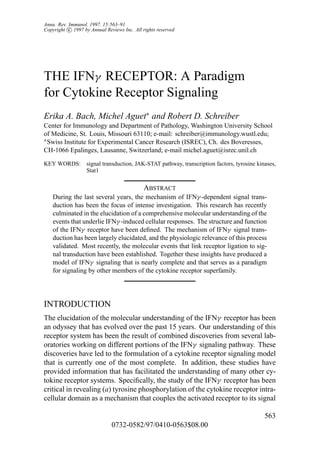
5. Figure 2 Crystal structure of a complex between IFNγ and the soluble IFNγ receptor α chain
extracellular domain. The two single chains that comprise the biologically active IFNγ
homodimer are shown in blue and magenta. Each IFNγ monomer contacts one soluble receptor,
shown in yellow and green, and thus one IFNγ homodimer dimerizes the IFNγ receptor α chain.
Upper panel: The view is perpendicular to the twofold axis of symmetry. The putative position
of the cell membrane is at the bottom of the page.
Lower panel: The complex as viewed parallel to the twofold axis of symmetry. The putative
site of interaction of the IFNγ receptor β chain indicated by arrows. [Reprinted with permis-
sion from Nature 376: 230-235, copyright 1995, Macmillian Magazines, Ltd (Reference 45).]
6. IFNγ RECEPTOR SIGNALING 567
action of mononuclear phagocytes. In addition, the cytokine regulates humoral
immune responses by effecting IgG heavy chain switching in either a direct or
an indirect manner. Finally, IFNγ regulates the production of a variety of other
immunomodulatory or proinflammatory cytokines such as IL-12 and TNFα.
GENERAL STRUCTURE OF THE IFNγ RECEPTOR
IFNγ Receptor Subunit Gene Structure, Regulation,
and Life Cycle
In the last decade the chromosomal locations of the genes encoding the human
and murine IFNγ receptor polypeptides have been identified and their structures
characterized. In addition, the expression patterns of these genes have been
defined. Recent studies indicate that the expression of the two IFNγ receptor
subunits differs significantly. Specifically, the receptor α chain is expressed
at moderate levels on the surface of nearly all cells. Receptor α chain gene
expression appears to be constitutive, and analysis of the promoter of this gene
reveals a structure resembling that found in housekeeping genes. In contrast,
the receptor β chain is constitutively expressed at extremely low levels, but
expression can be regulated in certain cell types by external stimuli. Regulation
of the receptor β chain gene thus becomes a critical factor in determining IFNγ
responsiveness in certain cells.
The human IFNγ receptor α chain is encoded by a 30-kb gene located on
the long arm of chromosome 6 (Table 1) (3). The murine homologue is a
22-kb gene present on chromosome 10 (27). The 5 flanking regions of these
genes contain a GC-rich region with no TATA box like that of promoters for
noninducible housekeeping genes (G Merlin, Z Dembic, personal communica-
tion). This observation suggests that expression of the IFNγ receptor α chain
is not regulated by external stimuli, a result that has been largely confirmed
experimentally. Both genes consist of 7 exons. Exons 1–5 encode the receptor
extracellular domain; exon 6 encodes a small portion of the membrane proximal
region of the extracellular domain and the transmembrane domain; and exon 7
encodes the entire intracellular domain. Transcription of the human and murine
IFNγ receptor α chain genes gives rise to mRNA transcripts of 2.3 kb (1). The
receptor α chain polypeptide is synthesized in the endoplasmic reticulum (ER)
and is posttranslationally modified as it moves from the ER to the Golgi by
the addition of N-linked carbohydrates (28, 29). Although expression of the
fully mature protein at the plasma membrane varies widely between tissues
(200–25,000 sites/cell), there does not appear to be a direct correlation between
the extent of receptor α chain expression and the magnitude of IFNγ -induced
responses in cells (1). Following IFNγ receptor ligation, the receptor-ligand
7. 568 BACH, AGUET & SCHREIBER
Table 1 Properties of the IFNγ receptor α and β subunits
α chain β chain
Property Human Murine Human Murine
Primary sequence
Signal peptide 17aa 26aa 21aa 18aa
Mature form 472aa 451aa 316aa 314aa
Homology 52% 58%
Chromosomal localization 6 10 21 16
Domain structure
Extracellular 228aa 228aa 226aa 224aa
Transmembrane 23aa 23aa 24aa 24aa
Intracellular 221aa 200aa 66aa 66aa
Potential N-linked glycosylation sites 5 5 5 6
Predicted Mr (kDa) 52.5 49.8 34.8 35.6
Mr (kDa) 90 90 61–67 60–65
Intracellular conserved tyrosines 5 3
complex is internalized and enters an acidified compartment. Within this com-
partment, the complex dissociates and free IFNγ is trafficked to the lysosome
where it is degraded. In many cells, such as fibroblasts and macrophages, the
uncoupled receptor α chain enters a large intracellular pool of α subunits and
eventually recycles back to the cell surface. In most cells, the size of the intra-
cellular pool is approximately 2–4 times that of the receptors expressed at the
cell surface (15, 28, 30–32).
The human IFNγ receptor β chain gene has been localized to chromosome
21q22.1 (17, 33). The murine homologue resides on chromosome 16 (Table 1)
(4). These syntenic chromosomal regions also contain the genes of several other
IFN receptor family members, including the subunits of the IFNα/β receptor
(IFNAR1 and IFNAR2) and the orphan IFN receptor family member denoted
CRF2–4 (33, 34). Transcriptional activation of the IFNγ receptor β chain gene
results in the generation of an mRNA transcript of 1.8 kb in human cells or 2 kb
in mouse cells (17, 18). At the present time, structural data is available only
for the mouse IFNγ receptor β chain gene. This 17-kb gene appears to consist
of 7 exons and contains, within the 5 flanking region, several potential binding
sites for a variety of externally regulated activated transcription factors.
The latter observation suggested that transcription of the β chain gene may
be tightly regulated, a hypothesis that has recently been strengthened experi-
mentally. Based on the observation that different CD4+ T helper cell subsets
differed in their ability to respond to IFNγ (35), two independent groups demon-
strated in 1995 that the IFNγ unresponsive state was due to a lack of cellular
expression of IFNγ receptor β chain (36, 37). Unresponsiveness was shown
8. IFNγ RECEPTOR SIGNALING 569
to be a result of IFNγ -dependent receptor β chain downregulation and was not
linked to T cell differentiation (37). In this system, Th1 cells, which produce
IFNγ , were found to lack the receptor β subunit and were IFNγ unresponsive.
In contrast, Th2 cells, which do not produce IFNγ , expressed the receptor β
chain and were IFNγ responsive (37). However, receptor β chain downregula-
tion was induced in murine Th2 cells, as well as in human peripheral blood T
cells, upon exposure to IFNγ (37, 38). Interestingly, ligand-induced receptor
β chain downregulation did not occur in certain fibroblast cell lines. Thus,
IFNγ appears to regulate expression of its own receptor β chain on certain cell
types and thereby determines the ability of these cells to respond to subsequent
exposure to IFNγ . Recently, treatment of T cells with phorbol esters or with
CD3 antibodies has been shown to effect induction of receptor β chain mRNA
(38). Taken together, these results demonstrate that β chain expression can be
regulated either positively or negatively in a stimulus-specific manner.
Structure of the IFNγ Receptor Polypeptides
The human and murine IFNγ receptor α chains are organized in a similar
manner and are symmetrically oriented around a single transmembrane domain
(Table 1 and Figure 1). However, despite this organizational similarity the two
polypeptides exhibit only 52.5% overall sequence identity. This modest level
of identity extends throughout both the extracellular and intracellular domains
of the polypeptides. The IFNγ receptor α chain is a member of the class 2
cytokine receptor family, which includes tissue factor, IFNAR1 and IFNAR2,
the ligand-binding component of the IL-10 receptor, and CRF2–4 (39). Like
all members of the class 2 cytokine receptor family, the intracellular domain
of this subunit is devoid of intrinsic kinase or phosphatase activities. Like the
IFNγ receptor α chain, the receptor β chain is also a member of the class 2
cytokine receptor family. The human and murine IFNγ receptor β subunits
are also structurally similar to one another (Table 1 and Figure 1). Although
human and murine receptor β chains exhibit 58% identity overall, this value
increases to 73% when their cytoplasmic domains are compared (17, 18).
Structure-Function Analyses of the IFNγ Receptor
Subunit Extracellular Domains
Immunochemical and radioligand binding experiments indicate that the IFNγ
receptor α chain binds ligand with a single high affinity (Ka) of 109 –1010 M−1
(1). Deletion mutagenesis analysis of the receptor soluble extracellular do-
main (sECD) showed that the majority of the extracellular domain (residues
6–227) was required for expression of ligand-binding activity (40). However,
by exchanging corresponding regions between the human and murine IFNγ re-
ceptor α chain extracellular domains, several important internal sequences were
9. 570 BACH, AGUET & SCHREIBER
Figure 1 Polypeptide chain structure of the human IFNγ receptor. The IFNγ receptor consists of
two species-matched polypeptides. The IFNγ receptor α chain is required for ligand binding and
signaling. The IFNγ receptor β chain is required primarily for signaling and plays only a minor
role in ligand binding. The intracellular domain of the receptor α chain contains two functionally
importance sequences: (1) an LPKS sequence required for α chain association with the tyrosine
kinase Jak1, and (2) a YDKPH sequence that, when phosphorylated, forms the docking site for
latent Stat1. The intracellular domain of the receptor β chain contains a functionally important
box1/box2 sequence required for Jak2 association.
10. IFNγ RECEPTOR SIGNALING 571
identified throughout the extracellular domain that contributed to the species
specificity of the ligand-binding process (41). Moreover, this study also re-
vealed the presence of distinct regions within the receptor α chain that played
an obligate role in biologic response induction but not in ligand binding. One ex-
planation for the latter observation is that the functionally important sequences
may contribute to the interaction between the IFNγ receptor α and β subunits.
Recent studies in the general field of receptor biology have established the
paradigm that a ligand can effect the activation of its cellular receptor by induc-
ing association or oligomerization of the appropriate receptor subunits. Among
cytokine receptors, this process was first described in studies of the receptor
for the monomeric ligand growth hormone (42). In the case of IFNγ , ligand-
induced receptor dimerization was anticipated due to the suspected bivalent
nature of the ligand. Experimental support for this possibility was provided
by studies that analyzed the ligand-binding characteristics of a soluble human
IFNγ receptor α subunit (43, 44). By means of ligand-binding assays, sucrose
density gradient ultracentrifugation, and HPLC gel filtration chromatography,
sECD and ligand were shown to form stable complexes in free solution that
consisted of one mole of ligand and two moles of soluble receptor. Formation
of the 2:1 (receptor : ligand) complex was also demonstrated on cell surfaces
using either chemical cross-linking or immunochemical approaches.
Structural confirmation of the nature of the IFNγ : IFNγ receptor complex
came in 1995 when the crystal structure of human IFNγ bound to the solu-
ble human IFNγ receptor α chain extracellular domain was solved to 2.9 A ˚
(Figure 2). This study confirmed the 2:1 stoichiometry of the receptor : IFNγ
complex and represented the first solved crystal structure of a ligand-occupied,
class 2 cytokine receptor (45). Within this complex, the core structure of bound
IFNγ was similar but not identical to that determined for the unbound cytokine.
The only major differences occurred within the AB loops and C-termini, which
are flexible and have little or no secondary structure in unbound IFNγ , but which
appear well ordered in receptor-bound IFNγ . The core structure of the ligated
IFNγ receptor α chain extracellular domain indicates that it forms a rod-like
molecule which is folded into two domains, denoted D1 (membrane distal) and
D2 (membrane proximal). Each domain is folded into two β-strands consisting
of β-pleated sheets. The domains are separated by an 11 amino acid linker
and are oriented at an angle of 120◦ relative to one another. The membrane
proximal D2 domain is positioned at a 60◦ angle relative to the cell membrane,
thereby causing the D1 domain to assume an angle that is complementary to
IFNγ . Receptor binding thus orients the symmetrical IFNγ molecule perpen-
dicular to the cell membrane and thereby allows for equivalent interactions to
occur between the second binding site of IFNγ and a second receptor α chain.
In their dimerized form, the IFNγ receptor α chains do not interact with one
11. 572 BACH, AGUET & SCHREIBER
˚
another and remain 27 A apart. This distance is much greater than would
have been predicted by the crystal structure of the complex of growth hormone
bound to its receptor (42). This characteristic becomes important because, un-
like the ligated growth hormone receptor, the ligated IFNγ receptor α chain
must interact with an additional receptor subunit in order to effect initiation of
the intracellular signaling process. Given the structural restraints apparent in
the core structure of the IFNγ /IFNγ receptor α chain complex, it is possible to
envision that symmetrical binding sites for the IFNγ receptor β chain are gener-
ated during ligand-induced receptor α chain dimerization. The crystal structure
thus supports the concept that ligand induces the assembly of an activated IFNγ
receptor complex that consists of two receptor α chains and two β chains.
Another study has defined the contribution of the IFNγ receptor β chain to
the ligand-binding process (46). Using an experimental system where the two
human IFNγ receptor subunits were expressed either individually or together
in murine fibroblasts, no direct interaction was detected between human IFNγ
and the human IFNγ receptor β subunit. However, when the human β subunit
was present at high levels on murine cells that also expressed the human IFNγ
receptor α subunit, IFNγ binding was increased fourfold over that observed on
cells expressing only the human receptor α chain. Thus, one function of the
IFNγ receptor β chain is to stabilize the complex formed between ligand and
the receptor α subunit.
Structure-Function Analyses of the IFNγ Receptor
Subunit Intracellular Domains
The human IFNγ receptor α chain was one of the first cytokine receptor subunits
subjected to a detailed structure-function analysis of its intracellular domain.
Using a combination of deletion and substitution mutagenesis approaches, three
distinct sequences within the subunit’s 221 amino acid intracellular domain were
identified as being required for specific receptor functions (15, 22, 47–49). The
first was a leucine-isoleucine sequence residing at positions 270 and 271 of
the mature polypeptide that plays a critical role in directing receptor trafficking
through the cell (1, 15). Deletion or alanine substitution of this sequence
resulted in a receptor mutant that was deficient in its ability to internalize ligand
and that accumulated at the cell surface. However, this receptor mutant was still
capable of supporting IFNγ -induced biologic responses, thereby dissociating
the ligand trafficking and signaling functions of this subunit.
More important, these analyses revealed the presence of two topographically
distinct, intracellular domain sequences that were required for induction of
IFNγ -dependent cellular responses. The first is a membrane proximal Leu-Pro-
Lys-Ser (LPKS) sequence residing at positions 266–269 (15, 22). On the basis
of alanine scanning analysis, the proline residue at position 267 was found to
12. IFNγ RECEPTOR SIGNALING 573
play the dominant functional role within this sequence (49). The second is a five
amino acid region that is located at positions 440–444 near the carboxy terminus
of the receptor α subunit and that contains the residues Tyr-Asp-Lys-Pro-His
(YDKPH) (15, 47). Mutational analysis of these five residues demonstrated
that only Y440 , D441 , and H444 were functionally important. Receptor α chains
harboring alanine substitutions at any of these three residues failed to induce
IFNγ -dependent cellular responses (47). The particular functional importance
of Y440 was confirmed by two additional observations. First, receptor α chains
that contained a conservative phenylalanine substitution at Y440 were also func-
tionally inactive (47). Second, mutation or deletion of any other tyrosine residue
within the receptor’s intracellular domain (denoted the 4XYF mutant) did not
ablate receptor activity (22). The physiologic importance of these two intra-
cellular domain regions was demonstrated in experiments in which receptor α
chains containing mutations within the LPKS and/or YDKPH sequences were
overexpressed either in cultured cell lines or in specific tissues of transgenic
mice. Analysis of these cells and/or tissues revealed that the overexpression
of functionally inactive mutant receptor α chains inhibited biologic responses
induced by endogenous IFNγ receptors in a dominant negative manner (50–52).
Recently, a structure-function analysis of the IFNγ receptor β chain intra-
cellular domain has been completed (53). This study, together with another
that used a cytoplasmically truncated form of the receptor β chain, showed
that only the membrane proximal half of the 66 amino acid intracellular do-
main was required for β chain function (53, 54). Moreover, within this portion
of the intracellular domain, two closely spaced sequences (263 PPSIP267 and
270 IEEYL274 ) were identified that played an obligate role in response induction
(53). Substitution of either of these five amino acid sequence blocks with ala-
nine residues abrogated receptor β chain function. However, no single amino
acid within this subregion of the receptor β chain intracellular domain could be
identified as playing a dominant functional role. Thus, only a restricted amount
of intracellular domain sequence within each of the two subunits of the IFNγ
receptor is required for induction of IFNγ -specific cellular responses.
SIGNALING
The JAK-STAT Pathway
Whereas the aforementioned experiments identified the IFNγ receptor subunits
on a molecular basis and characterized the key functional regions within these
proteins, they did not define the mechanism(s) of IFNγ receptor signaling.
This process was largely elucidated in experiments, conducted concomitantly
with the IFNγ receptor characterization studies, that identified two distinct
protein classes involved in mediating IFN-dependent cellular responses. The
13. 574 BACH, AGUET & SCHREIBER
Figure 3 Structure of STAT proteins and their utilization by cytokine receptors. There are currently
seven members of the signal transducers and activators of transcription (STAT) family that are
activated in a specific manner by distinct cytokine receptors. Receptor specificity has been defined
using gene targeted mice (76–82).
first class, termed STAT proteins, was initially identified in seminal biochemical
studies performed in the laboratory of James Darnell (Figure 3). The second
consisted of a group of unusual protein tyrosine kinases, termed Janus family
kinases (JAKs), the participation of which in IFN signaling was defined in
elegant genetic studies conducted in the laboratories of Ian Kerr and George
Stark (Figure 4). The combination of these two sets of observations led to the
definition of a novel signal transduction pathway, now known as the JAK-STAT
pathway, that is responsible for mediating the activation of many, if not all,
IFN-inducible genes (19–21).
This research was made possible by the identification of a family of genes
(termed interferon-stimulated genes or ISGs) that were induced rapidly (i.e.
within 15 to 30 min) in IFN-treated cells and the transcription of which was not
dependent on new protein synthesis (55). Analysis of the promoter regions of
these immediate-early ISGs revealed the presence of two classes of conserved
nucleotide sequences that directed the rapid transcriptional activation of IFN-
inducible genes (19, 20). The first element, termed the interferon-stimulated
response element (ISRE), was a 12–15 nucleotide site with a consensus se-
quence of AGTTTCNNTTTCNC/T that was responsible for driving expres-
sion of IFNα/β inducible genes. The second, termed the gamma-interferon
activation site (GAS), was a 9 nucleotide site with a consensus sequence of
TTNCNNNAA that effected transcriptional activation of IFNγ -induced genes.
14. IFNγ RECEPTOR SIGNALING 575
Figure 4 Structure of Janus family kinases and their utilization by cytokine receptors. There are
currently four members of the Janus (JAK) family of protein tyrosine kinases. They associate with
cytokine receptor intracellular domains and are required to form the STAT docking sites on ligated
cytokine receptors and to activate STAT proteins. Receptor utilization has been defined using gene
targeted mice (74, 75).
Synthetic oligonucleotides that contained ISRE elements were used to iso-
late and characterize two novel transcription factors of molecular masses 91
and 113 (p91 and p113) (56, 57). These transcription factors, now known as
Stat1α (p91) and Stat2 (p113), were highly unusual because they contained src
homology 2 (SH2) domains (19–21, 58). The importance of the SH2 domain
was revealed when the mechanism of IFN-induced STAT protein activation was
established. In unstimulated cells, STAT proteins were present in the cytosol
in a latent monomeric form (59). However, upon addition of IFN to cells, the
STATs were activated by tyrosine phosphorylation, formed homo- or hetero-
dimers, translocated to the nucleus, and bound to the promoter regions of ISGs,
effecting gene induction (59–62). Whereas IFNα was found to activate Stat1α
and Stat2 [which then combined with a 48-kDa protein to form the transcrip-
tionally active complex known as ISGF3 (57)], IFNγ effected activation only
of Stat1α (59). Subsequent studies revealed that Stat1 and Stat2 represent the
founding members of a larger protein family that currently consists of seven dis-
tinct gene products. These family members, Stat1, Stat2, Stat3, Stat4, Stat5a,
Stat5b, and Stat6, play key roles in mediating the biologic effects of a variety
of different cytokines and growth factors (Figure 3) (19–21) .
At the same time that these experiments were conducted, other labs
were pioneering the investigation of IFNα and IFNγ signaling mechanisms
predominantly using genetic approaches. These studies used a mutagenesis
15. 576 BACH, AGUET & SCHREIBER
protocol that involved both positive and negative selection and thereby gener-
ated eight cellular complementation groups that displayed distinctive combina-
tions of IFNα and/or IFNγ signaling defects (19). For one IFNα-unresponsive
complementation group (denoted U1A), a single gene was identified that com-
plemented the genetic deficiency (63). Partial sequence analysis of this gene
revealed that it encoded a previously identified protein tyrosine kinase of un-
known function known as Tyk2. Tyk2 was a structurally unusual protein ty-
rosine kinase because it contained two kinase-like domains. It belongs to a
small family of ubiquitously expressed kinases known as Janus kinases (Fig-
ure 4) (64). At the time, this family was thought to contain two additional
members known as Jak1 and Jak2 (64, 65). By means of cDNAs encoding
the three Janus family members and the panel of IFN-unresponsive cellular
mutants, it was shown that IFNα signaling required the concomitant presence
of both Tyk2 and Jak1, whereas IFNγ signaling required the dual presence
of Jak1 and Jak2 (66, 67). Recently, a fourth family member, Jak3, has been
identified and displays a restricted expression pattern largely limited to cells of
hematopoietic origin (68, 69). However, Jak3 does not play a role in mediating
IFN biologic responses. Additional work from several laboratories has demon-
strated that distinct combinations of Janus family members are involved in the
signaling pathways of many cytokine and growth factor receptors that possess
intracellular domains devoid of endogenous kinase activity (Figure 4) (19–21).
The functional connection between the JAK and STAT protein families was
made when it became apparent that the JAKs were the enzymes responsible for
effecting cytokine-dependent STAT phosphorylation and activation (70, 71).
This point was demonstrated by showing that cells deficient in either Jak1,
Jak2, or Tyk2 were unable to activate Stat1 following treatment with IFN (66,
67). These results demonstrated that these two classes of proteins formed a
regulated signal transduction pathway. The general physiologic relevance of
the JAK-STAT pathway has been unequivocally demonstrated by two sets of
observations. First, humans lacking Jak3 are unable to respond to lymphocyte
growth factors such as IL-7 and IL-2 and display severe defects in their ability
to produce T lymphocytes (72, 73). Second, mice with targeted disruptions of
specific JAK (74, 75) or STAT (76–82) genes display distinct defects in immune
system development and/or function.
The Link Between the IFNγ Receptor
and the JAK-STAT Pathway
THE JAK CONNECTION In 1991, studies on the signal transducing molecule
of the IL-6 receptor system, gp130, revealed that two functionally critical se-
quences within the membrane proximal intracellular domain of this polypeptide
were conserved among the members of the cytokine receptor superfamily.
16. IFNγ RECEPTOR SIGNALING 577
These sequences, termed box1 and box2, were originally defined as PXXPXP
and LEVL, respectively (83, 84). Subsequent studies revealed that box1/box2
sequences were important in mediating the interaction between certain cytokine
receptor cytoplasmic domains and Janus kinases (85, 86). This information
proved to be useful in defining the interactions of the IFNγ receptor subunits
with specific members of the JAK family.
Sequence comparisons indicated that the functionally critical, membrane
proximal, intracellular domain sequences within the IFNγ receptor α and β
subunits were similar to box1/box2 motifs. To examine whether these se-
quences functioned in a similar manner within the IFNγ receptor polypeptides,
coprecipitation studies were performed on cells treated with either buffer or
IFNγ . In unstimulated cells, the IFNγ receptor α chain was found to associate
with an inactive form of Jak1 (49, 87). This interaction was specific because
it did not occur with other Janus family members. Using an alanine scanning
mutagenesis approach, Jak1 binding to the IFNγ receptor α chain intracellular
domain was found to be dependent on the presence of the intact, function-
ally important 266 LPKS269 sequence (49). In IFNγ -treated cells, the receptor α
chain–associated Jak1 molecules became activated through tyrosine phosphory-
lation (49, 87). These results indicated that whereas inactive Jak1 constitutively
associates with the IFNγ receptor α chain, its activation is ligand dependent.
Similar studies have been conducted on the IFNγ receptor β subunit. Using
coprecipitation/western blot analyses, three groups have demonstrated that the
IFNγ receptor β chain cytoplasmic domain associates with Jak2 in a constitutive
and specific manner (53, 54, 88). Structure-function analysis of the receptor
β chain demonstrated that this association is mediated through a functionally
critical, 12 amino acid box1/box2-like sequence 263 PPSIPLQIEEYL274 located
13 amino acids away from the membrane in the β chain intracellular domain
(53). Mutation within this region produced a receptor β subunit that was unable
to interact with Jak2 and failed to support Stat1 activation or IFNγ -dependent
biologic response induction (53).
The elucidation of the structure-function relationships of the JAK association
sites present on the IFNγ receptor polypeptides led to subsequent experiments
that examined the issue of Janus kinase substrate specificity. A key question
that needed to be addressed was whether the apparent obligate pairing of Jak1
and Jak2 for IFNγ signaling reflected the specificity of the kinase-receptor sub-
unit interaction or the enzymatic substrate specificity of the kinases themselves.
Two approaches have been utilized to address this issue. In one study chimeric
molecules were generated in which the human IFNγ receptor β chain cytoplas-
mic domain was replaced by either Jak2 or c-src (53). These chimeras were then
expressed in murine cells that also expressed the human IFNγ receptor α sub-
unit, and the ability of these murine transfectants to respond to human IFNγ
17. 578 BACH, AGUET & SCHREIBER
was monitored. Cells expressing the human receptor β chain–Jak2 chimera
were capable of manifesting a full complement of biologic responses to hu-
man IFNγ (53). In contrast, cells expressing the receptor β chain–src chimera
were unresponsive to the human ligand. These results showed that the sole
obligate function of the intracellular domain of the IFNγ receptor β subunit is
to chaperone Jak2 into the ligand-activated receptor complex. The results also
suggested that Janus kinases may display a certain level of substrate specificity
that obligates their participation in the JAK-STAT pathway and that cannot
be replaced by other classes of protein tyrosine kinases. In a second study,
a different family of human IFNγ receptor β chain chimeras were generated,
in which the β chain cytoplasmic domain was replaced with other cytokine
receptor cytoplasmic domains that utilize Janus kinases other than Jak2 (89).
When expressed in hamster cells that also expressed the human IFNγ receptor
α chain, all chimeric molecules were functionally competent to respond to hu-
man IFNγ , although some quantitative differences were noted (89). This result
implies that the specificity displayed by the Janus family kinases resides at the
level of enzyme-receptor association. This concept has been strengthened by
the recent generation and characterization of mice with a targeted disruption
of the Jak1 gene (SJ Rodig, M Aguet, RD Schreiber, unpublished observa-
tions). Taken together these results suggest that the Janus kinases are critical
for JAK-STAT pathway activation but are not a source of pathway specificity.
LIGAND-INDUCED ASSEMBLY AND ACTIVATION OF THE IFNγ RECEPTOR Where-
as these studies defined the critical sites on the IFNγ receptor subunits that
are responsible for mediating the association between the receptor polypep-
tides, Jak1 and Jak2, they did not account for how the receptor-associated JAK
kinases were activated. One possible mechanism that could account for the
ligand-induced activation of IFNγ receptor-associated kinases was that ligand
could effect oligomerization of the two IFNγ receptor subunits and thereby
permit Jak1 and Jak2 to transactivate one another. However, until recently, data
supporting this concept has been difficult to obtain. One study suggested that
ligand induces the association of the IFNγ receptor α and β chains (46). Using
chemical cross-linking and immunoprecipitation approaches, a complex con-
taining two IFNγ receptor α chains and one to two β chains was detected only in
cells that had been exposed to IFNγ . In contrast, another study claimed that the
IFNγ receptor subunits preassociate with one another in the absence of ligand
(88). However, the latter study employed a digitonin lysis step that failed to fully
solubilize cellular membranes and generated large pieces of membrane that con-
tained a variety of irrelevant integral membrane proteins. The final resolution
of this issue came when ligand-induced complex formation was shown to oc-
cur under physiologic conditions in the absence of chemical cross-linkers (53).
18. IFNγ RECEPTOR SIGNALING 579
In this study, immunoprecipitates derived from untreated, detergent-solubilized
cells and formed with nonblocking monoclonal antibodies specific for the IFNγ
receptor α chain, contained the α subunit and Jak1 but not the β subunit, Jak2,
or irrelevant membrane proteins (53). In contrast, α chain immunoprecipitates
from IFNγ -treated cells contained both receptor subunits and activated forms
of both Jak1 and Jak2. These results unequivocally demonstrated that the re-
ceptor α and β subunits are not strongly preassociated with one another on the
surface of unstimulated cells but are induced to associate upon exposure to lig-
and. Thus, ligand-induced receptor subunit association leads to transactivation
of Jak1 and Jak2.
THE STAT CONNECTION Whereas the importance of Stat1 activation in medi-
ating many IFNγ -dependent responses had been established, the mechanism
that coupled ligation of the IFNγ receptor to the JAK-STAT pathway remained
ill defined. A clue that led to the resolution of this issue was derived from
the structure-function mutagenesis analyses described above that pointed to the
critical importance of a single tyrosine residue residing at position 440 in the hu-
man IFNγ receptor α chain cytoplasmic domain (47). Mechanistic insights into
the importance of Y440 came with the demonstration that ligation of the IFNγ
receptor leads to rapid and reversible tyrosine phosphorylation of the receptor α
chain intracellular domain (22, 87). Tyrosine phosphorylation was observed in
cells expressing the wild-type receptor α chain, the YF440 mutant, or the 4XYF
mutant (22). Thus, whereas none of the tyrosine residues were important for
ligand-induced JAK activation, one residue (Y440 ) was identified that served
both as a substrate site for these enzymes and as a key element in the induction of
IFNγ -dependent biologic responses. The breakthrough in this area came from
our observation that the YF440 mutant also failed to support IFNγ -dependent
Stat1 activation (22). This result suggested a direct link between ligand-induced
receptor tyrosine phosphorylation and activation of the JAK-STAT pathway.
Proof of this concept was provided by studies showing that small 5–12 amino
acid phosphopeptides derived from the IFNγ receptor α chain that contained
the minimal sequence 440 Y(PO4 )DKPH444 were capable of directly binding to
Stat1 and blocking its subsequent activation by IFNγ in a cell-free assay sys-
tem (22, 90). Importantly, these phosphopeptides not only precipitated a latent
form of Stat1 from crude cell lysates but also interacted with highly purified,
recombinant Stat1 in the absence of additional proteins. This interaction was of
moderate affinity (KD = 137 nM) and was specific because (a) Stat1 did not bind
either to nonphosphorylated forms of the peptides or to irrelevant phosphopep-
tides, and (b) Y440 -containing phosphopeptides did not interact strongly with
irrelevant STAT molecules, such as Stat2, that are not activated by IFNγ (22,
90). Additional experiments revealed that Stat1 binding to the phosphorylated
19. 580 BACH, AGUET & SCHREIBER
IFNγ receptor α chain sequence also required the presence of two additional
residues, D441 and H444 , the only two other residues in this region that are
also required for receptor function (47). No other receptor α chain residues
were identified that played an obligate role in formation of the Stat1 docking
site on the receptor. On the basis of surface plasmon resonance experiments,
Stat1 binding to its phosphorylated receptor docking site was determined to be
110 times stronger than its ability to bind to a Stat1 phosphopeptide contain-
ing Y701 (90). These results thus showed that IFNγ -induced Stat1 activation
was an ordered, affinity-driven process. Moreover, this study represented the
first demonstration that STAT proteins bind in a specific and direct manner to
distinct docking sites on ligated, tyrosine-phosphorylated cytokine receptors.
Parallel experiments revealed that the SH2 domain of STAT proteins was
responsible for directing STAT binding to the receptor. This concept was de-
rived from two types of experiments. First, antibodies specific for the Stat1
SH2 domain inhibited binding of Stat1 to the active IFNγ receptor α chain
phosphopeptide (90). Second, transfer of the SH2 domain of one STAT protein
to another transferred its ability to be recruited from one receptor to another
(91). In the latter experiments STAT recruitment by the IFNγ and IFNα recep-
tors could be exchanged by interchanging the SH2 domains of Stat1 and Stat2.
Specifically, a Stat1 chimera containing the Stat2 SH2 domain could be directly
recruited by the activated IFNα receptor, whereas a Stat2 chimera containing
the Stat1 SH2 domain was directly recruited by the activated IFNγ receptor
(91). Taken together, these data further refined the general model by showing
that STAT proteins were specifically recruited to activated cytokine receptors
based on the ability of the SH2 domain of the STAT protein to bind specifically
to a ligand-induced receptor docking site.
The aforementioned data suggested that similar STAT recruiting regions may
be present in the intracellular domains of other cytokine receptors. This possi-
bility was first confirmed in studies showing that IL-4 effects the recruitment
and activation of another STAT protein, Stat6, via the IL-4 receptor α chain
in a manner analogous to that shown for Stat1 recruitment by the IFNγ re-
ceptor (92). However, in the case of Stat6, two phosphorylated IL-4 receptor
docking sites were identified and had the sequences 577 GY(PO4 )KAFS582 and
605 GY(PO4 )KPFQ610 . Subsequent studies by several groups defined receptor
docking sites for Stats 2, 3, and 5 (93–96). Critical support for the physiologic
relevance of this process has come from the demonstration that STAT recruit-
ment by activated cytokine receptors can be transferred from one receptor to
another by transfer of STAT docking sites between receptors. This process was
illustrated in experiments in which Stat 3 recruiting activity was transferred
to a truncated erythropoietin receptor, which normally recruits Stat5, by trans-
fer of the Stat3 docking site from the IL-6 receptor family signal transducer
gp130 (94). Whereas these experiments were the first to show a transfer of
20. IFNγ RECEPTOR SIGNALING 581
STAT recruitment from one cytokine receptor to another, they did not show the
transfer of biologic function. Recently a concomitant transfer of both STAT
recruitment and biologic response induction was accomplished using a proto-
col whereby STAT docking sites from the IL-2 receptor β chain and the IL-4
receptor α chain were interchanged (97). Thus, the sum total of these exper-
iments, which grew out of the IFNγ receptor signaling studies, has led to the
development of a molecular model that explains, in part, the molecular basis of
signaling specificity of the members of the cytokine receptor superfamily.
PHYSIOLOGIC RELEVANCE OF IFNγ -DEPENDENT
STAT1 ACTIVATION
Although Stat1 was originally identified as a latent cytosolic transcription fac-
tor involved in IFNα and IFNγ signaling, it has subsequently been shown, by
in vitro gel shift analyses, to be activated by a wide variety of other cytokines
and growth factors, including IL-6, IL-10, EGF, PDGF, and growth hormone
(19–21). These observations have raised questions about the specificity of the
JAK-STAT signaling pathway, since many of these cytokines induce biologic
responses in cells that are opposite to those induced by the interferons. How-
ever, the specificity issue has largely been resolved by the recent generation
of mice with a targeted disruption of the Stat1 gene (76, 77). Stat1 knockout
mice were obtained in the expected Mendelian proportions and were able to
reproduce. Thus, like the IFNs, Stat1 is required neither for fertility nor em-
bryonic development. However, Stat1 gene-targeted mice displayed a global
deficiency in their ability to respond to either IFNγ or IFNα. Specifically, cells
derived from these animals were unable to initiate transcription of a variety of
IFN-inducible genes, such as interferon regulatory factor-1 (IRF-1), guanylate
binding protein-1 (GBP-1), MHC class II transactivator (CIITA), and the com-
plement protein C3, or to synthesize IFN-induced proteins. Moreover, Stat1
knockout mice were exquisitely sensitive to infection by a variety of micro-
bial pathogens (such as Listeria monocytogenes) and viruses (such as vesicular
stomatitis virus). Thus, the overall phenotype of the Stat1 knockout mice was
one that encompassed the combined phenotypes of mice that are unresponsive
to IFNγ and to IFNα (98–100). In contrast, Stat1 knockout mice displayed nor-
mal responses to IL-6, IL-10, EGF, and GH. These studies reveal that Stat1 is
required for the induction of most, if not all, IFN-dependent biologic responses
and demonstrate that, in a physiologic setting, Stat1 plays a dedicated role in
signaling only for interferon-mediated biologic effects. Thus, the specificity
shown by IFNγ in effecting biologic responses in cells is dependent on two
temporally and spatially distinct processes: the specific recruitment of Stat1 to
the activated IFNγ receptor at the plasma membrane and the specific induction
in the nucleus of IFNγ -activated gene transcription by Stat1 homodimers.
21. 582 BACH, AGUET & SCHREIBER
Figure 5 Proposed signaling mechanism of the IFNγ receptor. The details of this model are
described in the text.
22. IFNγ RECEPTOR SIGNALING 583
THE MODEL OF IFNγ RECEPTOR SIGNAL
TRANSDUCTION
The results discussed above can now be put together to form one of the most
complete models of cytokine receptor signaling to date (Figure 5) (53). In un-
stimulated cells, the IFNγ receptor α and β subunits are not preassociated with
each other but rather associate through their intracellular domains with inactive
forms of specific Janus family kinases. Jak1 and Jak2 constitutively associate
with the receptor α and β chains, respectively. Addition of IFNγ , a homod-
imeric ligand, to the cells induces the rapid dimerization of receptor α chains,
thereby forming a site that is recognized, in a species-specific manner, by the
extracellular domain of the receptor β subunit. The ligand-induced assembly
of the complete receptor complex containing two α and two β subunits brings
into close juxtaposition the intracellular domains of these proteins together
with the inactive JAK enzymes that they carry. In this complex, Jak1 and Jak2
transactivate one another and then phosphorylate the functionally critical Y440
residue on the receptor α subunit, thereby forming a paired set of Stat1 dock-
ing sites on the ligated receptor. Two Stat1 molecules then associate with the
paired docking sites, are brought into close proximity with receptor-associated
activated JAK enzymes, and are activated by phosphorylation of the Stat1 Y701
residue. Tyrosine-phosphorylated Stat1 molecules dissociate from their recep-
tor tether and form homodimeric complexes. The activated Stat1 complex is
then phosphorylated on a specific C-terminal serine residue (S723 ) (101). Re-
cent reports suggest that the serine phosphorylation is mediated by an as-yet-
undefined MAP-kinase-like enzyme (101, 102). Activated Stat1 translocates
to the nucleus and, after binding to a specific sequence in the promoter region
of immediate-early IFNγ -inducible genes, effects gene transcription. Thus,
IFNγ signaling is an ordered, affinity-driven process that derives its specificity
from (a) the specific binding of a particular STAT protein to a defined, ligand-
induced docking site on the activated receptor and (b) the ability of the Stat1
homodimer to specifically activate IFNγ -induced gene transcription.
PHYSIOLOGICAL CONSEQUENCES OF IFNγ
RECEPTOR DISREGULATION
IFNγ Receptor α Chain Deficiencies in Microbial Infection
The physiologic consequences of inactivating mutations within the genes en-
coding the IFNγ receptor subunits have become evident through the analysis
of experimental murine models and naturally occurring human genetic defects.
Mice carrying a disruption in the murine IFNγ receptor α chain gene displayed a
phenotype consistent with previous in vitro and in vivo studies of mice treated
23. 584 BACH, AGUET & SCHREIBER
with neutralizing IFNγ -specific monoclonal antibodies (99, 103). IFNγ re-
ceptor α chain−/− (IFNγ R−/− ) mice exhibited no overt defects in embryonic
development and showed normal development of lymphoid compartments and
the immune system. These animals displayed a greatly impaired ability to resist
infection by a variety of microbial pathogens including Listeria monocytogenes,
Leishmania major, and several mycobacteria species, including M. bovis and
M. avium, despite the fact that the mice developed normal helper and cytotoxic
T cell responses to these pathogens (99, 104). This result demonstrates that the
IFNγ receptor plays a critical role in the expression of innate host resistance to
microbial infection. In contrast, IFNγ R−/− mice were able to mount a curative
response to many viruses, indicating that this receptor system is not the major
mediator of antiviral effects in vivo (100).
On the basis of these results, the prediction was made that mutations in the
human IFNγ receptor α chain gene might result in individuals with recurrent
microbial but not viral infections. Recently two groups have simultaneously
identified such mutations in children who manifest a severe susceptibility to
weakly pathogenic mycobacterial species (105, 106). In one study, a group of
related Maltese children were identified that showed extreme susceptibility to
infection with M. fortuitum, M. avium and M. chelonei (105). Genetic analysis
of these children revealed an inactivating mutation in the IFNγ receptor α chain
gene. In the other study, a Tunisian child was identified with disseminated M.
bovis infection following Bacillus Calmette-Gu´ rin (BCG) vaccination (106).
e
BCG, an attenuated M. bovis strain, is used in many countries as a live vaccine
against human tuberculosis and leprosy. In most children, BCG vaccination is
innocuous. However, in an extremely small percentage of the population, BCG
vaccination results in a disseminated infection that is often fatal. A French na-
tional study found that approximately half of the individuals with disseminated
BCG infection have some form of classical immunodeficiency (106). However,
the other half of the population was regarded as idiopathic because no immun-
odeficiency could be identified as the causal agent underlying their condition.
Analysis of an idiopathic patient revealed the presence of an inactivating muta-
tion in the IFNγ receptor α chain gene (106). Subsequently, another unrelated
child was identified with a similar but not identical mutation (107). Genetic
analysis of the families of these patients has revealed that the mutations are in-
herited in an autosomal recessive manner (105–107). It is noteworthy that these
patients only showed enhanced susceptibility to mycobacterium, but not to typ-
ical bacteria or other more common microbial pathogens or fungi. Moreover,
in all three kindred, the patients were able to mount antibody and/or curative
responses to several different viruses (105–107). It will be important in the
future to determine why IFNγ receptor defects lead exclusively to enhanced
susceptibility to mycobacterial infection.
24. IFNγ RECEPTOR SIGNALING 585
IFNγ Receptor α Chain Deficiencies in Cancer
Overexpression of a truncated murine IFNγ receptor α chain in certain trans-
plantable murine tumors rendered the tumors unresponsive to IFNγ in a dom-
inant negative manner. These IFNγ unresponsive tumors resisted rejection
when transplanted into naive and immune syngeneic hosts (51). These studies
clearly identified an important role for IFNγ in promoting tumor immunogenic-
ity. These observations also raised the question of whether IFNγ plays a critical
role in promoting host surveillance that is capable of controlling the growth of
primary tumors. To address this issue, IFNγ receptor α chain knockout mice or
wild-type 129/Sv syngeneic control mice were treated with three doses of the
chemical carcinogen 3-Methylcolanthrene and were monitored over a 130-day
period for tumor development. At every dose tested, IFNγ insensitive animals
developed tumors more frequently than did wild-type controls, and at the low-
est carcinogen doses, only the knockout mice developed tumors (DH Kaplan,
AS Dighe, E Richards, LJ Old, RD Schreiber, unpublished observations). The
tumors originating from IFNγ R−/− mice, when explanted and reintroduced
into naive control and IFNγ R−/− mice, grew progressively in both types of
host. Moreover, when IFNγ signaling was restored in one tumor by expression
of the IFNγ receptor α chain, this tumor was now rejected by naive 129/Sv
hosts. These data indicate that tumors that develop from IFNγ unresponsive
tissues may be able to circumvent detection and rejection by the host immune
system. Thus, IFNγ plays a key role in a system of tumor surveillance in
immunocompetent hosts.
These results predict that some human tumors may develop spontaneous
mutations in the IFNγ signaling system that render them IFNγ insensitive.
This process would thereby contribute to successful tumor establishment in
a naive but immunocompetent host. This possibility has in fact been real-
ized in an examination of isolated human lung carcinomas that were tested
for defects in IFNγ and IFNα signaling capabilities (DH Kaplan, E Stock-
ert, LJ Old, RD Schreiber, unpublished observations). Several tumors of one
histologic classification (25% of adenocarcinomas) displayed insensitivity to
IFNγ , whereas only one tumor was insensitive to both IFNγ and IFNα. No
tumors were found that displayed a selective IFNα insensitivity. Analysis of
the different tumors showed that some lacked expression of the IFNγ receptor
α chain, some expressed an abnormally active Jak2 enzyme, and the tumor
with combined insensitivity to both IFNγ and IFNα did not express Jak1. Re-
constitution experiments showed that IFNγ responsiveness was restored in the
appropriate tumor cells following replacement of the missing or abnormal re-
ceptor α chain, Jak2, or Jak1. Thus, in contrast to the case of the human IFNγ
receptor mutations that resulted in decreased host effector function against cer-
tain mycobacterial infections, genetic defects within the IFNγ receptor system
25. 586 BACH, AGUET & SCHREIBER
that occur within developing tumor cells favor the pathologic outgrowth of the
neoplastic cells.
CONCLUDING REMARKS
In this review we have focused on the recent events that have led to the synthesis
of a comprehensive model of the IFNγ signaling pathway. Although this work
has been ongoing since the early 1980s, the most rapid advances have occurred
during the last five years. During this time, most if not all of the components
of the IFNγ receptor and IFNγ signal transduction system were identified and
the critical molecular interactions defined that established the temporal and
topographical relationships that make this an effective and specific signaling
pathway. This work has revealed the central role played by a single transcription
factor known as Stat1, which now is recognized as the direct link between
the IFNγ receptor and IFNγ -inducible genes. Undoubtedly, future work will
further refine the IFNγ signaling model. However, even now, this model serves
as the general paradigm for signaling through other members of the cytokine
receptor superfamily.
ACKNOWLEDGMENTS
The authors are particularly grateful to Dr. TL Nagabhushan for providing the
photographs of the IFNγ -IFNγ receptor crystal structure and, together, with
Schering-Plough Research Institute, for supplying the financial support for re-
producing this figure. We are also grateful to Drs. Gilles Merlin, Zlatko Dembic
and Jean-Laurent Casanova and to Dan Kaplan for sharing their unpublished
data. The authors also wish to thank Dan Kaplan and Keith Pinckard and
Drs. Paul Allen, Antonio Celada, and Tony Kossiakoff for critical comments.
Work in the Schreiber laboratory has been supported by grants from NIH and
Genentech, Inc.
Visit the Annual Reviews home page at
http://www.annurev.org.
Literature Cited
1. Farrar MA, Schreiber RD. 1993. The feron gamma. Proc. Natl. Acad. Sci. USA
molecular cell biology of interferon-γ 84:4151–55
and its receptor. Annu. Rev. Immunol. 3. Pfizenmaier K, Wiegmann K, Scheurich
11:571–611 P, Kr¨ nke M, Merlin G, Aguet M,
o
2. Jung V, Rashidbaigi A, Jones C, Tis- Knowles BB, Ucer U. 1988. High affin-
chfield JA, Shows TB, Pestka S. 1987. ity human IFN-gamma-binding capacity
Human chromosomes 6 and 21 are re- is encoded by a single receptor gene lo-
quired for sensitivity to human inter- cated in proximity to c-ras on human chro-
26. IFNγ RECEPTOR SIGNALING 587
mosome region 6q16 to 6q22. J. Immunol. human IFNγ receptor in hamster cells. J.
141:856–60 Biol. Chem. 265:1827–30
4. Hibino Y, Mariano TM, Kumar CS, 15. Farrar MA, Fernandez-Luna J, Schreiber
Kozak CA, Pestka S. 1991. Expression RD. 1991. Identification of two regions
and reconstitution of a biologically active within the cytoplasmic domain of the hu-
mouse interferon gamma receptor in ham- man interferon-gamma receptor required
ster cells. Chromosomal location of an ac- for function. J. Biol. Chem. 266:19626–
cessory factor. J. Biol. Chem. 266:6948– 35
51 16. Fischer T, Rehm A, Aguet M, Pfizenmaier
5. Novick D, Orchansky P, Revel M, Ru- K. 1990. Human chromosome 21 is neces-
binstein M. 1987. The human interferon- sary and sufficient to confer human IFNγ
gamma receptor. Purification, character- responsiveness to somatic cell hybrids ex-
ization, and preparation of antibodies. J. pressing the cloned human IFNγ receptor
Biol. Chem. 262:8483–87 gene. Cytokine 2:157–61
6. Aguet M, Merlin G. 1987. Purification of 17. Soh J, Donnelly RO, Kotenko S, Mari-
human gamma interferon receptors by se- ano TM, Cook JR, Wang N, Emanuel S,
quential affinity chromatography on im- Schwartz B, Miki T, Pestka S. 1994. Iden-
mobilized monoclonal anti-receptor anti- tification and sequence of an accessory
bodies and human gamma interferon. J. factor required for activation of the human
Exp. Med. 165:988–99 interferon γ receptor. Cell 76:793–802
7. Calderon J, Sheehan KCF, Chance C, 18. Hemmi S, Bohni R, Stark G, DiMarco F,
Thomas ML, Schreiber RD. 1988. Pu- Aguet M. 1994. A novel member of the
rification and characterization of the interferon receptor family complements
human interferon-gamma receptor from functionality of the murine interferon γ
placenta. Proc. Natl. Acad. Sci. USA receptor in human cells. Cell 76:803–10
85:4837–41 19. Darnell JE Jr, Kerr IM, Stark GR. 1994.
8. Aguet M, Dembic Z, Merlin G. 1988. Jak-STAT pathways and transcriptional
Molecular cloning and expression of activation in response to IFNs and other
the human interferon-γ receptor. Cell extracellular signaling proteins. Science
55:273–80 264:1415–21
9. Gray PW, Leong S, Fennie EH, Far- 20. Schindler C, Darnell JE Jr. 1995. Tran-
rar MA, Pingel JT, Fernandez-Luna J, scriptional responses to polypeptide lig-
Schreiber RD. 1989. Cloning and expres- ands: the JAK-STAT pathway. Annu. Rev.
sion of the cDNA for the murine inter- Biochem. 64:621–51
feron gamma receptor. Proc. Natl. Acad. 21. Ihle JN, Witthuhn BA, Quelle FW, Ya-
Sci. USA 86:8497–501 mamoto K, Silvennoinen O. 1995. Signal-
10. Hemmi S, Peghini P, Metzler M, Merlin ing through the hematopoietic cytokine
G, Dembic Z, Aguet M. 1989. Cloning receptors. Annu. Rev. Immunol. 13:369–
of murine interferon gamma receptor 98
cDNA: Expression in human cells medi- 22. Greenlund AC, Farrar MA, Viviano BL,
ates high-affinity binding but is not suffi- Schreiber RD. 1994. Ligand-induced
cient to confer sensitivity to murine inter- IFNγ receptor phosphorylation couples
feron gamma. Proc. Natl. Acad. Sci. USA the receptor to its signal transduction sys-
86:9901–5 tem (p91). EMBO J. 13:1591–600
11. Kumar CS, Muthukumaran G, Frost LJ, 23. Gray PW, Leung DW, Pennica D, Yelver-
Noe M, Ahn YH, Mariano TM, Pestka S. ton E, Najarian R, Simonsen CC, Derynck
1989. Molecular characterization of the R, Sherwood PJ, Wallace DM, Berger
murine interferon-γ receptor cDNA. J. SL, Levinson AD, Goeddel DV. 1982.
Biol. Chem. 264:17939–46 Expression of human immune interferon
12. Munro S, Maniatis T. 1989. Expression cDNA in E. coli and monkey cells. Nature
and cloning of the murine interferon-γ re- 295:503–8
ceptor cDNA. Proc. Natl. Acad. Sci. USA 24. Gray PW, Goeddel DV. 1983. Cloning
86:9248–52 and expression of murine immune inter-
13. Cofano F, Moore SK, Tanaka S, Yuhki feron cDNA. Proc. Natl. Acad. Sci. USA
N, Landolfo S, Applella E. 1990. Affinity 80:5842–46
purification, peptide analysis, and cDNA 25. Kelker HC, Le J, Rubin BY, Yip YK, Na-
sequence of the mouse interferon-γ re- gler C, Vilcek J. 1984. Three molecular
ceptor. J. Biol. Chem. 265:4064–71 weight forms of natural human interferon-
14. Jung V, Jones C, Kumar CS, Stefanos S, gamma revealed by immunoprecipitation
O’Connell S, Pestka S. 1990. Expression with monoclonal antibody. J. Biol. Chem.
and reconstitution of a biologically active 259:4301–4
27. 588 BACH, AGUET & SCHREIBER
26. Ealick SE, Cook WJ, Vijay-Kumar S, Car- 37. Bach EA, Szabo SJ, Dighe AS, Ashkenazi
son M, Nagabhushan TL, Trotta PP, Bugg A, Aguet M, Murphy KM, Schreiber RD.
CE. 1991. Three-dimensional structure of 1995. Ligand-induced autoregulation of
recombinant human interferon-γ . Science IFN-γ receptor β chain expression in T
252:698–702 helper cell subsets. Science 270:1215–18
27. Mariano TM, Kozak CA, Langer JA, 38. Sakatsume M, Finbloom DS. 1996. Mod-
Pestka S. 1987. The mouse immune in- ulation of the expression of the IFN-γ re-
terferon receptor gene is located on chro- ceptor β-chain controls responsiveness to
mosome 10. J. Biol. Chem. 262:5812– IFN-γ in human peripheral blood T cells.
14 J. Immunol. 156:4160–66
28. Hershey GK, Schreiber RD. 1989. 39. Bazan JF. 1990. Structural design and
Biosynthetic analysis of the human molecular evolution of a cytokine recep-
interferon-γ receptor. Identification of tor superfamily. Proc. Natl. Acad. Sci.
N-linked glycosylation intermediates. J. USA 87:6934–38
Biol. Chem. 264:11,981–88 40. Fountoulakis M, Lahm H-W, Maris
29. Mao C, Aguet M, Merlin G. 1989. A, Friedlein A, Manneberg M, Stue-
Molecular characterization of the human ber D, Garotta G. 1991. A 25-kDa
interferon-gamma receptor: analysis of stretch of the extracellular domain of
polymorphism and glycosylation. J. In- the human interferon γ receptor is
terferon Res. 9:659–69 required for full ligand binding ca-
30. Anderson P, Yip YK, Vilcek J. 1983. Hu- pacity. J. Biol. Chem. 266:14,970–
man interferon-gamma is internalized and 77
degraded by cultured fibroblasts. J. Biol. 41. Axelrod A, Gibbs VC, Goeddel DV. 1994.
Chem. 258:6497–502 The interferon-γ receptor extracellular
31. Celada A, Schreiber RD. 1987. Internal- domain. Non-identical requirements for
ization and degradation of receptor-bound ligand binding and signaling. J. Biol.
interferon-γ by murine macrophages. Chem. 269:15533–39
Demonstration of receptor recycling. J. 42. De Vos AM, Ultsch M, Kossiakoff AA.
Immunol. 139:147–53 1992. Human growth hormone and ex-
32. Finbloom DS. 1988. Internalization tracellular domain of its receptor: crys-
and degradation of human recombinant tal structure of the complex. Science
interferon-gamma in the human histo- 255:306–12
cytic lymphoma cell line, U937, re- 43. Greenlund AC, Schreiber RD, Goeddel
lationship to Fc receptor enhancement DV, Pennica D. 1993. Interferon-γ in-
and anti-proliferation. Clin. Immunol. Im- duces receptor dimerization in solution
munopathol. 47:93–105 and on cells. J. Biol. Chem. 268:18103–10
33. Cook JR, Emanuel SL, Donnelly RJ, Soh 44. Fountoulakis M, Zulauf M, Lustig A,
J, Mariano TM, Schwartz B, Rhee S, Garotta G. 1992. Stoichiometry of inter-
Pestka S. 1994. Sublocalization of the action between inferferon-γ and its recep-
human interferon-gamma receptor acces- tor. Eur. J. Biochem. 208:781–87
sory factor gene and characterization of 45. Walter MR, Windsor WT, Nagabhushan
accessory factor activity by yeast artifi- TL, Lundell DJ, Lunn CA, Zauodny
cial chromosomal fragmentation. J. Biol. PJ, Narula SW. 1995. Crystal structure
Chem. 269:7013–18 of a complex between interferon-γ and
34. Lutfalla G, Gardiner K, Uz´ G. 1993.
e its soluble high-affinity receptor. Nature
A new member of the cytokine receptor 376:230–35
gene family maps on chromosome 21 at 46. Marsters S, Pennica D, Bach E, Schreiber
less than 35 kb from IFNAR. Genomics RD, Ashkenazi A. 1995. Interferon γ sig-
16:366–73 nals via a high-affinity multisubunit re-
35. Gajewski TF, Fitch FW. 1988. Anti- ceptor complex that contains two types of
proliferative effect of IFN-gamma in im- polypeptide chain. Proc. Natl. Acad. Sci.
mune regulation. I. IFN-gamma inhibits USA 92:5401–5
the proliferation of Th2 but not Th1 47. Farrar MA, Campbell JD, Schreiber RD.
murine helper T lymphocyte clones. J. Im- 1992. Identification of a functionally im-
munol. 140:4245–52 portant sequence motif in the carboxy ter-
36. Pernis A, Gupta S, Gollob KJ, Garfein minus of the interferon-γ receptor. Proc.
E, Coffman RL, Schindler C, Rothman Natl. Acad. Sci. USA 89:11706–10
P. 1995. Lack of interferon-γ receptor β 48. Cook JR, Jung V, Schwartz B, Wang
chain and the prevention of interferon-γ P, Pestka S. 1992. Structural analysis of
signaling in TH 1 cells. Science 269:245– the human interferon-gamma receptor: a
47 small segment of the intracellular domain
28. IFNγ RECEPTOR SIGNALING 589
is specifically required for class I major 258:1808–12
histocompatibility complex antigen in- 60. Schindler C, Shuai K, Prezioso VR, Dar-
duction and antiviral activity. Proc. Natl. nell JE Jr. 1992. Interferon-dependent ty-
Acad. Sci. USA 89:11317–21 rosine phosphorylation of a latent cy-
49. Kaplan DH, Greenlund AC, Tanner JW, toplasmic transcription factor. Science
Shaw AS, Schreiber RD. 1996. Identifi- 257:809–13
cation of an interferon-γ receptor α chain 61. Shuai K, Stark GR, Kerr IM, Darnell JE Jr.
sequence required for JAK-1 binding. J. 1993. A single phosphotyrosine residue
Biol. Chem. 271:9–12 of stat 91 required for gene activation by
50. Dighe AS, Farrar MA, Schreiber RD. interferon-γ . Science 261:1744–46
1993. Inhibition of cellular responsive- 62. Shuai K, Horvath CM, Huang LHT,
ness to interferon-γ (IFNγ ) induced by Qureshi SA, Cowburn D, Darnell JE Jr.
overexpression of inactive forms of the 1994. Interferon activation of the tran-
IFNγ receptor. J. Biol. Chem. 268:10645– scription factor stat91 involves dimeriza-
53 tion through SH2-phosphotyrosyl peptide
51. Dighe AS, Richards E, Old LJ, Schreiber interactions. Cell 76:821–28
RD. 1994. Enhanced in vivo growth and 63. Velazquez L, Fellous M, Stark GR, Pel-
resistance to rejection of tumor cells ex- legrini S. 1992. A protein tyrosine kinase
pressing dominant negative IFNγ recep- in the interferon α/β signaling pathway.
tors. Immunity 1:447–56 Cell 70:313–22
52. Dighe AS, Campbell D, Hsieh C-S, 64. Wilks AF, Harpur AG, Kurban RR, Ralph
Clarke S, Greaves DR, Gordon S, Murphy SJ, Zurcher G, Ziemiecki A. 1991. Two
KM, Schreiber RD. 1995. Tissue-specific novel protein-tyrosine kinases, each with
targeting of cytokine unresponsiveness in a second phosphotransferase-related cat-
transgenic mice. Immunity 3:657–66 alytic domain, define a new class of pro-
53. Bach EA, Tanner JW, Marsters SA, tein kinase. Mol. Cell. Biol. 11:2057–65
Ashkenazi A, Aguet M, Shaw AS, 65. Harpur AG, Andres AC, Ziemiecki A, As-
Schreiber RD. 1996. Ligand-induced as- ton RR, Wilks AF. 1992. JAK2, a third
sembly and activation of the gamma in- member of the JAK family of protein ty-
terferon receptor in intact cells. Mol. Cell. rosine kinases. Oncogene 7:1347–53
Biol. 16:3214–21 66. M¨ ller M, Briscoe J, Laxton C, Guschin
u
54. Kotenko S, Izotova L, Pollack B, Mariano D, Ziemiecki A, Silvennoinen O, Harpur
T, Donnelly R, Muthukumaran G, Cook J, AG, Barbier G, Witthuhn BA, Schindler
Garotta G, Silvennoinen O, Ihle J, Pestka C, Pellegrini S, Wilks AF, Ihle JN, Stark
S. 1995. Interaction between the compo- GR, Kerr IM. 1993. The protein tyrosine
nents of the interferon γ receptor com- kinase JAK1 complements a mutant cell
plex. J. Biol. Chem. 270:20915–21 line defective in the interferon-α/β and
55. Kerr IM, Stark GR. 1991. The control -γ signal transduction pathways. Nature
of interferon-inducible gene expression. 366:129–35
FEBS Lett. 285:194–98 67. Watling D, Guschin D, M¨ ller M, Sil-
u
56. Schindler C, Fu X-Y, Improta T, Aeber- vennoinen O, Witthuhn BA, Quelle FW,
sold R, Darnell JE Jr. 1992. Proteins of Rogers NC, Schindler C, Stark GR, Ihle
transcription factor ISGF-3: One gene en- JN, Kerr IM. 1993. Complementation by
codes the 91- and 84-kDa ISGF-3 proteins the protein tyrosine kinase JAK2 of a mu-
that are activated by interferon α. Proc. tant cell line defective in the interferon-
Natl. Acad. Sci. USA 89:7836–39 γ signal transduction pathway. Nature
57. Fu X-Y, Schindler C, Improta T, Aeber- 366:166–70
sold R, Darnell JE Jr. 1992. The proteins 68. Witthuhn BA, Silvennoinen O, Miura O,
of ISGF-3, the interferon α-induced tran- Lai KS, Cwik C, Liu ET, Ihle JN. 1994.
scriptional activator, define a gene fam- Involvement of the Jak-3 janus kinase in
ily involved in signal transduction. Proc. signalling by interleukins 2 and 4 in lym-
Natl. Acad. Sci. USA 89:7840–43 phoid and myeloid cells. Nature 370:153–
58. Fu X-Y. 1992. A transcription factor with 57
SH2 and SH3 domains is directly acti- 69. Kawamura M, McVicar DW, Johnston
vated by an interferon α-induced cyto- JA, Blake TB, Chen YQ, Lal BK, Lloyd
plasmic protein tyrosine kinase(s). Cell AR, Kelvin DJ, Staples JE, Ortlaldo JK,
70:323–35 O’Shea JJ. 1994. Molecular cloning of L-
59. Shuai K, Schindler C, Prezioso VR, Dar- JAK, a janus family protein-tyrosine ki-
nell JE Jr. 1992. Activation of transcrip- nase expressed in natural killer cells and
tion by IFN-γ : tyrosine phosphorylation activated leukocytes. Proc. Natl. Acad.
of a 91-kD DNA binding protein. Science Sci. USA 91:6374–78
29. 590 BACH, AGUET & SCHREIBER
70. Shuai K, Ziemiecki A, Wilks AF, Harpur Quelle FW, Nosaka T, Vignali DAA, Do-
AG, Sadowski HB, Gilman MZ, Darnell herty PC, Grosveld G, Paul WE, Ihle JN.
JE Jr. 1993. Polypeptide signalling to the 1996. Lack of IL-4-induced Th2 response
nucleus through tyrosine phosphorylation and IgE class switching in mice with dis-
of Jak and Stat proteins. Nature 366:580– rupted Stat6 gene. Nature 380:630–33
83 81. Takeda K, Tanaka T, Shi W, Matsumoto
71. Silvennoinen O, Ihle JN, Schlessinger J, M, Minami M, Kashiwamura S, Nakan-
Levy DE. 1993. Interferon-induced nu- ishi K, Yoshida N, Kishimoto T, Akira S.
clear signalling by Jak protein tyrosine 1996. Essential role of Stat6 in IL-4 sig-
kinases. Nature 366:583–85 nalling. Nature 380:627–30
72. Russell SM, Tayebi N, Nakajima H, Riedy 82. Kaplan MH, Schindler U, Smiley ST,
MC, Roberts JL, Aman MJ, Migone T, Grusby MJ. 1996. Stat6 is required for
Noguchi M, Markert ML, Buckley RH, mediating responses to IL-4 and for
O’Shea JJ, Leonard WJ. 1995. Mutation the development of Th2 cells. Immunity
of Jak3 in a patient with SCID: essen- 4:313–19
tial role of Jak3 in lymphoid development. 83. Murakami M, Narazaki M, Hibi M,
Science 270:797–800 Yawata H, Yasukawa K, Hamaguchi M,
73. Macchi P, Villa A, Gillani S, Sacco MG, Kishimoto T. 1991. Critical cytoplasmic
Frattini A, Porta F, Ugazio AG, John- region of the interleukin 6 signal trans-
ston JA, Candotti F, O’Shea JJ, Vez- ducer gp130 is conserved in the cytokine
zoni P, Notarangelo LD. 1995. Mutations receptor family. Proc. Natl. Acad. Sci.
of Jak-3 gene in patients with autoso- USA 88:11349–53
mal severe combined immune deficiency 84. Miura O, Cleveland JL, Ihle JN. 1993. In-
(SCID). Nature 377:65–68 activation of erythropoietin receptor func-
74. Thomis DC, Gurniak CB, Tivol E, Sharpe tion by point mutation in a region having
AH, Berg LJ. 1995. Defects in B lym- homology with other cytokine receptors.
phocyte maturation and T lymphocyte ac- Mol. Cell. Biol. 13:1788–95
tivation in mice lacking Jak3. Science 85. Tanner JW, Chen W, Young RL, Long-
270:794–97 more GD, Shaw AS. 1995. The conserved
75. Nosaka T, van Deursen JMA, Tripp RA, box 1 motif of cytokine receptors is re-
Thierfelder WE, Witthuhn BA, McMickle quired for association with JAK kinases.
AP, Doherty PC, Grosveld GC, Ihle JN. J. Biol. Chem. 270:6523–30
1995. Defective lymphoid development in 86. VanderKurr JA, Wang X, Zhang L,
mice lacking Jak3. Science 270:800–2 Campbell GS, Allevato G, Billestrup N,
76. Meraz MA, White JM, Sheehan KCF, Norstedt G, Carter-Su C. 1994. Domains
Bach EA, Rodig SJ, Dighe AS, Kaplan of the growth hormone receptor required
DH, Riley JK, Greenlund AC, Campbell for association and activation of JAK2 ty-
D, Carver-Moore K, DuBois RN, Clark R, rosine kinase. J. Biol. Chem. 269:21709–
Aguet M, Schreiber RD. 1996. Targeted 17
disruption of the Stat1 gene in mice re- 87. Igarashi K, Garotta G, Ozmen L,
veals unexpected physiologic specificity Ziemiecki A, Wilks AF, Harpur AG,
in the Jak-STAT signaling pathway. Cell Larner AC, Finbloom DS. 1994.
84:431–42 Interferon-γ induces tyrosine phospho-
77. Durbin JE, Hackenmiller R, Simon MC, rylation of interferon γ receptor and
Levy DE. 1996. Targeted disruption of the regulated association of protein tyrosine
mouse Stat1 gene results in compromised kinases, Jak1 and Jak2 with its receptor.
innate immunity to viral infection. Cell J. Biol. Chem. 269:14333–36
84:443–50 88. Sakatsume M, Igarashi K, Winestock KD,
78. Thierfelder WE, van Deursen JM, Ya- Garotta G, Larner AC, Finbloom DS.
mamoto K, Tripp RA, Sarawar SR, Car- 1995. The Jak kinases differentially asso-
son RT, Sangster MY, Vignali DAA, Do- ciate with the α and β (accessory factor)
herty PC, Grosveld GC, Ihle JN. 1996. chains of the interferon γ receptor to form
Requirement for Stat4 in interleukin-12- a functional receptor unit capable of acti-
mediated responses of natural killer and vating STAT transcription factors. J. Biol.
T cells. Nature 382:171–74 Chem. 270:17528–34
79. Kaplan MH, Sun Y-L, Hoey T, Grusby 89. Kotenko SV, Izotova LS, Pollack BP,
MJ. 1996. Impaired IL-12 responses and Muthukumaran G, Paukku K, Silven-
enhanced development of Th2 cells in noinen O, Ihle JH, Pestka S. 1996. Other
Stat4-deficient mice. Nature 382:174–77 kinases can substitute for Jak2 in signal
80. Shimoda K, van Deursen J, Sangster MY, transduction by interferon-γ . J. Biol.
Sarawar SR, Carson RT, Tripp RA, Chu C, Chem. 271:17174–82
30. IFNγ RECEPTOR SIGNALING 591
90. Greenlund AC, Morales MO, Viviano S, Bluethmann H, Kamijo R, Vilcek J,
BL, Yan H, Krolewski J, Schreiber RD. Zinkernagel R, Aguet M. 1993. Immune
1995. STAT recruitment by tyrosine- response in mice that lack the interferon-
phosphorylated cytokine receptors: an or- γ receptor. Science 259:1742–45
dered reversible affinity-driven process. 100. M¨ ller U, Steinhoff U, Reis LFL, Hemmi
u
Immunity 2:677–87 S, Pavlovic J, Zinkernagel RM, Aguet M.
91. Heim MH, Kerr IM, Stark GR, Dar- 1994. Functional role of type I and type
nell JE Jr. 1995. Contribution of STAT II interferons in antiviral defense. Science
SH2 groups to specific interferon signal- 264:1918–21
ing by the Jak-STAT pathway. Science 101. Wen Z, Zhong Z, Darnell JE Jr. 1995.
267:1347–49 Maximal activation of transcription by
92. Hou J, Schindler U, Henzel WJ, Ho T, Stat1 and Stat3 requires both tyrosine
Brasseur M, McKnight SL. 1994. An and serine phosphorylation. Cell 82:241–
interleukin-4-induced transcription fac- 50
tor: IL-4 stat. Science 265:1701–6 102. David M, Petricoin E III, Bejamin C, Pine
93. Yan H, Krishnan K, Greenlund AC, Gupta R, Weber MJ, Larner AC. 1995. Require-
S, Lim JTE, Schreiber RD, Schindler ment of MAP kinase (ERK2) activity in
CW, Krolewski JJ. 1996. Phosphorylated interferon α- and interferon β-stimulated
interferon-α receptor 1 subunit (IFNaR1) gene expression through STAT proteins.
acts as a docking site for the latent form Science 269:1721–23
of the 113 kDa STAT2 protein. EMBO J. 103. Buchmeier NA, Schreiber RD. 1985.
15:1064–74 Requirement of endogenous interferon-
94. Stahl N, Farrugglella TJ, Boulton TG, gamma production for resolution of Liste-
Zhong Z, Darnell JE Jr, Yancopoulos ria monocytogenes infection. Proc. Natl.
GD. 1995. Choice of STATs and other Acad. Sci. USA 82:7404–8
substrates specified by modular tyrosine- 104. Kamijo R, Le J, Shapiro D, Havell
based motifs in cytokine receptors. Sci- EA, Huang S, Aguet M, Bosland M,
ence 267:1349–53 Vilcek J. 1993. Mice that lack that
95. Weber-Nordt RM, Riley JK, Green- interferon-γ receptor have profoundly al-
lund AC, Moore KW, Darnell JE, tered responses to infection with Bacil-
Schreiber RD. 1996. Stat3 recruitment lus Calmette-Gu´ rin and subsequent chal-
e
by two distinct ligand-induced, tyrosine- lenge with lipopolysaccharide. J. Exp.
phosphorylated docking sites in the Med. 178:1435–40
interleukin-10 receptor intracellular do- 105. Newport MJ, Huxley CM, Huston
main. J. Biol. Chem. 271:27954–61 S, Hawrylowicz CM, Oostra BA,
96. Lin JX, Migone TS, Tsang M, Friedmann Williamson R, Levin M. 1996. A mu-
M, Weatherbee JA, Zhou L, Yamauchi A, tation in the interferon-γ receptor gene
Bloom ET, Mietz J, John S, Leonard WJ. causes susceptibility to mycobacterial
1995. The role of shared receptor motifs infection in man. N. Engl. J. Med. In
and common stat proteins in the gener- press
ation of cytokine pleiotropy and redun- 106. Jouanguy E, Altare F, Lamhamedi S,
dancy by IL-2, IL-4, IL-7, IL-13, and IL- Revy P, Newport M, Levin M, Blanche S,
15. Immunity 2:331–39 Fischer A, Casanova J-L. 1996. Interferon
97. Ryan JJ, McReynolds LJ, Keegan A, gamma receptor deficiency associated
Wang LH, Garfein E, Rothman P, Nelms with idiopathic lethal Bacillus Calmette-
K, Paul WE. 1996. Growth and gene ex- Gu´ rin (BCG) infection. N. Engl. J. Med.
e
pression are predominantly controlled by In press
distinct regions of the human IL-4 recep- 107. Pierre-Audigier C, Jouanguy E, Lam-
tor. Immunity 4:123–32 hamedi S, Altare F, Rauzier J, Vincent
98. Dalton DK, Pitts-Meek S, Keshav S, Fi- V, Canioni D, Emile J-F, Fischer A,
gari IS, Bradley A, Stewart TA. 1993. Blanche S, Gaillard J-L, Casanova J-
Multiple defects of immune function in L.1996. Lethal disseminated Mycobac-
mice with disrupted interferon-γ genes. terium smegmatis infection in a child with
Science 259:1739–42 inherited interferon γ receptor deficiency.
99. Huang S, Hendriks W, Althage A, Hemmi Clin. Infect. Dis. In press