2. DEFINITIONS AND CLASSIFICATION
Definition of Heart Failure
Heart failure is a complex clinical syndrome, with
multiple etiologies and diverse clinical manifestations.
Many definitions have been offered, but we prefer that
set forth by Arnold Katz, which not only describes the
clinical aspects of HF, but also reflects a growing
understanding of the cellular processes which accom-
pany this condition:4
“ѧheart failure is a clinical syndrome in which heart
disease reduces cardiac output, increases venous pres-
sures, and is accompanied by molecular abnormalities
that cause progressive deterioration of the failing heart
and premature myocardial cell death.”
It is important to note that at present, this definition
is not clinically applicable as a diagnostic roadmap. In
fact, there is no gold standard diagnostic approach to
HF. Rather, the recognition of HF depends on a thor-
ough characterization of the patient from a clinical,
hemodynamic, and – increasingly – neurohumoral per-
spective. In specific cases, the weight of the diagnosis
may stem from elements of the medical history, while in
other cases, echocardiography or cardiac catheteriza-
tion may provide essential data. Perturbations in circu-
lating hormones such as the natriuretic peptides are
coming to play a substantial role in the diagnosis of HF
in the adult population, but are less widely used for this
purpose in children.
Additionally, there is often ambiguity concerning the
use of the term HF for children with uncorrected
structural lesions resulting in left to right shunting with
preserved systolic function. In this manuscript, we do
not address the clinical issues posed by such patients,
which are very different from HF associated with
myocyte dysfunction.
NYHA Classification
The New York Heart Association (NYHA) classification
is widely used for grading HF in adult patients because
of its simplicity in providing a practical assessment of
functional limitation. It is an ordinal scale defined by the
degree to which symptoms of HF limit a patient’s
physical activity. However, the applicability to younger
children and infants is limited.
Ross Classification
The Ross Classification was developed for grading HF in
infants and younger children (Table 1).5
In 1994 the
Ross Classification was adopted by the Canadian Car-
diovascular Society as their official system for grading
HF in children,6
and the system is currently used in the
national Cardiomyopathy Registry and in a multicenter
study of carvedilol. A direct correlation between the
Ross class and plasma norepinephrine concentrations5
and an inverse relationship between the Ross class and
-receptor density support the validity of the Ross
classification system.7
Other Scoring Systems
Several other scoring systems have been proposed for
grading HF in children. One such system developed for
infants has a 12-point scale based on variables assigned
by 4 pediatric cardiologists blinded to the patient’s
diagnosis.8
These variables were: quantity and duration
of feeding, respiratory rate and pattern, heart rate,
peripheral perfusion, presence of a diastolic filling
sound, and degree of hepatomegaly. Another recently
proposed system is the New York University Pediatric
Heart Failure Index.9
In this system, a total score from 0
to 30 is obtained by adding together points based on
physiologic indicators and the patient’s specific medical
regimen. Items scored are signs and symptoms, HF
medications, and ventricular pathophysiology. None of
these systems have been validated in large numbers of
children nor tested against biological markers of HF or
exercise capabilities.10
Ohuchi and colleagues have
recently published a detailed analysis of the relationship
between changes in neurohumoral indices and clinical
status of children and young adults with congenital
heart disease.11
Staging System
Both the NYHA and Ross HF scales concentrate on
current symptomatology. Neither of these scales dis-
criminates well among patients with early stages of
disease, nor between stable and decompensated stages
of illness. Overt HF symptoms occur late in the disease
process, indicating a failure of compensatory mecha-
nisms. The ACC/AHA 2002 HF guidelines therefore
advocate a HF classification schema that addresses these
deficiencies and complements the NYHA scale.12
The
ACC/AHA staging identifies patients at risk for HF who
require early intervention to prolong the symptom-free
state; it also delineates patients who require aggressive
management of symptoms once they become manifest.
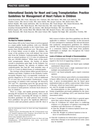
Table 1. Ross Classification
Class Interpretation
I Asymptomatic
II Mild tachypnea or diaphoresis with feeding in infants.
Dyspnea on exertion in older children.
III Marked tachypnea or diaphoresis with feeding in infants.
Prolonged feeding times with growth failure due to
heart failure. In older children, marked dyspnea on
exertion.
IV Symptoms such as tachypnea, retractions, grunting, or
diaphoresis at rest.
1314 Rosenthal et al. The Journal of Heart and Lung Transplantation
December 2004
3. The system advocated by the ACC/AHA for HF stag-
ing in adults can be readily applied to infants and
children as well, with minor modifications as shown in
Table 2.12
The writing committee of this document has
adopted this nomenclature due to the advantages enu-
merated above.
CHRONIC HEART FAILURE IN THE BIVENTRICULAR
CIRCULATION
Introduction
The spectrum of etiologies for HF in children is consid-
erable, and a discussion of the diagnostic approach to
children with HF is beyond the scope of this manu-
script. This topic has been addressed thoroughly in a
number of excellent publications, to which the reader
is referred for further detail.13–15
HF in children may develop from myocyte dysfunc-
tion (such as is seen in idiopathic cardiomyopathy, or
post-operative forms of cardiac dysfunction), or on the
basis of congenital disorders resulting in volume or
pressure overload. In the current era, both volume
loading and pressure loading are typically – but not
invariably – addressed surgically or in the catheteriza-
tion laboratory.
Chronic volume overload associated with mitral or
aortic insufficiency may be well tolerated for a pro-
longed period of time. In contrast to adult patients,
mitral valve replacement for infants and children is
often delayed because of technical difficulties inherent
to the patient size and anticoagulation requirements.
Limited data suggest that although early ventricular
dysfunction after surgery is common after correction of
chronic mitral insufficiency in children, left ventricular
function frequently normalizes over time.16,17
Left Ventricular Systolic Dysfunction
Pharmacotherapy.
Digitalis.
Clinical Data in Adult Patients with HF. Digoxin
improves symptoms in adults with HF.18
However,
despite large study cohorts, digoxin has not been
shown to improve survival in HF. Adams and colleagues
have recently shown that low doses of digoxin are as
effective as higher doses in preventing further HF, and
may reduce the incidence of side effects and toxicity,
especially when other medications are instituted which
can increase digoxin levels.19
In one recent post-hoc
analysis of the DIG trial, higher serum digoxin levels
were associated with increased mortality in men with
HF, independent of glomerular filtration rate.20
Clinical Data in Pediatric Patients. Although only
scant pediatric data are available, digoxin is widely used
to treat HF in infants and children. Recommendations
can only be extrapolated from those for adult patients
with left ventricular systolic dysfunction and HF.
Diuretics.
Clinical Data in Adult Patients. Few data are available
concerning the appropriate use of diuretics, but their
use is widespread. Current guidelines in adult patients
recommend the use of diuretics in all patients with HF
and fluid retention in order to achieve a euvolemic
state.
Spironolactone, specifically, has been shown to im-
prove survival in adults with advanced HF.21
This does
not appear to represent a diuretic effect, but rather is
specifically due to blockade of aldosterone; the benefit
has also been demonstrated with another aldosterone
antagonist, eplerenone.22
The activation of the renin-
aldosterone-angiotensin system (RAAS) is thought to be
critical in the pathogenesis of HF, and interruption of
the RAAS is a foundation of modern HF therapy.23
Although this is primarily accomplished in current
management by administration of ACE inhibitors, spi-
ronolactone has an additive effect in adults with severe
HF.
Clinical Data in Pediatric Patients. No published
clinical studies are available concerning the effective-
ness of diuretics in reducing mortality or improving
symptoms in pediatric patients.
Angiotensin Converting Enzyme Inhibitors and
Angiotensin Receptor Blockers.
Clinical Data in Adult Patients. HF is associated with
chronic activation of the RAAS and increased sympa-
Table 2. Proposed Heart Failure Staging for Infants and Children*
Stage Interpretation
A Patients with increased risk of developing HF, but who have
normal cardiac function and no evidence of cardiac
chamber volume overload. Examples: previous exposure to
cardiotoxic agents, family history of heritable
cardiomyopathy, univentricular heart, congenitally corrected
transposition of the great arteries.
B Patients with abnormal cardiac morphology or cardiac
function, with no symptoms of HF, past or present.
Examples: aortic insufficiency with LV enlargement, history
of anthracycline with decreased LV systolic function.
C Patients with underlying structural or functional heart disease,
and past or current symptoms of HF.
D Patients with end-stage HF requiring continous infusion of
intropic agents, mechanical circulatory support, cardiac
transplantation or hospice care.
*HF, heart failure; LV, left ventricular.
The Journal of Heart and Lung Transplantation Rosenthal et al. 1315
Volume 23, Number 12
4. thetic drive.24
These alterations, which may be benefi-
cial acutely, contribute to the progression of HF over
time. Increased adrenergic tone increases afterload and
myocardial oxygen demand. Angiotensin II is a vasocon-
strictor which causes myocyte hypertrophy and fibrosis
as well as aldosterone secretion. Increased concentra-
tions of both aldosterone and angiotensin II are associ-
ated with a poor outcome in HF patients.25
The efficacy
of ACE inhibitor therapy in HF is related to disruption of
the activation of the renin-angiotensin axis and to
decreased cardiac adrenergic drive.23,24,26
Multiple large clinical trials have shown that therapy
with ACE inhibitors improves symptoms and survival in
adult patients (primarily middle-aged men) with HF, and
reduces the rate of disease progression in asymptomatic
patients.27–30
In the majority of trials, ACE inhibitors
were well tolerated. Symptomatic hypotension can
occur; careful uptitration of these agents and adjust-
ment of diuretic dosages is necessary. Cough, which is
often associated with pulmonary edema, is present
more frequently in patients treated with ACE
inhibitors.28
A frequent observation in the literature evaluating
treatment of patients with HF concerns the underutili-
zation of ACE inhibitors. On the one hand, only a
minority of eligible adults receives an ACE inhibitor.31
Of equal importance, those patients who are treated
with ACE inhibitors are often treated with doses con-
siderably lower than the doses for which efficacy has
been established in clinical trials.31
Although there is
some controversy regarding appropriate dosing of ACE
inhibitors for HF, most studies suggest a more robust
response to higher doses of ACE inhibitors when hemo-
dynamics, symptoms or neurohumoral profiles are con-
sidered.32–35
The ATLAS trial compared the effects of
low and high dose lisinopril on the morbidity and
mortality of 3164 subjects with NYHA class II-IV.35
No
survival benefit was seen from high dose therapy as
compared to low dose, but high dose therapy reduced
the composite outcome of death or hospitalization by
12%, and reduced hospitalization by 13%. Although
dizziness, hyperkalemia and hypotension were all more
common in the high dose group, these side effects did
not require withdrawal from therapy more commonly
than in the lower dose group. The ATLAS trial con-
firmed that certain benefits of ACE inhibition are greater
at higher doses, and that these agents are clinically
tolerated at higher doses. It is also noteworthy that the
effect on symptoms was not associated with dose level,
suggesting that titration of ACE inhibitors according to
clinical endpoints is not a viable strategy.
Data concerning administration of ACE inhibitors to
patients with structural heart disease is understandably
limited. However, the beneficial effects of ACE inhibi-
tion on LV volume, dimensions, mass index, wall stress
and reduction in the volume-loaded ventricle have been
demonstrated for adult patients with aortic
regurgitation.36
There are important differences between the ACE
inhibitors and the angiotensin receptor blockers
(ARB’s). The ARB’s are competitive antagonists for the
angiotensin II receptor that is responsible for mediating
all the known actions of angiotensin II. ARB’s block the
cell surface receptor37
for angiotensin, rather than
acting through blockade of angiotensin converting en-
zyme. Unlike ACE inhibitors, ARB’s do not inhibit
bradykinin breakdown, which has been implicated in
causing the troublesome cough that is a prominent side
effect of ACE inhibitors. ARB’s are not nephrotoxic,
affording these drugs a theoretical advantage over ACE
inhibitors.
Despite these mechanistic differences, trials in adult
patients with HF have not shown any important differ-
ences in hemodynamic effects, efficacy and safety be-
tween ARB’s and ACE inhibitors.38,39
Currently ARB’s
are recommended in adults intolerant to ACE inhibitors.
The addition of ARB’s to ACE inhibitor therapy may
increase efficacy40
and is often recommended in adult
HF patients intolerant to -blockade.
Clinical Data in Pediatric Patients. Although ACE
inhibitors have been used in the pediatric population
for two decades, relatively few studies concerning the
administration of ACE inhibitors to children with HF are
available. Numerous small observational studies have
shown that ACE inhibitors benefit children with HF
caused by systemic ventricular systolic dysfunc-
tion.41–46
Effects on mortality have not been described,
with the exception of one retrospective report, in
which survival was improved by administration of ACE
inhibitors during the first year of treatment but not
subsequently.46
The use of ACE inhibitors in patients with structural
heart disease is less well understood. However, a single
controlled study in children with preserved ventricular
function and volume-overloaded ventricles from valvar
insufficiency showed a reduction in both LV volume
overload as well as hypertrophy in the ACE inhibitor
treated group over an average of 3 years of follow-up.47
No safety or efficacy data regarding the use of ARB in
children with HF are available. There is very limited
experience with ARB therapy for pediatric patients
with hypertension.48
 adrenergic Blockers.
General Remarks. Initial compensatory mechanisms in
the failing heart include activation of the sympathetic
nervous system and increased levels of circulating
catecholamines. In the long term the increased levels of
1316 Rosenthal et al. The Journal of Heart and Lung Transplantation
December 2004
5. catecholamines, particularly norepinephrine, contrib-
ute to the progression of HF through multiple mecha-
nisms, including myocardial fibrosis and apoptosis,
peripheral vasoconstriction, and salt/water retention by
the kidneys.49–51
The rationale for the use of adrenergic
antagonists in HF is to antagonize the deleterious effects
of sympathetic activation on the myocardium.
The risks associated with the use of beta-blockers
include hypotension and worsening HF.52–55
Usually
these symptoms occur in the first 48 hours after initia-
tion of treatment or at the time of up-titration of the
drug. Fluid retention may cause an increase in symp-
toms56–58
and adjustment in diuretic dosage may be
needed. Bradycardia is seen with the use of -blockers,
is usually asymptomatic, but may require dose reduc-
tion if there is associated hypotension.52
-blockers are
contraindicated in patients with severe bradycardia,
sick sinus syndrome and second or third degree heart
block, unless a pacemaker is in place. -blockers are
also contraindicated in patients with bronchial asthma
and in patients with cardiogenic shock.
Clinical Data in Adult Patients. Metoprolol was the
first -blocker studied in placebo-controlled trials in
patients with HF. Early studies failed to show a statisti-
cally significant decrease in the risk of death or listing
for transplantation.59,60
Eventually, in a trial involving
3991 patients with ischemic and non-ischemic cardio-
myopathy and mild to moderate CHF, administration of
metoprolol reduced mortality by 34%, which was sta-
tistically significant.61
In addition to metoprolol, carve-
dilol has been studied extensively in adult patients with
HF resulting from left ventricular dysfunction. Several
placebo-controlled trials have shown that carvedilol
therapy decreases the risk of clinical progression of HF
and decreases all-cause mortality.62–64
Of particular
interest, the COPERNICUS study (Carvedilol Prospec-
tive Randomized Cumulative Survival Trial)65
evaluated
the effects of carvedilol in 2289 patients with severe HF
from either ischemic or non-ischemic cardiomyopathy.
Patients treated with carvedilol had a 24% reduction in
combined risk of death and hospitalization for any
reason and a 35% reduction in mortality, showing that
even these severely compromised patients benefited
from carvedilol therapy.
Clinical Data in Pediatric Patients. The reported use
of -blockers in children with HF is very limited, and no
large placebo-controlled trials are available.
In a multi-institutional experience Shaddy, et al.
reviewed the results with metoprolol in 15 children
with cardiomyopathy of different etiologies.66
Metopro-
lol was started at 0.2-0.4 mg/kg/day and slowly in-
creased to a maximum dose of 1.1 mg/kg/day. There
was no significant difference in ejection fraction on
conventional therapy from the time of diagnosis to the
time when metoprolol was started, but after a mean of
23 months on metoprolol there was a statistically
significant and clinically important increase in ejection
fraction from 27% to 41%.
The experience with carvedilol in pediatric patients
with HF is similarly limited. Bruns, et al.53
reviewed the
use of carvedilol in 46 infants and children with cardio-
myopathy (80%) or congenital heart disease (20%) at 6
centers. Patients were on standard treatment with
digoxin, diuretics, and ACE inhibitors for at least three
months before the start of -blocker therapy. Carvedilol
was initiated at an average dose of 0.08 mg/Kg/day and
titrated to a mean maximum dose of 0.92 mg/kg/day.
After 3 months of therapy, modified NYHA class im-
proved in 67% of patients and worsened in 11%.
Shortening fraction improved slightly, from 16.2% to
19.0%. Side effects, mainly dizziness, hypotension, and
headache, occurred in 54% of patients and were well
tolerated overall.
In a single center study Rusconi, et al.54
reviewed the
results in 24 pediatric patients with dilated cardiomy-
opathy. Carvedilol was added to standard treatment at a
mean of 14 months after the diagnosis of cardiomyop-
athy was made, with an average maximum dose of 1.0
mg/kg/day. Adverse effects occurred in 5 patients. The
medication was tolerated in 22 patients. The mean left
ventricular ejection fraction improved from 25% to 42%
(pϽ0.001). The NYHA class improved in 15 patients, 1
patient died and 3 were transplanted. Lower doses of
carvedilol may also be effective, as suggested by Azeka
and colleagues.67
In this study of 22 children with DCM,
improvement of both ejection fraction and clinical
status was seen at 6 months with treatment dose of 0.2
mg/kg day of carvedilol, which was tolerated in all
patients.
Therapeutic Recommendations: No structural
Disease.
Recommendation 1: The underlying cause of new-
onset ventricular dysfunction (HF Stages B, C or D)
should be evaluated thoroughly in all patients. The
evaluation may include metabolic and genetic evalua-
tion in selected cases, as indicated by the available
history and physical findings. Invasive assessment, in-
cluding myocardial biopsy, may be considered in se-
lected cases. In infants, particular care should be paid to
the exclusion of coronary artery anomalies and other
anatomic causes. (Level of Evidence C; Strength of
Recommendation I)
Recommendation 2: Screening of first-degree rela-
tives should be considered in patients with new-onset
ventricular dysfunction due to DCM (HF Stages B, C or D).
(Level of Evidence C; Strength of Recommendation I)
Recommendation 3: Patients with fluid retention
The Journal of Heart and Lung Transplantation Rosenthal et al. 1317
Volume 23, Number 12
6. associated with ventricular dysfunction (HF stage C)
should be treated with diuretics to achieve a euvolemic
state using clinical criteria of fluid status and cardiac
output. (Level of Evidence C; Strength of Recommen-
dation I)
Recommendation 4: Digoxin is not currently rec-
ommended for patients with asymptomatic forms of left
ventricular dysfunction (HF Stage B) because this agent
did not alter survival in large trials of adult patients with
HF. (Level of Evidence C; Strength of Recommendation
IIb)
Recommendation 5: Digoxin should be employed
for patients with ventricular dysfunction, and symp-
toms of HF (HF Stage C), for the purpose of relieving
symptoms. Lower doses of digoxin are preferred for
this purpose. (Level of Evidence B; Strength of Recom-
mendation I)
Recommendation 6: For the treatment of moderate
or severe degrees of left ventricular dysfunction with or
without symptoms (HF Stage B and C), ACE inhibitors
should be routinely employed unless there is a specific
contraindication. These medications should be started
at low doses, and should be up-titrated to a maximum
tolerated safe dose. Uptitration may require a reduction
in the dose of diuretics. (Level of Evidence B; Strength
of Recommendation I)
Recommendation 7: For the treatment of decom-
pensated left ventricular dysfunction (HF Stage D), the
use of ACE inhibitors as initial therapy is not recom-
mended. (Level of Evidence C; Strength of Recommen-
dation IIb)
Recommendation 8: Patients who have an indica-
tion for ACE inhibitors therapy, but are intolerant of
ACE inhibitors should be considered for ARB therapy.
(Level of Evidence C; Strength of Recommendation IIa)
Recommendation 9: Given the limited information
available concerning the efficacy and safety of  agonist
receptor blockade in infants and children with HF, no
recommendation is made concerning the use of this
therapy for patients with left ventricular dysfunction
(HF Stage B or C). If a decision is made to initiate
-blocker therapy, consultation or co-management with
a heart failure or heart transplantation referral center
may be desirable. (Level of Evidence B; Strength of
Recommendation IIa)
Recommendation 10: Use of -blocker therapy is
not indicated for patients in HF Stage D. (Level of
Evidence C; Strength of Recommendation IIb)
Therapeutic Recommendations: Volume or Pres-
sure Overload Conditions. Recommendation 11: In
all cases of HF associated with structural heart disease
(HF Stage B, C or D), consideration should be given to
surgical repair of significant lesions, as the long-term
outlook may be more favorable than with medical
management alone. (Level of Evidence C; Strength of
Recommendation I)
Recommendation 12: In pressure-induced left ven-
tricular hypertrophy, with normal myocardial function,
ACE inhibitors are not recommended in the absence of
a non-cardiac indication such as hypertension. (Level of
Evidence C; Strength of Recommendation III)
Left Ventricular Diastolic Dysfunction
Overview. Diastolic dysfunction is a syndrome charac-
terized by impaired filling of one or both ventricles.68,69
Diastolic heart failure refers to a clinical syndrome of HF
with preserved systolic function.70–72
Diastolic dysfunc-
tion is the sole or primary cause of HF in as many as 1/3
of adult patients with HF.12,70–74
No published esti-
mates of the prevalence of diastolic dysfunction in the
pediatric population are available.
There are 2 fundamental types of diastolic abnormal-
ity: impaired ventricular relaxation (affecting early dias-
tole), and increased myocardial stiffness (affecting late
diastolic filling).70
The hemodynamic consequences of
diastolic dysfunction include increased ventricular fill-
ing pressures leading to elevation in atrial and venous
pressures, and in the case of left ventricular dysfunc-
tion, leading also to an increase in pulmonary arterial
pressure.
Conditions that cause diastolic dysfunction are varied
and include pericardial as well as myocardial etiolo-
gies.75–78
Of note, patients with chronic diastolic dys-
function are at risk for sudden death, as well as for
developing pulmonary hypertension, which further
complicates treatment and limits survival.75–78
Pharmacotherapy. There are no large-scale, random-
ized controlled trials of diastolic HF therapy in the adult
or pediatric population.70,71
Diuretics.
Clinical Data. Diuretics are the first line of therapy for
diastolic dysfunction. Diuretics reduce pulmonary con-
gestion and relieve symptoms such as orthopnea, cough
and dyspnea. Injudicious or excessive use will reduce
preload and result in diminished cardiac output.73–75
ACE Inhibitors and Angiotensin Receptor Block-
ers.
Clinical Data in Adult Patients. Therapy with ACE
inhibitors may benefit some forms of diastolic dysfunc-
tion as tissue ACE levels are increased in models of
hypertrophy,70,79,80
and angiotensin II is known to
induce myocyte hypertrophy as well as fibrosis. ACE
inhibitors may be employed to obtain regression of left
ventricular hypertrophy, reverse vascular hypertrophy
and fibrosis, and improve endothelial function. Theo-
1318 Rosenthal et al. The Journal of Heart and Lung Transplantation
December 2004
7. retically, ACE inhibitors and perhaps angiotensin recep-
tor blockers may be a reasonable treatment option.70
In the PRESERVE study, enalapril had moderately
beneficial and statistically indistinguishable effects on
regression of left ventricular hypertrophy compared
with nifedipine.81
There are currently no mortality data
to support the use of ACE inhibitors for patients with
chronic HF and preserved ejection fraction in the
absence of another indication for ACE inhibitors ther-
apy (such as hypertension).
Limited experience in the adult population demon-
strates improved exercise tolerance with ARB therapy
in patients with diastolic dysfunction.82
Clinical Data in Pediatric Patients. Captopril was
deleterious in 1 small study with 4 pediatric patients
with restrictive cardiomyopathy who demonstrated sys-
temic hypotension without an improvement in cardiac
output.41
Calcium Channel Blockers.
Clinical Data in Adult Patients. Much of the literature
regarding calcium channel blockers is in the elderly
population and in patients with hypertrophic cardiomy-
opathy. There are no prospective randomized trials.
Benefit may be achieved by cardiac slowing, prolonging
filling time, and improving myocardial relaxation.73,79
Prolonged administration of calcium channel blockers
may lead to regression of left ventricular hypertrophy.81
Calcium channel blockers may also directly improve
ventricular relaxation or compliance, but there are few
data to indicate that this is a clinically important
effect.74
When compared to enalapril, nifedipine had moder-
ately beneficial and statistically indistinguishable effects
on regression of left ventricular hypertrophy compared
with enalapril.81
Some patients may deteriorate after
administration of calcium channel blockers; this is likely
the result of a decrease in afterload.73
Clinical Data in Pediatric Patients. There are no data
on the use of calcium channel blockers for the treat-
ment of diastolic dysfunction in children.
 Blockers.
Clinical Data. Use of -blockers has been reported for
diastolic dysfunction in elderly patients with hypertro-
phic cardiomyopathy but no prospective randomized
study has been performed. The rationale for use in-
cludes reduced heart rate leading to a prolonged filling
time and relief of cardiac ischemia.73,74
It is recognized
that some patients deteriorate with institution of 
blockade.73
-blockers are proposed to directly im-
prove diastolic dysfunction by augmenting ventricular
relaxation or improving compliance, but there are few
data to indicate that these agents exert a clinically
important effect by this mechanism.
Nitrates.
Clinical Data. Nitric oxide or drugs that enhance NO
release (sodium nitroprusside, nitroglycerin) improve
diastolic dysfunction when administered locally to the
coronary arteries.69,83
Systemic use of nitrates may
result in decreased systemic venous preload, increased
pulmonary venous filling, reduced systemic afterload,
hypotension and clinical deterioration.73
Patients with
chronic restrictive disease may have typical or atypical
anginal pain, which may respond to nitrate therapy.
Therapeutic Recommendations. Recommenda-
tion 13: Clinical management of diastolic dysfunction
should address symptoms and attempt to address the
underlying cause of the diastolic dysfunction, if known.
This should include a careful evaluation for pericardial
disease, and coronary insufficiency with attendant myo-
cardial ischemia. Systemic hypertension, if present,
should be controlled aggressively. (Level of Evidence C;
Strength of Recommendation I)
Recommendation 14: Fluid management to control
symptoms remains a cornerstone in the management of
symptomatic diastolic dysfunction (HF Stage C). Diuret-
ics can be very useful to control symptoms but must be
used cautiously as cardiac output depends on the
elevated filling pressures. Renal function should be
followed closely with care taken not to over-diurese a
patient. Finally, sodium and fluid restriction may be
helpful in controlling symptoms. (Level of Evidence C;
Strength of Recommendation I)
Recommendation 15: Patients with marked atrial
dilatation due to diastolic dysfunction (HF stage B or C)
have a propensity for the formation of thrombi and
prophylactic anticoagulation may be considered in this
setting. (Level of Evidence C; Strength of Recommen-
dation IIa)
Recommendation 16: Atrial arrhythmias are not
infrequent in patients with diastolic dysfunction due to
atrial enlargement. However, atrial contribution to ven-
tricular filling is particularly important for this group of
patients (HF Stage B and C). Therefore, efforts should
be made to maintain sinus rhythm by use of antiarrhyth-
mic therapy and pacemaker therapy. (Level of Evidence
C; Strength of Recommendation I)
Recommendation 17: Asymptomatic diastolic dys-
function (HF Stage B) should be followed closely, but
does not require pharmacotherapy. (Level of Evidence
C; Strength of Recommendation IIa)
Recommendation 18: Treatment with agents
which reduce afterload, such as ACE inhibitors, 
The Journal of Heart and Lung Transplantation Rosenthal et al. 1319
Volume 23, Number 12
8. blockers, calcium channel blockers and nitrates, should
be cautiously undertaken with close monitoring.
Abrupt decreases in afterload in patients with restric-
tive or constrictive disease may be deleterious. (Level of
Evidence C; Strength of Recommendation IIb)
Recommendation 19: Patients with diastolic dys-
function that is refractory to optimal medical/surgical
management should be evaluated for heart transplanta-
tion as they are at high risk of developing secondary
pulmonary hypertension, and of sudden death (HF
Stage C). (Level of Evidence B; Strength of Recommen-
dation I)
The Systemic Right Ventricle
Overview. The morphologic right ventricle (RV) is
connected to the aorta and is thus the systemic ventri-
cle in 2 main groups of patients with biventricular
circulation: patients born with d-transposition of the
great arteries who were treated with atrial baffle sur-
gery (Mustard or Senning repair) and patients born with
congenitally-corrected transposition of the great arter-
ies. RV dysfunction has been described in both these
groups of patients and may lead to HF. Although RV
performance at rest is often normal,84
exercise ability is
often limited in patients who have undergone atrial
baffle surgery because of both chronotropic incompe-
tence and limited stroke volume reserve.85–92
The mechanism of systemic right ventricular dysfunc-
tion is poorly understood. Various theories include: (a)
a sub-optimal myocardial fiber arrangement and me-
chanics in the right ventricle, (b) adverse pattern and
reduced heterogeneity of ventricular strain, (c) tricus-
pid insufficiency and, (d) myocardial fibrosis secondary
to prolonged hypoxemia during infancy while awaiting
atrial baffle surgery.93
Perfusion defects with associated
wall motion abnormalities have been described using
single photon emission computed tomography,94–96
suggesting a role for chronic subendocardial ischemia.
The clinical course in patients with congenitally-
corrected transposition of the great arteries is often
determined by associated structural (ventricular septal
defect, pulmonary stenosis) or electrophysiologic ab-
normalities (AV block). Although there are reports of
patients without such abnormalities whose right ventri-
cles have continued to function normally well into
adulthood, systemic RV dysfunction and HF occur with
increasing prevalence with advancing age. By 45 years
of age, 25% of patients without and 67% of patients with
associated lesions have HF.97
There is a limited surgical experience with conver-
sion of the atrial baffle to a systemic left ventricle
(“double-switch”), with baffle takedown accompanied
by arterial switch procedure. Early results indicate that
surgery is feasible, but surgical risk may be substantial
for this approach, and the long-term results are not
known.98
A similar procedure has been performed in
small numbers of patients with corrected transposition,
although again, long-term outcomes are not yet defined.
Pharmacotherapy.
Clinical Data. Data specifically addressing the medical
therapy of systemic right ventricular dysfunction are
exceptionally scanty. Administration of ACE inhibitors
to 14 survivors of the Mustard operation did not affect
exercise tolerance or right ventricular ejection frac-
tion.99
However, selected patients did show benefit. In
the absence of more specific data, the management
guidelines below are based on clinical experience and
the effectiveness of these regimens in adult patients
with systemic LV failure.
Special Considerations. Although -blockers are now
considered an important part of HF therapy in adult
patients with cardiomyopathy, specific problems may
be encountered in administration of these agents to
patients with a systemic RV. These difficulties relate to
the high prevalence of sinus node dysfunction in survi-
vors of atrial baffle repair and AV node dysfunction in
patients with congenitally corrected transposition of
the great arteries. The administration of -blockers in
these patients may be particularly problematic, and may
result in symptomatic bradycardia and exacerbation of
HF.
Therapeutic Recommendations. Recommenda-
tion 20: Patients with a right ventricle in the systemic
position are at risk of developing systemic ventricular
dysfunction (HF Stage A) and should undergo periodic
evaluation of ventricular function. (Level of Evidence B;
Strength of Recommendation I)
Recommendation 21: Patients with fluid retention
associated with systemic right ventricular dysfunction
(HF stage C) should be treated with diuretics to achieve
a euvolemic state. In patients receiving chronic diuretic
therapy, electrolyte balance and renal function should
be evaluated periodically. (Level of Evidence C;
Strength of Recommendation I)
Recommendation 22: Digoxin should be employed
for patients with symptomatic systemic RV dysfunction
(HF Stage C), for the purpose of relieving symptoms.
Digoxin is not currently recommended for patients
with asymptomatic systemic right ventricular dysfunc-
tion (HF Stage B). (Level of Evidence C; Strength of
Recommendation IIa)
Recommendation 23: For the treatment of asymp-
tomatic systemic right ventricular dysfunction (HF
Stage B), ACE inhibitors should be routinely employed
unless there is a specific contraindication. These medi-
cations should be employed at standard doses. (Level of
Evidence C; Strength of Recommendation IIa)
1320 Rosenthal et al. The Journal of Heart and Lung Transplantation
December 2004
9. Recommendation 24: For the treatment of symp-
tomatic systemic right ventricular dysfunction (HF
Stage C), ACE inhibitors should be routinely employed,
as above. (Level of Evidence C; Strength of Recommen-
dation I)
Recommendation 25: Patients who have an indica-
tion for ACE inhibitors therapy, but are intolerant of
ACE inhibitors should be considered for ARB therapy.
(Level of Evidence C; Strength of Recommendation IIa)
Recommendation 26: Consideration of surgical re-
vision of the tricuspid valve should be given where both
systemic right ventricular dysfunction and severe tricus-
pid valve regurgitation are present, particularly when
the regurgitation is due to an intrinsic abnormality of
the tricuspid valve (HF Stage B and C). (Level of
Evidence C; Strength of Recommendation IIa)
Recommendation 27: Anatomic revision (“double-
switch”) or cardiac transplantation should be consid-
ered for patients with advanced systemic right ventric-
ular failure (HF Stage C) that is refractory to medical
therapy. (Level of Evidence C; Strength of Recommen-
dation IIa)
CHRONIC HEART FAILURE IN THE UNIVENTRICULAR
CIRCULATION
Overview
For most patients with single ventricle anatomy, surgi-
cal management is the primary treatment path, and may
include a variety of early palliative procedures followed
by bi-directional Glenn (superior cavopulmonary con-
nection, SCPC), and ultimately the Fontan procedure
(total cavopulmonary connection, TCPC). Early pallia-
tion with complete mixing of systemic and pulmonary
venous flow requires that ventricular output must be
maintained at a level that is 2 to 3 times normal.100,101
This chronic volume overload places the myocardium
at considerable risk.100
Ventricular anatomic type and
the specifics of early management are important deter-
minants of ventricular outcome after the SCPC or TCPC.
In particular, patients with a prior aortopulmonary
shunt typically have larger postoperative ventricular
size than those with a prior pulmonary artery band.102
In some series,102
systemic left ventricles are more
dilated than systemic right ventricles at the time of
volume-unloading surgery, although other reports have
found no difference in size or function between mor-
phologically single right or left ventricles.103
The dura-
bility of the systemic right ventricle is in question, with
reports suggesting this may104–106
or may not107
be a
problem. In one series, early postoperative survival was
better in patients with a morphologic left ventricle but
by 6 months after completion of the Fontan procedure,
ventricular morphology did not influence survival.107
In patients who have completed the bi-directional
Glenn or Fontan palliation, the manifestations of HF do
not include the typical symptoms that occur in patients
with 2 ventricles. After SCPC, TCPC, or Fontan pallia-
tion, systemic venous pressure is by necessity higher
than pulmonary. These patients experience peripheral
edema, pleural and pericardial effusions, cyanosis, and
symptoms related to reduced cardiac output such as
chronic fatigue, loss of appetite, and extreme exercise
intolerance. These symptoms can clearly be related to
factors other than myocardial dysfunction, many of
which are common in this patient population. The
basic physiology of the post-Fontan circulation is not
intrinsically distinguishable from HF: activation of the
renin-angiotensin system is usual if not universal, neu-
rohumoral activation does not correlate with hemody-
namic variables, and increased levels of catecholamines
do not track well with severity of symptoms.108,109
Reduction in exercise capacity may be helpful in
making this distinction; however exercise performance
in children with successful or optimal Fontan palliation
may be quantitatively identical to that in children with
mild HF.110
Reduced exercise capacity may in part be
explained by diminished resting stroke volume and
stroke volume reserve111
compared with normal hearts.
The impact of reduced stroke volume in patients who
have had the Fontan operation is compounded by
chronotropic incompetence, with most series reporting
maximum achieved heart rate to be about 75% of the
age-predicted normal value,112–114
and in some se-
ries,115,116
though not all,117
peak heart rate correlates
with the percent of predicted VO2max that is achieved.
Exercise-induced hypoxia may also be a limiting factor.
Arterial saturation typically falls during exercise in
Fontan patients113,118
and correlates with exercise ca-
pacity.116
The cause of the exercise-related hypoxia is
not clear. Vital capacity and functional residual capacity
are reduced,119
consistent with a restrictive pattern of
pulmonary abnormality.114
Adult patients after Fontan
have been reported to have sufficient restriction so as to
manifest severely diminished maximum ventilation
with secondary reduction in aerobic capacity.114
The role of ventricular dysfunction and myocardial
insufficiency in limiting exercise capacity in these pa-
tients remains unclear. This highlights the importance
of identifying other potentially treatable sources of
exercise intolerance before classifying the patient as
having cardiogenic HF.
Diagnostic Approach. In patients who have previ-
ously undergone Fontan-type repair, thorough evalua-
tion and management of circulatory insufficiency is
essential. Significant systemic or pulmonary venous
obstruction is unlikely to be tolerated. Valve regurgita-
tion is common and may be progressive, causing per-
sistent volume overload, and is associated with a poorer
outcome.105
The Journal of Heart and Lung Transplantation Rosenthal et al. 1321
Volume 23, Number 12
10. Atrial arrhythmias increase in frequency with age120
and may be associated with tachycardia-induced ven-
tricular dysfunction. Chronotropic incompetence may
be more poorly tolerated than in other forms of heart
disease.121
Residual right-to-left shunts may augment
during exercise, severely limiting exercise capacity. In
general, invasive hemodynamic investigation is needed
both to document that myocardial dysfunction is pri-
marily responsible and that other potentially treatable
contributing factors are not present.
Pharmacotherapy.
Diuretics. Use of diuretics is common in the early
post-operative period and in many instances becomes a
permanent component of therapy, even in the absence
of specific findings that clearly point to fluid overload.
Peripheral edema, pleural and pericardial effusions, and
other evidence of elevated systemic venous pressure
occur frequently and often respond favorably to diuret-
ics. However, the absence of a pulmonary pumping
chamber renders the Fontan circulation particularly
vulnerable to an inability to maintain an adequate
systemic ventricular preload. Reduction of systemic
venous pressure through diuresis reduces systemic
ventricular preload in a more direct fashion than in
patients with a biventricular circulation. Although the
beneficial use of diuretics for HF invariably represents a
balancing act with regard to maintenance of cardiac
output, the margin of safety is far narrower in these
patients and requires a greater degree of vigilance with
regard to electrolyte disturbances, pre-renal azotemia,
and exacerbation of exercise intolerance.
Digoxin. Digoxin is in common use for post-Fontan
patients despite the absence of specific evidence of
safety or benefit in this setting. There are no theoretical
concerns that would make this population more or less
likely to benefit from this agent than other groups with
ventricular dysfunction.
ACE Inhibitors. In patients with single ventricles,
activation of the RAAS contributes to the increased
vascular resistance that typically occurs after the Fontan
procedure, and a favorable response to ACE inhibitors
or ARB might therefore be anticipated.108
However, in
a study of Fontan patients who did not have HF,
administration of enalapril for 6 weeks did not alter
systemic resistance, resting cardiac index, or exercise
capacity.122,123
Although these agents were well toler-
ated, and clinical experience confirms that ACE inhibi-
tors can be safely administered to patients who have
had the Fontan operation, the efficacy and risks of
long-term ACE inhibitor therapy in this setting are not
known.
 Blockers. The utility of -blockers in patients after
Fontan-type operations is unknown. Specific clinical
data in this population are absent. There is a reasonable
probability that -blocker therapy may impair exercise
tolerance in these patients because of the pervasive
finding of chronotropic incompetence in post-Fontan
patients and the generally adverse impact of -blocker
therapy on VO2max.
Therapeutic Recommendations. Recommenda-
tion 28: In patients with a univentricular circulation
who undergo deterioration in clinical status (HF Stage B
and C), consideration should be given to invasive
assessment of hemodynamics, due to the heightened
sensitivity of this group to elevations of filling pressure.
(Level of Evidence C; Strength of Recommendation I)
Recommendation 29: A clinical decision as to the
severity or presence of HF symptoms in patients with a
univentricular circulation must include consideration of
the patient’s prior status and underlying anatomic de-
fects. Longitudinal evaluation is of great importance in
this regard. (Level of Evidence C; Strength of Recom-
mendation I)
Recommendation 30: Maintenance of atrioventric-
ular synchrony should be a priority in the management
of patients with a univentricular circulation and HF (HF
Stages B and C). (Level of Evidence C; Strength of
Recommendation I)
Recommendation 31: Patients with fluid retention
associated with systemic ventricular dysfunction in a
univentricular circulation (HF stage C) should be
treated with diuretics to achieve a euvolemic state. In
patients receiving chronic diuretic therapy, electrolyte
balance and renal function should be periodically eval-
uated. (Level of Evidence C; Strength of Recommenda-
tion I)
Recommendation 32: Digoxin should be employed
for patients with systemic ventricular dysfunction in a
univentricular circulation (HF Stage C), for the purpose
of relieving symptoms. Digoxin is not currently recom-
mended for patients with asymptomatic systemic ven-
tricular dysfunction in a univentricular circulation (HF
Stage B). (Level of Evidence C; Strength of Recommen-
dation IIa)
Recommendation 33: For patients with a univen-
tricular circulation after SCPC or TCPC, in whom
systolic function is normal (HF Stage A), ACE inhibitors
should not be employed routinely. (Level of Evidence
C; Strength of Recommendation III).
Recommendation 34: For the treatment of asymp-
tomatic systemic ventricular dysfunction in a univen-
tricular circulation after SCPC or TCPC (HF Stage B),
ACE inhibitors should be employed unless there is a
specific contraindication. These medications should be
1322 Rosenthal et al. The Journal of Heart and Lung Transplantation
December 2004
11. employed at standard doses (Level of Evidence C;
Strength of Recommendation IIa).
Recommendation 35: For the treatment of symp-
tomatic systemic ventricular dysfunction in a univen-
tricular circulation after SCPC or TCPC (HF Stage C),
ACE inhibitors should be employed, as above. (Level of
Evidence C; Strength of Recommendation IIa)
Recommendation 36: Use of -blocker therapy is
not routinely recommended for patients with systemic
ventricular dysfunction in a univentricular circulation
after SCPC or TCPC (HF Stage B and C). (Level of
Evidence C; Strength of Recommendation IIb)
ACUTE HEART FAILURE
Pharmacologic Support
In adult studies, it has been recognized that inotropes
may be required in the management of refractory acute
HF exacerbations that are accompanied by hypoperfu-
sion and hypotension.124
Currently available inotropic
agents increase contractility through a common path-
way of increasing intracellular levels of cyclic adenylate
monophosphate (cAMP). Increased cytoplasmic levels
of cAMP cause increased calcium release from the
sarcoplasmic reticulum and increased contractile force
generation by the contractile apparatus. Increases in
cAMP occur by two different mechanisms: -adrener-
gic-mediated stimulation (increased production) or
phosphodiesterase III (PDE III) inhibition (decreased
degradation).125
The catecholamines are the most potent positive
inotropic agents available; however, effects are not
limited to inotropy. They also possess chronotropic
properties and complex effects on vascular beds of the
various organs of the body. Consequently, the choice of
an agent may depend as much on the state of the
circulation as it does on the myocardium.
Amrinone and milrinone belong to a class of nongly-
coside, nonsympathomimetic inotropic agents (phos-
phodiesterase III inhibitors). Intravenous administration
of amrinone or milrinone increases cardiac output and
reduces cardiac filling pressures, pulmonary vascular
resistance, and systemic vascular resistance with mini-
mal effect on the heart rate and systemic blood pressure
of adult patients.126
These drugs are particularly useful
in the treatment of cardiogenic shock because they
increase contractility and reduce afterload by periph-
eral vasodilatation without a consistent increase in
myocardial oxygen consumption. Milrinone has been
well studied in the pediatric population.126–128
A re-
cently completed randomized, controlled trial demon-
strated that milrinone infusion reduced the incidence of
low cardiac output following cardiac surgery.129
Both of these agents require careful bolus dosing
prior to initiating an infusion – a rapid infusion of the
bolus dose may cause hypotension. An alternative ap-
proach, which may be preferred in unstable patients, is
to begin infusion without an initial bolus.130
Since both
of these drugs have relatively long half-lives, they
should be used cautiously in the presence of significant
hypotension. However, recent data indicate that the
half-life of milrinone may be shorter than originally
extrapolated from adult data.129
Nesiritide, a recombinant B-type natriuretic peptide,
is the first new drug approved by the U.S. Food and
Drug Administration in 14 years for the treatment of
acutely decompensated HF in adults. Endogenous B-
type natriuretic peptide is a cardiac hormone produced
by the failing heart to counteract the maladaptive
hemodynamic, neural, and hormonal compensations
associated with the syndrome of CHF. Nesiritide is
identical to the naturally occurring peptide, and like
this peptide, Nesiritide reduces preload and afterload,
leading to increases in cardiac index without reflex
tachycardia or direct inotropic effect, increased diuresis
and natriuresis, and suppression of the renin-angioten-
sin-aldosterone axis and sympathetic system. In a large
randomized controlled clinical trial of 489 patients,
nesiritide was found to be faster and more effective
than IV nitroglycerin plus standard care at improving
hemodynamic and symptomatology in patients with
acutely decompensated CHF who have dyspnea at
rest.131–133
Because of its unique mechanism of action
and greater safety compared to both standard inotropic
therapy with dobutamine and vasodilator therapy with
nitroglycerin, the availability of nesiritide may alter the
current algorithms for CHF management. However, no
pediatric data are currently available.
Infants and children with low blood pressure and
adequate cardiac function after cardiac surgery refrac-
tory to standard cardiopressors respond well to the
pressor action of exogenous vasopressin and permit
withdrawal or significant lowering of epinephrine dose.
In a study of 11 children with vasodilatory shock after
cardiac surgery, all 9 children with adequate cardiac
function improved with vasopressin and survived; the 2
patients that received vasopressin in the setting of poor
cardiac function died, despite transient improvement in
blood pressure.134
Plasma vasopressin levels were de-
creased before treatment in 3 patients in whom blood
levels were tested indicating that deficiency of the
hormone may contribute to this hypotensive condition.
Mechanical Support
Overview. Mechanical circulatory support has become
an important addition to the treatment armamentarium
for the infant or child with acutely decompensated HF
and low cardiac output unresponsive to pharmacologic
maneuvers. Options for mechanical circulatory support
include total heart-lung bypass (Extracorporeal Mem-
brane Oxygenation [ECMO]), or more limited cardiac
The Journal of Heart and Lung Transplantation Rosenthal et al. 1323
Volume 23, Number 12
12. support with a left ventricular assist device (LVAD),
right ventricular assist device (RVAD), or intra-aortic
balloon pump (IABP). In pediatric patients, most expe-
rience has been with ECMO, primarily because size
limitations have generally precluded extensive use of
other modalities. However, with the development of
smaller ventricular assist devices (primarily in Europe)
and the adaptation of existing devices, mechanical
ventricular assistance has been applied to infants as
young as 5 days of age.135–145
The choice of modality in
a given patient will depend on the specific pathophys-
iology of HF in that patient, on the availability of
support devices and on the expertise of the child’s
physicians.
Risks and Benefits. Mechanical assist in children can
be life saving in low cardiac output situations where
there is a reversible underlying abnormality that would
benefit from short-term cardiac support.146
Mechanical
support maintains end-organ function and reduces myo-
cardial oxygen requirements during a critical period of
recovery from a cardiac insult. Preliminary evidence
indicates that cardiac remodeling, on both a structural
and molecular level, occurs during mechanical support.
Studies with small number of patients have demon-
strated rates of survival to hospital discharge ranging
from 25 to 65%.147–150
Ibrahim and colleagues reviewed
a 10-year experience in 96 cardiac patients requiring
ECMO (67 patients) or ventricular assist devices (29
patients).147
Of these patients, 40% and 41%, respec-
tively, survived to hospital discharge. The use of me-
chanical support in patients with single ventricle phys-
iology has been associated in some studies with a
poorer outcome, although recent data from Jaggers et
al. and others support the use of ECMO after Norwood
palliation.151
Overall hospital survival in the 35 patients
in that series was 61%.151
Mechanical assist can also be used a bridge to trans-
plant when cardiac recovery is not expected.152
In this
case, mechanical support extends the period over
which a patient can wait for a donor organ. End-organ
function may be preserved, or perturbations in function
may reverse with improved perfusion, thus improving
transplant suitability and outcome.153
Therapeutic Recommendations. Recommenda-
tion 37: Institution of mechanical cardiac support
should be considered in patients without structural
congenital heart disease, who manifest acute low car-
diac output or who have intractable arrhythmias during
a presumably temporary condition that is refractory to
medical therapy (HF Stage D) such as myocarditis,
septic shock, or acute rejection following cardiac trans-
plantation. (Level of Evidence C; Strength of Recom-
mendation I)
Recommendation 38: Institution of mechanical car-
diac support should be considered in patients with or
without structural congenital heart disease, who have
acute decompensation of end-stage HF, primarily as a
bridge to cardiac transplantation (HF Stage D). (Level of
Evidence B; Strength of Recommendation I)
Recommendation 39: Institution of mechanical car-
diac support may be considered in patients who have
experienced cardiac arrest, hypoxia with pulmonary
hypertension, or severe ventricular dysfunction with
low cardiac output after surgery for congenital heart
disease, including “rescue” of patients who fail to wean
from cardiopulmonary bypass or who have myocarditis
(HF Stage D). However, the outcomes in this group are
less satisfactory than for other indications for mechan-
ical support. (Level of Evidence B; Strength of Recom-
mendation IIa)
Recommendation 40: Mechanical cardiac support
is not indicated in those cases in which there is
evidence of a severe and irreversible defect (e.g. cata-
strophic intracranial hemorrhage, or advanced multisys-
tem organ failure). However, in practice, the determi-
nation of the severity and/or irreversibility of the
associated condition may be difficult to determine, so
the decision concerning eligibility for mechanical sup-
port is a difficult clinical judgment. (Level of Evidence
C; Strength of Recommendation IIb)
ELECTROPHYSIOLOGY CONSIDERATIONS
Overview
Arrhythmia is a major cause of morbidity and mortality
in pediatric patients with end-stage HF.154–161
Myocar-
dial scars and stretching associated with pressure or
volume overload, previous heart surgery or intrinsic
myocardial disease provide a ripe substrate for arrhyth-
mogenesis.162,163
Multiple triggers for arrhythmia are
prevalent in patients with HF and include electrolyte
imbalance, blood gas and pH abnormalities, ischemia
and administration of potentially pro-arrhythmic drugs
such as digoxin, positive inotropes, phosphodiesterase
inhibitors and anti-arrhythmic drugs themselves.164,165
Although it is controversial whether ventricular arrhyth-
mias are independent risk factors for sudden death in
this population, sustained tachycardia may cause hemo-
dynamic collapse, and complex non-sustained ventric-
ular ectopy reflects potential electrical instability of the
myocardium.157
Atrial arrhythmias may also impair car-
diac output and aggravate HF by eliminating the atrial
contribution to filling and/or causing a rapid ventricular
response.
In patients with dilated cardiomyopathy, between 50
and 63% of those patients who die have ventricular
arrhythmias at presentation.154,155
In pediatric patients
awaiting transplantation, 62% had life-threatening ar-
rhythmias, most commonly ventricular tachycardia.166
1324 Rosenthal et al. The Journal of Heart and Lung Transplantation
December 2004
13. Arrhythmia management is an important component in
the overall care of the pediatric patient with HF.
Treatment can be broken down into the management
of acute arrhythmias causing hemodynamic collapse
and chronic arrhythmias requiring long-term suppres-
sion because they lead to impaired cardiac performance
or may pose a significant risk factor for sudden death.
Pharmacotherapy
Acute Arrhythmia Management. Arrhythmias likely
to require acute treatment in patients with HF include
atrial tachyarrhythmias with a rapid ventricular rate,
paroxysmal supraventricular tachycardia, junctional ec-
topic tachycardia and sustained ventricular tachycardia.
Synchronized DC cardioversion/defibrillation should be
considered for treatment of either supraventricular or
ventricular arrhythmias if hemodynamic collapse is
imminent.167,168
Atrial tachycardia, flutter or fibrillation can be initially
managed by controlling the ventricular rate with AV
nodal blocking agents. For rate control, digoxin is not
likely to be useful primarily because of its delayed onset
of action. Intravenous diltiazem is a reasonable first
choice, but hypotension is a major concern in a setting
of impaired ventricular function. An intravenous
-blocker, such as esmolol can be useful in decreasing
the ventricular rate but hypotension is likely to pre-
clude its use.
The treatment of the atrial rhythm itself includes
intravenous amiodarone, procainamide or ibutilide.
However, side effects such as hypotension and torsade
de pointes occur frequently and elective cardioversion
is often preferable. There is very little experience using
IV ibutilide for the acute conversion of atrial flutter or
fibrillation in pediatric patients with significant myocar-
dial dysfunction. Paroxysmal supraventricular tachycar-
dia is best treated with intravenous adenosine, but a
longer acting agent such as procainamide or amioda-
rone may be necessary for control of recurrent
episodes.
Junctional ectopic tachycardia responds best to IV
amiodarone and to minimizing the dose of any intrave-
nous inotropes or phosphodiesterase inhibitors. The
pharmacologic treatments for ventricular tachycardia,
include intravenous lidocaine, amiodarone and procain-
amide.169,170
Lidocaine is the first line drug because of
its rapid onset of action and limited toxicity if drug
levels are kept in the therapeutic range. If this is
unsuccessful, either intravenous amiodarone or pro-
cainamide can be utilized, but amiodarone is preferable
because of a lower incidence of hypotension and
tendency for arrhythmia aggravation.
Chronic Arrhythmia Management. Chronic drug
suppression of arrhythmias in patients with HF can be
very problematic. For atrial arrhythmias, many drugs
have been shown to have a modest success rate includ-
ing those in the category of IA, IC and III. However,
amiodarone stands out as the most appropriate drug
because of its somewhat higher success rate and lesser
tendency for pro-arrhythmia.171,172
For ventricular
tachycardias, the superiority of amiodarone over the
other drugs is even more evident. This medication is
usually well tolerated, even in patients with very poor
contractility. The major limiting factor in the short-term
is bradycardia and hypotension, while longer use re-
quires monitoring for thyroid and liver dysfunction as
well as pulmonary interstitial disease.
As an alternative to pharmacotherapy in the setting of
chronic atrial arrhythmias, ablation therapy can be
considered. The safety of radiofrequency or surgical
ablation is well established, and there are significant
data attesting to its efficacy.172–174
Triedman and col-
leagues report a 73% procedural success rate for radio-
frequency ablation, with approximately 50% success at
1 year, for a mixed population including Fontan and
Mustard patients.172
In a population of Fontan patients,
short-term control of atrial tachycardia was achieved in
14/15 patients using a modified RA maze intraopera-
tively, with no recurrences after 43 months of
follow-up.173
Therapeutic Recommendations. Recommenda-
tion 41: In patients with significant arrhythmias in the
setting of HF associated with structural heart disease
(HF Stages B, C or D), consideration should be given to
surgical repair of important uncorrected or residual
defects, as this is likely to be essential to achieve
adequate rhythm control. (Level of Evidence C;
Strength of Recommendation I)
Recommendation 42: In patients with significant
arrhythmias in the setting of HF associated with struc-
tural heart disease (HF Stages B, C or D), consideration
should be given to improving or optimizing the medical
treatment for HF and correcting aggravating factors
such as electrolyte abnormalities, as this is likely to be a
key determinant of the successful control of arrhyth-
mias. (Level of Evidence C; Strength of Recommenda-
tion I)
Recommendation 43: In patients with significant
arrhythmias in the setting of HF associated with struc-
tural heart disease (HF Stages B, C or D), maintenance of
atrio-ventricular synchrony is of great importance in
optimizing hemodynamics, and management of intra-
atrial arrhythmias should be oriented towards restora-
tion of sinus rhythm rather than on ventricular rate
control alone. (Level of Evidence C; Strength of Recom-
mendation I)
Recommendation 44: In patients with impaired
ventricular function (HF Stages B, C or D), many forms
The Journal of Heart and Lung Transplantation Rosenthal et al. 1325
Volume 23, Number 12
14. of tachycardia can precipitate acute hemodynamic col-
lapse, and many of the available pharmacotherapies can
precipitate hypotension. Therefore, consideration
should be given to early utilization of electrical cardio-
version/defibrillation for treatment of both atrial and
ventricular tachycardias. (Level of Evidence C; Strength
of Recommendation IIa)
Recommendation 45: For acute treatment of clini-
cally significant junctional ectopic tachycardia, the first
line of therapy should usually be amiodarone because of
its superior efficacy as compared to alternative medica-
tions. (Level of Evidence C; Strength of Recommenda-
tion IIa)
Recommendation 46: For chronic suppression of
atrial arrhythmias in children with HF (HF Stages B, C or
D), the first line of therapy should usually be amioda-
rone because of its superior efficacy as compared to
alternative medications. (Level of Evidence C; Strength
of Recommendation IIa)
Recommendation 47: Patients with potentially sig-
nificant atrioventricular tachycardias who are sched-
uled to undergo completion of the Fontan procedure,
should be considered for definitive ablation therapy,
prior to the Fontan surgery, to reduce the risk of serious
arrhythmia during the postoperative period and be-
yond. Ablation, in these circumstances, might be ac-
complished by transcatheter technique or by intra-
operative Maze procedure. (Level of Evidence C;
Strength of Recommendation IIa)
Device Therapy.
Pediatric ICD and Resynchronization Experience.
The development of the ICD has improved survival in
the adult HF population.175
Recent multicenter trials in
adult HF patients have shown that ICD therapy is more
beneficial than antiarrhythmics in both ischemic and
idiopathic cardiomyopathies.176,177
Data from Bocker
and colleagues suggest that ICD therapy prolongs life in
NYHA classes I–III, with greatest initial benefit in class
II and III but longest benefit in class I.178
However no
multicenter prospective trials of ICD therapy in chil-
dren with any heart disease have been performed.
In a study of the use of defibrillator therapy in adult
patients awaiting cardiac transplant, 71% received ap-
propriate discharge with no inappropriate discharg-
es.179
In a multicenter retrospective review of ICD
therapy in pediatric patients awaiting heart transplanta-
tion, 45% of the patients had appropriate discharges
with an inappropriate discharge rate of 27%. Freedom
from appropriate discharge at 100 days was 63%.180
In pediatric practice it appears that ICD are com-
monly used in older HF patients with missed sudden
death episodes, but not as commonly used in younger
patients, or those with syncope.181
This may be due to
the higher rate of complications and the technical
difficulties associated with placing ICD in the pediatric
population. Berul and colleagues found a significantly
higher complication and infection rate when compar-
ing a pediatric group of ICD recipients to an adult
group.182
However, this issue may be diminishing with
the development of newer leadless ICD systems for
smaller children.183
In adult patients with HF associated with LV dysfunc-
tion and left bundle branch block, LV resynchronization
has proven valuable in improving hemodynamics and
clinical status, which is thought to be the result of
improved mechanical efficiency within the left ventri-
cle.184–187
This therapy has not been validated in chil-
dren, but Dubin and colleagues have shown that resyn-
chronization of the right ventricle in patients with right
ventricular dysfunction can produce favorable hemody-
namic changes in an acute setting.188
In addition,
Janousek et al have shown improved blood pressure in
patients with acute heart failure following repair of
congenital heart disease, by combining atrioventricular
optimization with ventricular resynchronization of the
right ventricle.189
While these results are intriguing,
their implications are not yet clear.
Special Considerations in Structural Heart Disease.
Patients with structural heart disease offer unique chal-
lenges in arrhythmia management that may not need to
be considered when treating the patient with dilated
cardiomyopathy alone. The negative inotropic proper-
ties of anti-arrhythmic therapy may be accentuated in
patients with congenital heart disease such as those
with single ventricle physiology, multiple ventricular
scars, or significant dysfunction of either the atrioven-
tricular or semilunar valves. A second area of concern is
that patients with significant ventricular dysfunction
may require an elevated heart rate to help maintain an
adequate cardiac output. Antiarrhythmic medications
may lead to a relative bradycardia, and impair cardiac
output. This is compounded in patients with congenital
heart disease who have underlying sinus or atrioventric-
ular node dysfunction.
There are also issues encountered with the use of
device therapy in patients with congenital heart disease
that are not seen in those with structurally normal
hearts. The use of transvenous pacing and defibrillator
leads is impossible in some forms of palliated congenital
heart disease. Patients who have undergone superior
vena cava to pulmonary artery anastomosis have no
access to the heart from above. In patients who have
undergone a Fontan procedure there is typically no
direct venous access to the heart. Transvenous pacing
and defibrillator leads are contraindicated in patients
with residual right to left shunting such as seen in
Eisenmenger’s syndrome. The inability to use trans-
venous systems in these patients necessitates the place-
1326 Rosenthal et al. The Journal of Heart and Lung Transplantation
December 2004
15. ment of epicardial systems with their risks of morbidity
and mortality in a hemodynamically compromised pa-
tient.
Although ventricular arrhythmias are the most worri-
some type of arrhythmias seen in patients with HF,
supraventricular arrhythmias also can lead to significant
hemodynamic compromise. Patients with significant
myocardial dysfunction will be compromised with rel-
atively modest increases in their heart rates and are at
risk for the rhythm to degenerate into a life threatening
arrhythmia. In a study by Rosenthal et al. of 96 patients
listed for cardiac transplantation, 2 of 5 patients with
SVT degenerated into ventricular fibrillation.166
Atrial
tachyarrhythmias are frequently seen in patients with
congenital heart disease that have had atrial level sur-
gery such as Fontan, Senning, or Mustard procedures.
Ventricular arrhythmias are common in patients with
HF. It is well known that patients who have undergone
surgery for congenital heart disease will often develop
ventricular arrhythmias from scars and suture lines in
the heart. Although there would seem to be an in-
creased preponderance of these arrhythmias in patients
with congenital heart disease and HF, Rosenthal and
colleagues found that of 8 pre-transplant patients with
VT only 1 had congenital heart disease.166
In the same
study one patient with congenital heart disease devel-
oped VF.
Therapeutic Recommendations. Recommenda-
tion 48: At this time, clinical criteria for ICD placement
in children are in flux and are not well defined. ICD
placement may be considered as a bridge to cardiac
transplantation in selected patients with advanced HF
(HF Stage C and D), on a case-by-case basis. (Level of
Evidence C; Strength of Recommendation IIa)
Recommendation 49: In patients with structural
heart disease, particularly including those with a uni-
ventricular circulation, pacemaker implantation should
be considered as possible adjunctive therapy, to main-
tain atrioventricular synchrony and chronotropic com-
petence, and to permit administration of other pharma-
cotherapies that are needed for treatment of HF. (Level
of Evidence C; Strength of Recommendation IIa)
REFERENCES
1. Lipshultz SE, Sleeper LA, Towbin JA, et al. The incidence
of pediatric cardiomyopathy in two regions of the
United States. N Engl J Med 2003;348:1647–55.
2. Boucek MM, Edwards LB, Keck BM, et al. The Registry of
the International Society for Heart and Lung Transplan-
tation: Sixth Official Pediatric Report–2003. J Heart Lung
Transplant 2003;22:636–52.
3. Heart Failure Society of America (HFSA) practice guide-
lines. HFSA guidelines for management of patients with
heart failure caused by left ventricular systolic dysfunc-
tion–pharmacological approaches. J Card Fail 1999;5:
357–82.
4. Katz AM: Heart Failure Pathophysiology, Molecular Biol-
ogy and Clinical Management. Philadelphia, Pa, Lippin-
cott, Williams & Wilkins, 2000.
5. Ross RD, Daniels SR, Schwartz DC, Hannon DW, Shukla
R, Kaplan S. Plasma norepinephrine levels in infants and
children with congestive heart failure. Am J Cardiol
1987;59:911–4.
6. Johnstone DE, Abdulla A, Arnold JM, et al. Diagnosis and
management of heart failure. Canadian Cardiovascular
Society. Can J Cardiol 1994;10:613–31, 35–54.
7. Wu JR, Chang HR, Huang TY, Chiang CH, Chen SS.
Reduction in lymphocyte beta-adrenergic receptor den-
sity in infants and children with heart failure secondary
to congenital heart disease. Am J Cardiol 1996;77:
170–4.
8. Ross RD, Bollinger RO, Pinsky WW. Grading the severity
of congestive heart failure in infants. Pediatr Cardiol
1992;13:72–5.
9. Connolly D, Rutkowski M, Auslender M, Artman M. The
New York University Pediatric Heart Failure Index: a
new method of quantifying chronic heart failure severity
in children. J Pediatr 2001;138:644–8.
10. Ross RD. Grading the graders of congestive heart failure
in children. J Pediatr 2001;138:618–20.
11. Ohuchi H, Takasugi H, Ohashi H, et al. Stratification of
pediatric heart failure on the basis of neurohormonal
and cardiac autonomic nervous activities in patients
with congenital heart disease. Circulation 2003;108:
2368–76.
12. Hunt SA, Baker DW, Chin MH, et al. ACC/AHA guidelines
for the evaluation and management of chronic heart
failure in the adult: executive summary. A report of the
American College of Cardiology/American Heart Associ-
ation Task Force on Practice Guidelines (Committee to
revise the 1995 Guidelines for the Evaluation and Man-
agement of Heart Failure). J Am Coll Cardiol 2001;38:
2101–13.
13. Bonnet D, de Lonlay P, Gautier I, et al. Efficiency of
metabolic screening in childhood cardiomyopathies. Eur
Heart J 1998;19:790–3.
14. Schwartz ML, Cox GF, Lin AE, et al. Clinical approach to
genetic cardiomyopathy in children. Circulation 1996;
94:2021–38.
15. Kohlschutter A, Hausdorf G. Primary (genetic) cardio-
myopathies in infancy. A survey of possible disorders
and guidelines for diagnosis. Eur J Pediatr 1986;145:
454–9.
16. Krishnan US, Gersony WM, Berman-Rosenzweig E, Apfel
HD. Late left ventricular function after surgery for
children with chronic symptomatic mitral regurgitation.
Circulation 1997;96:4280–5.
17. Skudicky D, Essop MR, Sareli P. Time-related changes in
left ventricular function after double valve replacement
for combined aortic and mitral regurgitation in a young
rheumatic population. Predictors of postoperative left
ventricular performance and role of chordal preserva-
tion. Circulation 1997;95:899–904.
The Journal of Heart and Lung Transplantation Rosenthal et al. 1327
Volume 23, Number 12
16. 18. Packer M, Gheorghiade M, Young JB, et al. Withdrawal
of digoxin from patients with chronic heart failure
treated with angiotensin-converting-enzyme inhibitors.
RADIANCE Study. N Engl J Med 1993;329:1–7.
19. Adams Jr, KF, Gheorghiade M, Uretsky BF, Patterson JH,
Schwartz TA, Young JB. Clinical benefits of low serum
digoxin concentrations in heart failure. J Am Coll Cardiol
2002;39:946–53.
20. Rathore SS, Curtis JP, Wang Y, Bristow MR, Krumholz
HM. Association of serum digoxin concentration and
outcomes in patients with heart failure. Jama 2003;
289:871–8.
21. Pitt B, Zannad F, Remme WJ, et al. The effect of
spironolactone on morbidity and mortality in patients
with severe heart failure. Randomized Aldactone Evalu-
ation Study Investigators. N Engl J Med 1999;341:709–
17.
22. Pitt B, Remme W, Zannad F, et al. Eplerenone, a
selective aldosterone blocker, in patients with left ven-
tricular dysfunction after myocardial infarction. N Engl
J Med 2003;348:1309–21.
23. Brunner-La Rocca HP, Vaddadi G, Esler MD. Recent
insight into therapy of congestive heart failure: focus on
ACE inhibition and angiotensin-II antagonism. J Am Coll
Cardiol 1999;33:1163–73.
24. Braunwald E, Bristow MR. Congestive heart failure: fifty
years of progress. Circulation 2000;102:IV14–23.
25. Swedberg K, Eneroth P, Kjekshus J, Wilhelmsen L.
Hormones regulating cardiovascular function in patients
with severe congestive heart failure and their relation to
mortality. CONSENSUS Trial Study Group. Circulation
1990;82:1730–6.
26. Gilbert EM, Sandoval A, Larrabee P, Renlund DG,
O’Connell JB, Bristow MR. Lisinopril lowers cardiac
adrenergic drive and increases beta-receptor density in
the failing human heart. Circulation 1993;88:472–80.
27. Effects of enalapril on mortality in severe congestive
heart failure. Results of the Cooperative North Scandi-
navian Enalapril Survival Study (CONSENSUS). The CON-
SENSUS Trial Study Group. N Engl J Med 1987; 316:1429-
35.
28. Cohn JN, Johnson G, Ziesche S, et al. A comparison of
enalapril with hydralazine-isosorbide dinitrate in the
treatment of chronic congestive heart failure. N Engl
J Med 1991;325:303–10.
29. Effect of enalapril on survival in patients with reduced
left ventricular ejection fractions and congestive heart
failure. The SOLVD Investigators. N Engl J Med 1991;
325:293–302.
30. Effect of enalapril on mortality and the development of
heart failure in asymptomatic patients with reduced left
ventricular ejection fractions. The SOLVD Investigattors.
N Engl J Med 1992; 327:685–91.
31. Packer M. Do angiotensin-converting enzyme inhibitors
prolong life in patients with heart failure treated in
clinical practice? J Am Coll Cardiol 1996;28:1323–7.
32. Uretsky BF, Shaver JA, Liang CS, et al. Modulation of
hemodynamic effects with a converting enzyme inhibi-
tor: acute hemodynamic dose-response relationship of a
new angiotensin converting enzyme inhibitor, lisinopril,
with observations on long-term clinical, functional, and
biochemical responses. Am Heart J 1988;116:480–8.
33. Pacher R, Globits S, Bergler-Klein J, et al. Clinical and
neurohumoral response of patients with severe conges-
tive heart failure treated with two different captopril
dosages. Eur Heart J 1993;14:273–8.
34. Riegger GA. Effects of quinapril on exercise tolerance in
patients with mild to moderate heart failure. Eur Heart J
1991;12:705–11.
35. Packer M, Poole-Wilson PA, Armstrong PW, et al. Com-
parative effects of low and high doses of the angiotensin-
converting enzyme inhibitor, lisinopril, on morbidity
and mortality in chronic heart failure. ATLAS Study
Group. Circulation 1999;100:2312–8.
36. Lin M, Chiang HT, Lin SL, et al. Vasodilator therapy in
chronic asymptomatic aortic regurgitation: enalapril ver-
sus hydralazine therapy. J Am Coll Cardiol 1994;24:
1046–53.
37. Pitt B, Konstam MA. Overview of angiotensin II-receptor
antagonists. Am J Cardiol 1998;82:47S–9S.
38. Pitt B, Segal R, Martinez FA, et al. Randomised trial of
losartan versus captopril in patients over 65 with heart
failure (Evaluation of Losartan in the Elderly Study,
ELITE). Lancet 1997;349:747–52.
39. Pitt B, Poole-Wilson P, Segal R, et al. Effects of losartan
versus captopril on mortality in patients with symptom-
atic heart failure: rationale, design, and baseline charac-
teristics of patients in the Losartan Heart Failure Survival
Study–ELITE II. J Card Fail 1999;5:146–54.
40. Hamroff G, Katz SD, Mancini D, et al. Addition of
angiotensin II receptor blockade to maximal angioten-
sin-converting enzyme inhibition improves exercise ca-
pacity in patients with severe congestive heart failure.
Circulation 1999;99:990–2.
41. Bengur AR, Beekman RH, Rocchini AP, Crowley DC,
Schork MA, Rosenthal A. Acute hemodynamic effects of
captopril in children with a congestive or restrictive
cardiomyopathy. Circulation 1991;83:523–7.
42. Leversha AM, Wilson NJ, Clarkson PM, Calder AL, Ram-
age MC, Neutze JM. Efficacy and dosage of enalapril in
congenital and acquired heart disease. Arch Dis Child
1994;70:35–9.
43. Seguchi M, Nakazawa M, Momma K. Effect of enalapril
on infants and children with congestive heart failure.
Cardiol Young 1992;2:14–19.
44. Stern H, Weil J, Genz T, Vogt W, Buhlmeyer K. Captopril
in children with dilated cardiomyopathy: acute and
long-term effects in a prospective study of hemodynamic
and hormonal effects. Pediatr Cardiol 1990;11:22–8.
45. Schiffman H, Gawlik L, Wessel A. Treatment effects of
captopril in neonates, infants and young children with
congestive heart failure. Z Kardiol 1996;85:709.
46. Lewis AB, Chabot M. The effect of treatment with
angiotensin-converting enzyme inhibitors on survival of
pediatric patients with dilated cardiomyopathy. Pediatr
Cardiol 1993;14:9–12.
47. Mori Y, Nakazawa M, Tomimatsu H, Momma K. Long-
term effect of angiotensin-converting enzyme inhibitor
in volume overloaded heart during growth: a controlled
pilot study. J Am Coll Cardiol 2000;36:270–5.
1328 Rosenthal et al. The Journal of Heart and Lung Transplantation
December 2004
17. 48. Marino MR, Vachharajani NN. Pharmacokinetics of irbe-
sartan are not altered in special populations. J Cardio-
vasc Pharmacol 2002;40:112–22.
49. Smith KM, Macmillan JB, McGrath JC. Investigation of
alpha1-adrenoceptor subtypes mediating vasoconstric-
tion in rabbit cutaneous resistance arteries. Br J Pharma-
col 1997;122:825–32.
50. Elhawary AM, Pang CC. Alpha 1b-adrenoceptors mediate
renal tubular sodium and water reabsorption in the rat.
Br J Pharmacol 1994;111:819–24.
51. Communal C, Singh K, Pimentel DR, Colucci WS. Nor-
epinephrine stimulates apoptosis in adult rat ventricular
myocytes by activation of the beta-adrenergic pathway.
Circulation 1998;98:1329–34.
52. Packer M. Current role of beta-adrenergic blockers in the
management of chronic heart failure. Am J Med 2001;
110(Suppl 7A):81S–94S.
53. Bruns LA, Chrisant MK, Lamour JM, et al. Carvedilol as
therapy in pediatric heart failure: an initial multicenter
experience. J Pediatr 2001;138:505–11.
54. Rusconi P, Gomez-Marin O, Rossique-Gonzalez M, et al.
Carvedilol in children with cardiomyopathy: 3 years
experience in a single institution. J Heart Lung Trans-
plant 2003; in press.
55. Packer M, Bristow MR, Cohn JN, et al. The effect of
carvedilol on morbidity and mortality in patients with
chronic heart failure. U.S. Carvedilol Heart Failure Study
Group. N Engl J Med 1996;334:1349–55.
56. Waagstein F, Caidahl K, Wallentin I, Bergh CH, Hjalmar-
son A. Long-term beta-blockade in dilated cardiomyopa-
thy. Effects of short- and long-term metoprolol treatment
followed by withdrawal and readministration of meto-
prolol. Circulation 1989;80:551–63.
57. Epstein SE, Braunwald E. The effect of beta adrenergic
blockade on patterns of urinary sodium excretion. Stud-
ies in normal subjects and in patients with heart disease.
Ann Intern Med 1966;65:20–7.
58. Weil JV, Chidsey CA. Plasma volume expansion resulting
from interference with adrenergic function in normal
man. Circulation 1968;37:54–61.
59. Waagstein F, Bristow MR, Swedberg K, et al. Beneficial
effects of metoprolol in idiopathic dilated cardiomyop-
athy. Metoprolol in Dilated Cardiomyopathy (MDC) Trial
Study Group. Lancet 1993;342:1441–6.
60. Effects of metoprolol CR in patients with ischemic and
dilated cardiomyopathy : the randomized evaluation of
strategies for left ventricular dysfunction pilot study.
Circulation 2000;101:378-84.
61. Hjalmarson A, Goldstein S, Fagerberg B, et al. Effects of
controlled-release metoprolol on total mortality, hospi-
talizations, and well-being in patients with heart failure:
the Metoprolol CR/XL Randomized Intervention Trial in
congestive heart failure (MERIT-HF). MERIT-HF Study
Group. Jama 2000;283:1295–302.
62. Colucci WS, Packer M, Bristow MR, et al. Carvedilol
inhibits clinical progression in patients with mild symp-
toms of heart failure. US Carvedilol Heart Failure Study
Group. Circulation 1996;94:2800–6.
63. Packer M, Colucci WS, Sackner-Bernstein JD, et al.
Double-blind, placebo-controlled study of the effects of
carvedilol in patients with moderate to severe heart
failure. The PRECISE Trial. Prospective Randomized
Evaluation of Carvedilol on Symptoms and Exercise.
Circulation 1996;94:2793–9.
64. Bristow MR, Gilbert EM, Abraham WT, et al. Carvedilol
produces dose-related improvements in left ventricular
function and survival in subjects with chronic heart failure.
MOCHA Investigators. Circulation 1996;94:2807–16.
65. Packer M, Coats AJ, Fowler MB, et al. Effect of carvedilol
on survival in severe chronic heart failure. N Engl J Med
2001;344:1651–8.
66. Shaddy RE, Tani LY, Gidding SS, et al. Beta-blocker
treatment of dilated cardiomyopathy with congestive
heart failure in children: a multi-institutional experience.
J Heart Lung Transplant 1999;18:269–74.
67. Azeka E, Franchini Ramires JA, Valler C, Alcides Bocchi
E. Delisting of infants and children from the heart
transplantation waiting list after carvedilol treatment.
J Am Coll Cardiol 2002;40:2034–8.
68. Grossman W. Diastolic dysfunction in congestive heart
failure. N Engl J Med 1991;325:1557–64.
69. Grossman W. Defining diastolic dysfunction. Circulation
2000;101:2020–1.
70. Sweitzer NK, Stevenson LW. Diastolic heart failure:
miles to go before we sleep. Am J Med 2000;109:683–5.
71. Vasan RS, Benjamin EJ, Levy D. Prevalence, clinical
features and prognosis of diastolic heart failure: an
epidemiologic perspective. J Am Coll Cardiol 1995;
26:1565–74.
72. Vasan RS, Levy D. Defining diastolic heart failure: a call
for standardized diagnostic criteria. Circulation 2000;
101:2118–21.
73. Tardif JC, Rouleau JL. Diastolic dysfunction. Can J Car-
diol 1996;12:389–98.
74. Guidelines for the evaluation and management of heart
failure. Report of the American College of Cardiology/
American Heart Association Task Force on Practice
Guidelines (Committee on Evaluation and Management
of Heart Failure). Circulation 1995;92:2764–84.
75. Lewis AB. Clinical profile and outcome of restrictive
cardiomyopathy in children. Am Heart J 1992;123:
1589–93.
76. Rivenes SM, Kearney DL, Smith EO, Towbin JA, Denfield
SW. Sudden death and cardiovascular collapse in chil-
dren with restrictive cardiomyopathy. Circulation 2000;
102:876–82.
77. Kushwaha SS, Fallon JT, Fuster V. Restrictive cardiomy-
opathy. N Engl J Med 1997;336:267–76.
78. Chen SC, Balfour IC, Jureidini S. Clinical spectrum of
restrictive cardiomyopathy in children. J Heart Lung
Transplant 2001;20:90–2.
79. Lee RT, Lord CP, Plappert T, Sutton MS. Effects of
nifedipine on transmitral Doppler blood flow velocity
profile in patients with concentric left ventricular hyper-
trophy. Am Heart J 1990;119:1130–6.
80. Lorell BH, Grossman W. Cardiac hypertrophy: the con-
sequences for diastole. J Am Coll Cardiol 1987;
9:1189–93.
81. Devereux RB, Palmieri V, Sharpe N, et al. Effects of
once-daily angiotensin-converting enzyme inhibition and
The Journal of Heart and Lung Transplantation Rosenthal et al. 1329
Volume 23, Number 12
18. calcium channel blockade-based antihypertensive
treatment regimens on left ventricular hypertrophy and
diastolic filling in hypertension: the prospective random-
ized enalapril study evaluating regression of ventricular
enlargement (preserve) trial. Circulation 2001;104:1248–
54.
82. Warner Jr., JG Metzger DC, Kitzman DW, Wesley DJ,
Little WC. Losartan improves exercise tolerance in pa-
tients with diastolic dysfunction and a hypertensive
response to exercise. J Am Coll Cardiol 1999;33:
1567–72.
83. Paulus WJ, Vantrimpont PJ, Shah AM. Acute effects of
nitric oxide on left ventricular relaxation and diastolic
distensibility in humans. Assessment by bicoronary so-
dium nitroprusside infusion. Circulation 1994;89:
2070–8.
84. Redington AN, Rigby ML, Shinebourne EA, Oldershaw
PJ. Changes in the pressure-volume relation of the right
ventricle when its loading conditions are modified. Br
Heart J 1990;63:45–9.
85. Hagler DJ, Ritter DG, Mair DD, et al. Right and left
ventricular function after the Mustard procedure in
transposition of the great arteries. Am J Cardiol 1979;44:
276–83.
86. Benson LN, Bonet J, McLaughlin P, et al. Assessment of
right ventricular function during supine bicycle exercise
after Mustard’s operation. Circulation 1982;65:1052–9.
87. Murphy JH, Barlai-Kovach MM, Mathews RA, et al. Rest
and exercise right and left ventricular function late after
the Mustard operation: assessment by radionuclide ven-
triculography. Am J Cardiol 1983;51:1520–6.
88. Mathews RA, Fricker FJ, Beerman LB, et al. Exercise
studies after the Mustard operation in transposition of
the great arteries. Am J Cardiol 1983;51:1526–9.
89. Ensing GJ, Heise CT, Driscoll DJ. Cardiovascular re-
sponse to exercise after the Mustard operation for
simple and complex transposition of the great vessels.
Am J Cardiol 1988;62:617–22.
90. Paridon SM, Humes RA, Pinsky WW. The role of chro-
notropic impairment during exercise after the Mustard
operation. J Am Coll Cardiol 1991;17:729–32.
91. Reybrouck T, Gewillig M, Dumoulin M, van der Hau-
waert LG. Cardiorespiratory exercise performance after
Senning operation for transposition of the great arteries.
Br Heart J 1993;70:175–9.
92. Matthys D, De Wolf D, Verhaaren H. Lack of increase in
stroke volume during exercise in asymptomatic adoles-
cents in sinus rhythm after intraatrial repair for simple
transposition of the great arteries. Am J Cardiol 1996;
78:595–6.
93. Fogel MA, Weinberg PM, Fellows KE, Hoffman EA. A
study in ventricular-ventricular interaction. Single right
ventricles compared with systemic right ventricles in a
dual-chamber circulation. Circulation 1995;92:219–30.
94. Millane T, Bernard EJ, Jaeggi E, et al. Role of ischemia
and infarction in late right ventricular dysfunction after
atrial repair of transposition of the great arteries. J Am
Coll Cardiol 2000;35:1661–8.
95. Lubiszewska B, Gosiewska E, Hoffman P, et al. Myocar-
dial perfusion and function of the systemic right ventri-
cle in patients after atrial switch procedure for complete
transposition: long-term follow-up. J Am Coll Cardiol
2000;36:1365–70.
96. Singh TP, Humes RA, Muzik O, Kottamasu S, Karpawich
PP, Di Carli MF. Myocardial flow reserve in patients with
a systemic right ventricle after atrial switch repair. J Am
Coll Cardiol 2001;37:2120–5.
97. Graham Jr, TP, Bernard YD, Mellen BG, et al. Long-term
outcome in congenitally corrected transposition of the
great arteries: a multi-institutional study. J Am Coll
Cardiol 2000;36:255–61.
98. Imamura M, Drummond-Webb JJ, Murphy Jr, DJ, et al.
Results of the double switch operation in the current
era. Ann Thorac Surg 2000;70:100–5.
99. Hechter SJ, Fredriksen PM, Liu P, et al. Angiotensin-
converting enzyme inhibitors in adults after the Mustard
procedure. Am J Cardiol 2001;87:660–3, A11.
100. Sluysmans T, Sanders SP, van der Velde M, et al. Natural
history and patterns of recovery of contractile function
in single left ventricle after Fontan operation. Circula-
tion 1992;86:1753–61.
101. Tanoue Y, Sese A, Ueno Y, Joh K, Hijii T. Bidirectional
Glenn procedure improves the mechanical efficiency of
a total cavopulmonary connection in high-risk fontan
candidates. Circulation 2001;103:2176–80.
102. Berman NB, Kimball TR. Systemic ventricular size and
performance before and after bidirectional cavopulmo-
nary anastomosis. J Pediatr 1993;122:S63–7.
103. Graham Jr, TP, Johns JA. Pre-operative assessment of
ventricular function in patients considered for Fontan
procedure. Herz 1992;17:213–9.
104. Matsuda H, Kawashima Y, Kishimoto H, et al. Problems
in the modified Fontan operation for univentricular
heart of the right ventricular type. Circulation 1987;76:
III45–52.
105. Uemura H, Yagihara T, Kawashima Y, et al. What factors
affect ventricular performance after a Fontan-type oper-
ation? J Thorac Cardiovasc Surg 1995;110:405–15.
106. Mahle WT, Coon PD, Wernovsky G, Rychik J. Quantita-
tive echocardiographic assessment of the performance
of the functionally single right ventricle after the Fontan
operation. Cardiol Young 2001;11:399–406.
107. Julsrud PR, Weigel TJ, Van Son JA, et al. Influence of
ventricular morphology on outcome after the Fontan
procedure. Am J Cardiol 2000;86:319–23.
108. Hjortdal VE, Stenbog EV, Ravn HB, et al. Neurohormonal
activation late after cavopulmonary connection. Heart
2000;83:439–43.
109. Mainwaring RD, Lamberti JJ, Moore JW, Billman GF,
Nelson JC. Comparison of the hormonal response after
bidirectional Glenn and Fontan procedures. Ann Thorac
Surg 1994;57:59–63; discussion 64.
110. Buheitel G, Hofbeck M, Gerling S, Koch A, Singer H.
Similarities and differences in the exercise performance
of patients after a modified Fontan procedure compared
to patients with complete transposition following a
Senning operation. Cardiol Young 2000;10:201–7.
111. Miyairi T, Kawauchi M, Takamoto S, Morizuki O, Furuse
A. Oxygen utilization and hemodynamic response dur-
1330 Rosenthal et al. The Journal of Heart and Lung Transplantation
December 2004