Hemostasis Physiology and Clinical correlations by Dr Faiza.pdf
Bacillary necrosis in farmed pangasius hypophthalmus
1. Journal of Fish Diseases 2001, 24, 509±513
Bacillary necrosis in farmed Pangasius hypophthalmus
(Sauvage) from the Mekong Delta, Vietnam
H W Ferguson1, J F Turnbull1, A Shinn1, K Thompson1, T T Dung2 and M Crumlish1
1 Institute of Aquaculture, University of Stirling, Scotland
2 Department for Freshwater Aquaculture, College of Agriculture, CanTho, Vietnam
(Sauvage) is also grown in cage systems but is
Abstract
destined mainly for export to European markets
This is the ®rst description of an economically (Singh 1990). There is little available published
important disease of intensively reared Pangasius information about systemic diseases of farmed
hypophthalmus from the Mekong Delta. Major Pangasius spp. Aeromonads, notably Aeromonas
presenting signs included an increase in mortality hydrophila, are the signi®cant pathogens noted in
that would not respond to treatment, and at the literature (Thanh Hung, Subagja, Slembrouck
necropsy, the presence of numerous white necrotic & Legendre 1998; Subagja, Slembrouck, Hung
and pyogranulomatous foci throughout the viscera, & Legendre 1999).
notably in spleen, liver and kidney. Despite the During routine examination of farmed
confounding presence in earlier samples of numer- P. hypophthalmus submitted to a diagnostic laborat-
ous, primarily renal, myxosporean parasites, the ory from two locations in Vietnam, a diagnosis was
common denominator amongst these and samples established of a disseminated severe multi-focal
from eight subsequently sampled sites was the necrotizing and granulomatous bacterial infection,
presence of moderate-to-large numbers of bacilli in with a range of concurrent parasitic infections,
all typical lesions. These bacteria were subsequently mostly myxosporean. A follow-up survey of ®sh with
isolated and tentatively identi®ed as a Bacillus sp. similar clinical disease in eight different sites, which
Pilot studies experimentally reproduced similar included a limited number of P. bocourti, con®rmed
lesions approximately 7 days following intraperito- this systemic bacterial infection as the primary, and
neal injection of the bacteria. sometimes the only disease in P. hypophthalmus, but
not in P. bocourti. This preliminary paper describes
Keywords: Bacillus sp., Pangasius hypophthalmus,
the pathology of the condition and provides early
pathology, Vietnam.
bacteriological ®ndings in an attempt to de®ne this
disease which we call `bacillary necrosis'.
Introduction
Pangasius spp. are frequently cultured in freshwater Materials and methods
systems throughout the Mekong Delta (Cacot
Clinical history
1999). In Vietnam, P. hypophthalmus (Sauvage) is
common both in large cages (monoculture) and in The ®rst group of samples (Batch 1) comprised nine
smaller, family-unit sized ponds (mono and poly- P. hypophthalmus. Three ®sh were brought to the
culture). It is sold locally on the domestic market or diagnostic laboratory by a farmer worried about
used for family consumption. Pangasius bocourti higher-than-acceptable mortality. The other six
were submitted from another farm for similar
reasons, although in this latter case, three ®sh were
considered to be disease-free controls. The lesions
Correspondence Dr M Crumlish, Institute of Aquaculture,
University of Stirling, Stirling FK9 4LA, UK found in these ®sh prompted a more detailed
(e-mail: mc3@stir.ac.uk) investigation. Accordingly, the second group of
Ó 2001
Blackwell Science Ltd 509
2. Journal of Fish Diseases 2001, 24, 509±513 H W Ferguson et al. Bacillary necrosis in Pangasius
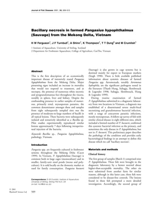
sampled ®sh (Batch 2) was from eight different sites held on TSA slopes for later identi®cation using
from various parts of the country (Fig. 1) and primary tests (Gram stain, shape, motility, oxida-
included both P. hypophthalmus and P. bocourti, as tion/fermentation, oxidase, and temperature pref-
both species are found in the same geographical erences) and secondary biochemical tests (API 20E
locations, although they are farmed separately. kit, Biomeureux, Basingstoke, UK).
Once again, samples comprised ®sh from farms
with higher-than-acceptable mortalities that had
Results
not responded to treatment.
Pathology
Pathology Clinically sick ®sh were emaciated, and several had
a swollen abdomen and petechial haemorrhages on
Fish were killed by anaesthetic overdose using 10%
the tail, ®ns, and sometimes over the rest of the
(w/v) benzocaine solution (Sigma, Poole, Dorset,
body. Internally, white 1±3 mm diameter miliary
UK). Samples from kidney, spleen, liver, gills,
lesions were observed under the capsular surface and
muscle and skin were taken and ®xed in 10%
throughout the parenchyma in the liver, kidney and
buffered formalin for routine processing to paraf®n
spleen of diseased ®sh. On cut surface the muscle
wax sections. These were stained with haematoxylin
appeared yellow, often with petechial haemor-
and eosin (H & E), although selected sections were
rhages. The gills were pale and viscous because of
also stained with Gram's stain, Giemsa and Ziehl-
the presence of increased mucus.
Neelsen.
Histopathology
Bacteriology
Lesions in Batch 1 ®sh involved kidney, liver and
Samples of the liver, kidney and spleen were taken
spleen, with a few multi-focal lesions in muscle.
aseptically and plated directly onto tryptone soya
Renal lesions were characterized by severe, acute to
agar (TSA, Becton Dickinson, Oxford, UK) plates.
sub-acute multiple focally extensive areas of necro-
These were then incubated at 15±37 °C and colon-
sis, progressing to pyogranulomatous lesions
ies observed after 24 h incubation. The predomin-
(Fig. 2). The lesions were centred primarily on
ant colony was sub-cultured to pure growth and
Figure 1 Map of Vietnam showing area involved. *Sampling sites.
Ó 2001
Blackwell Science Ltd 510
3. Journal of Fish Diseases 2001, 24, 509±513 H W Ferguson et al. Bacillary necrosis in Pangasius
Figure 2 Spleen from P. hypophthalmus showing focally-exten- Figure 4 Liver from P. hypophthalmus showing clump of bacilli
sive to con¯uent areas of necrosis within parenchyma (H & E, at margin of lesion (Gram stain, ´350).
´ 15).
bundles, giving the appearance of being intracellular
(Fig. 4). Special-stained sections showed that the
bacteria were Gram-negative and non acid-fast, and
although visible on H & E sections, they were most
easily observed on special-stained sections.
Based on the above, it was felt that the multi-
focal necrosis was the most signi®cant lesion, and
that the disease, therefore, was primarily bacterial.
There was little doubt, however, that the parasites,
especially the renal myxosporea, were also having a
signi®cant effect on the ®sh.
Lesions in Batch 2 ®sh were very similar in
P. hypophthalmus from all eight sites. Little of
Figure 3 Kidney from P. hypophthalmus showing numerous signi®cance was found in P. bocourti, even in
Myxobolus-like myxosporeans within glomerulus, virtually oblit- clinically `sick' ®sh. In affected P. hypophthalmus,
erating normal architecture (H & E, ´ 350).
however, diagnosis was more clear-cut than in
Batch 1 ®sh. Yet again the main lesion was acute
the interstitium, but they extended out to encom- severe multi-focal necrosis in kidney, spleen and
pass surrounding tubules which had varying degrees liver, but this time there was much less complica-
of degeneration and necrosis. These lesions were tion of the overall picture because of concurrent
present in all six clinically sick ®sh from both sites, disease, especially myxosporeans; this was partic-
and even in one of the apparently clinically healthy ularly true for the visceral organs. Some `sick'
®sh. In all nine ®sh there were also varying degrees P. hypophthalmus did have mild to moderate gill
of nephrosis, tubular necrosis, and glomerulo- disease (multi-focal branchial lamellar epithelial
nephritis associated with various prespore and hyperplasia and fusion), associated with monogen-
sporulated Myxobolus- and Sphaerospora-like eans, trichodinids and Myxobolus-like myxospore-
myxosporean parasites (Fig. 3). Similar severe acute ans, but there was no obvious difference in severity
to sub-acute multi-focal areas of necrosis were also between those ®sh adjudged to be clinically healthy
present in the spleen and liver of most ®sh, except and those adjudged clinically to be sick. Conse-
for two from the clinically healthy group. Muscle quently, the gill lesions were considered to be of
lesions were characterized by the presence of a few reduced signi®cance. Out of a total of 43 ®sh
small aggregates of microsporidian or myxosporean examined, there was disagreement in ®ve cases
spores accompanied by limited in¯ammation. between the clinical assessment and the pathological
Associated with the necrotic lesions in kidney, ®ndings. In one case, a P. hypophthalmus that was
spleen and liver, were moderate to large numbers of judged to be healthy turned out to have severe
medium-to-long bacilli, usually situated around the lesions, while in four others (three of which were
periphery, and often aggregated together into P. bocourti ), the converse was true, but with no
Ó 2001
Blackwell Science Ltd 511
4. Journal of Fish Diseases 2001, 24, 509±513 H W Ferguson et al. Bacillary necrosis in Pangasius
satisfactory alternative explanation for their clinical archipelago and Antarctica (Logan, Lebbe, Hoste,
condition. Overall the lesions in P. hypophthalmus Goris, Forsyth, Heyndrickx, Murray, Syme, Wynn-
were very similar to those encountered in Batch 1, Williams & De Vos 2000). Much more work is
but were mostly acute multi-focal necrosis, with less needed on characterizing the isolates and establish-
obvious progression to granulomatous in¯amma- ing their precise role in the pathogenesis of the
tion. Kidney and spleen tended to be more often disease, but pilot studies (2 ´ 107 organisms
and more severely affected than liver; indeed in injected intraperitoneally) have already shown that
some ®sh, lesions were very large, virtually com- the bacteria will reproduce morphologically similar
pletely obliterating normal architecture. Large early lesions in experimentally inoculated ®sh. We can
thrombi were sometimes seen in major vessels. ®nd no records in the literature of other Bacillus
Once again, bacteria morphologically identical to spp. causing systemic disease in ®sh although B.
those seen in Batch 1 ®sh were much in evidence mycoides will cause dermal ulceration (Goodwin,
throughout all lesions in all organs. Roy, Grizzle & Goldsby 1994). Bacillus anthracis is
responsible for anthrax in man and other mammals
(Mesolson, Guillemin, Hugh-Jones, Langmuir,
Bacteriology
Popova, Shelokov & Yampolskaya 1994) and B.
Bacterial colonies were 1 mm in diameter and cream cereus is associated with food-poisoning and other
when grown on TSA for 24 h at 28 °C. They were syndromes in man. Some Bacillus spp. are used as
Gram-variable, long slender bacilli, motile, weakly growth promoters in the aquatic environment,
oxidase positive, and had no reaction with sugars while the toxins of the soil-inhabiting B. thuringi-
(O/F, Becton Dickinson, Oxford, UK). Positive ensis are used in insect control (Entwistle, Cory,
enzymatic reactions were found only for the pro- Bailey & Higgins 1993). The ability of our isolates
duction of hydrogen sulphide and gelatinase when to thrive in vitro at 37 °C, and possibly beyond,
tested using the API 20E-identi®cation-kit. The raises the disturbing possibility that they may have
bacteria grew at a temperature range from 15 to zoonotic potential. Certainly the type of aqua-
37 °C on TSA, but optimal growth was found culture practised on the Mekong, with animal and
between 28 and 37 °C. The bacteria could not be human ef¯uent deliberately used to fertilize the
identi®ed. The closest match was Bacillus fumarioli receiving waters, lends itself very well to the
with 95% con®dence using 16S rRNA analysis from promotion of cross-species transfer.
Genebank (MIDI Laboratories, Delaware, USA). There are several systemic bacterial infections in
®sh that usually present as chronic granulomatous
disease, but under some circumstances can present
Discussion
as acute necrosis, the type of host response and
Although the results are clearly preliminary, on the presentation seemingly dependent on accompanying
basis of the ®ndings from these two separate batches environmental or other stress factors. Examples
of submissions, totalling 10 farms in all, it seems include pasteurellosis (Photobacterium damsela
reasonable to suggest that this disease has a bacterial subsp. piscicida), bacterial kidney disease (Renibac-
aetiology. Accordingly we are naming this condition terium salmoninarum), and even mycobacteriosis
`bacillary necrosis of Pangasius'. The disease was (Ferguson 1989). In this investigation there seems
not seen in P. bocourti, only in P. hypophthalmus. little doubt that the concurrent parasitic disease,
We do not know if this is the result of a difference notably the renal myxosporean infection(s), could
in species susceptibility, of some variation in have had a considerable debilitating effect in some
husbandry practices, or merely the result of individuals, and would have contributed substan-
insuf®cient sample size. This is obviously an area tially to the clinical picture overall. Indeed in many
that demands further investigation, as it may have a cases their presence may have been responsible for
major impact on choosing which species to farm. some of the variation in in¯ammatory response
The only isolate that resembled the morphology noted between Batch 1 and Batch 2 ®sh.
of the bacteria in sections was identi®ed as a Bacillus Nevertheless, the overriding common ®nding was
sp., with a 95% similarity to B. fumarioli, the bacterial-associated necrosis, and it is the
an endospore-forming soil-inhabiting bacterium consequences of these lesions that should probably
encountered in geothermal environments in various direct investigations into the cause of death in
parts of the world including the South Sandwich these animals. The relative importance of the
Ó 2001
Blackwell Science Ltd 512
5. Journal of Fish Diseases 2001, 24, 509±513 H W Ferguson et al. Bacillary necrosis in Pangasius
interstitial and tubular components of the renal Logan N.A., Lebbe L., Hoste B., Goris J., Forsyth G.,
lesions with regard to immune responsiveness and Heyndrickx M., Murray B.L., Syme N., Wynn-Williams D.D.
& De Vos P. (2000) Aerobic endospore-forming bacteria
osmoregulation respectively, and the signi®cance from geothermal environments in northern Victoria Land,
of the thrombosis in relation to possible toxin release Antarctica, and Candlemas Island, South Sandwich
and terminal disseminated intravascular coagulation, archipelago, with the proposal of Bacillus fumarioli sp. nov.
are all avenues that remain to be explored. International Journal of Systematic and Evolutionary
Microbiology 50, 1741±1753.
Mesolson M., Guillemin J., Hugh-Jones M., Langmuir A.,
Acknowledgments Popova I., Shelokov A. and Yampolskaya O. (1994) The
Sverdlovsk anthrax outbreak of 1979. Science 266,
We would like to thank DFID for ®nancial support, 1202±1208.
and the College of Agriculture, Can Tho, Vietnam. Singh S.B. (1990) Status of aquaculture in the Socialist Republic
of Vietnam. In: Aquaculture in Asia (ed. by M.M. Joseph),
pp. 371±384. Asian Fisheries Society, Indian Branch,
References
Mangalore.
Cacot P. (1999) Etude du cycle sexuel et maitrise de la reproduction Subagja J., Slembrouck J., Hung L.T. & Legendre M. (1999)
de Pangasius bocourti. (Sauvage, 1880 ) et Pangasius hypoph- Larval rearing of an Asian cat®sh Pangasius hypophthalmus
thalmus (Sauvage, 1878 ) dans le delta due Mekong au (Siluroidei Pangasiidae): analysis of precocious mortality and
Viet-Nam. PhD Thesis, CanTho University, Vietnam. proposition of appropriate treatments. Aquatic Living Resources
Entwistle P.F., Cory J.S., Bailey M.J. & Higgins S. eds (1993) 12, 37±44.
Bacillus thuringiensis, An Environmental Biopesticide: Theory Thanh Hung Le., Subagja J., Slembrouck J. & Legendre M.
and Practice. John Wiley & Sons, New York, USA. (1998) Study on mass mortality of Pangasius hypophthalmus
Ferguson H.W. (1989) Systemic Pathology of Fish. A Text and during larval rearing and its control and prevention. Abstract
Atlas of Comparative Tissue Responses in Diseases of Teleosts. 27, National Workshop on Aquaculture, September 29±30,
Iowa State University Press, Ames, Iowa, USA. 1998, Bac Ninh, Vietnam.
Goodwin A.E., Roy J.S., Grizzte J.M. & Goldsby M.T. (1994)
Received: 27 February 2001
Bacillus Mycoides- a bacterial pathogen of channel cat®sh.
Accepted: 10 May 2001
Diseases of Aquatic Organisms 18, 173±179.
Ó 2001
Blackwell Science Ltd 513