2. OOOOE
e74 Kim et al. March 2009
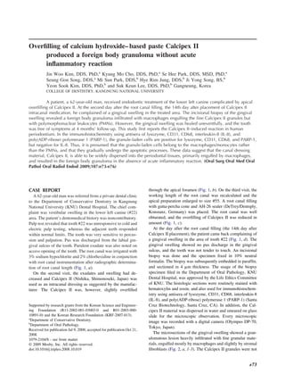
Fig. 1. Standard radiographs during the endodontic treatment of the lower left canine. a, X-ray for initial working length
determination. b, On the second visit, Calcipex II was overfilled into the periapical area (arrows). c, After root canal filling, some
Calcipex II remained periapical but reduced in amount (arrows). d, At the second day after canal filling, when the gingival
swelling occurred, the canal filling looked fine and there was no change in periapical area (arrows).
localized in the granuloma lesion but gradually dispersed root canal is filled in a single-session treatment.1-3
across the border of the granuloma into the adjacent connec- Calcipex II is a calcium hydroxide water-based paste
tive tissue (Fig. 2, a, 4 and 5). The fine granular materials for root canal dressing, which is easy to handle and the
were identical to the Calcipex II granules smeared on the
most easily removed. Great care had to be taken to
glass slide, exhibiting slightly translucent and fine granular
avoid extrusion via the apical foramen compared with
aggregation but no calcification or polymerization (Fig. 2, b).
The Calcipex II granules were mainly engulfed by macro- the silicon oil– based Vitapex.4 In the present study, the
phages, and also slightly localized at the cytoplasms of fibro- Calcipex II used as an intracanal dressing material was
blasts. Still a lot of the fine Calcipex II granules were freely overfilled into the periapical area, and its fine granular
dispersed in the stromal fibrous tissue, but rarely in the lumen materials were rapidly migrated up through the gingival
of capillaries. In the immunohistochemistry using antisera of tissue. The Calcipex II granules were not calcified nor
lysozyme, CD31, CD68, IL-8, and PARP-1, the Calcipex II polymerized in the gingival tissue; rather, they were
granule–laden cells were positive for lysozyme, CD31, and diffusely scattered throughout the collagenous fibrous
CD68, indicating that the cells belonged to the lineage of
tissue, were rapidly engulfed by the macropahges, and
macrophage/histiocyte/monocyte and were in the apoptotic
resulted in a foreign body granuloma. The granuloma
processes (Fig. 2, c-e). On the other hand, the Calcipex II
granule–laden cells were negative for IL-8, which is a che- was mainly infiltrated with macrophages, and only a
mokine usually positive in neutrophils (Fig. 2, f), similar to few lymphocytes were found, but exclusive of PMNs.
the negative control staining (Fig. 2, h). Under the micro- In the immunohistochemistry, the Calcipex II granule–
scopic observation in routine hematoxylin and eosin stain, the laden cells were strongly positive for lysozyme, CD31,
macrophages filled with the Calcipex II granules could not and CD68, which are markers for the macrophages,
readily dissolve the fine granules; rather, they were gradually including histiocytes and monocytes,5-11 whereas they
destroyed by the rupture of their cell membrane. Nevertheless were negative for the antibody of IL-8, which is a
there appeared no acute inflammatory reaction. The Calcipex
marker for PMNs.12 However, the inflammatory degen-
II–laden macrophages were strongly positive for PARP-1,
indicating that the cells were undergoing the apoptotic pro-
eration or necrosis by cytotoxic chemokine IL-8 was
cesses, possibly due to the overload of Calcipex II (Fig. 2, g). not observed in the Calcipex II–induced foreign body
Eventually, the Calcipex II induced a foreign body granuloma granuloma.
by macrophages, but no acute inflammation by PMNs. The Calcipex II granules were too small, 500 nm,
to be localized in the root canal, so that they were easily
DISCUSSION overflowed and dispersed into the periapical tissues
The use of a calcium hydroxide– based intracanal depending on the dynamic pressure that occurred dur-
dressing is important for periapical repair in teeth with ing canal filling and biting. The fine Calcipex II gran-
periapical lesion. Dressing with calcium hydroxide ules were rapidly engulfed by macrophages and even
paste results in better periapical repair than when the endocytosed by stromal fibroblasts. However, the Cal-
3. OOOOE
Volume 107, Number 3 Kim et al. e75
Fig. 2. Photomicrographs of the Calcipex II-induced foreign body granuloma. a, Hematoxylin and eosin stain. a1, Low
magnification of main lesion: The Calcipex II granules were diffusely observed in the stromal cells. a2-a5, High magnification.
a2, Most of the Calcipex II granules were engulfed by macrophages (arrows), only a few lymphocytes (arrowheads) were
infiltrated but not PMN. a3, Some Calcipex II granules were also endocytosed by stromal fibroblasts (arrowheads); no Calcipex
II granules were found in the lumen of capillaries (C). a4, Note the massive dispersion of the Calcipex II granules in the fibrous
connective tissue; many Calcipex II granules were found in the cytoplasms of fibroblasts (arrowheads). a5, Border area between
the fibrous connective tissue and granulomatous lesion. The Calcipex II granules were diffusely dispersed; note many macro-
phages infiltrated into the border area. b, Calcipex II paste smeared on the slide glass. b1, Low magnification: The Calcipex II
granules were well dispersed and aggregated (arrows). b2, High magnification: The fine granules of Calcipex II was very small,
measuring 500 nm in diameter. c-g, Immunohistochemistry. c, Lysozyme antibody, positive in the Calcipex II granule–laden
cells (arrows). d, CD31 antibody, positive in the Calcipex II granule–laden cells (arrows). e, CD68 antibody, positive in the
Calcipex II granule–laden cells (arrows). f, Interleukin-8 antibody, negative in the Calcipex II granule–laden cells (arrows). g,
Poly(ADP-ribose) polymerase 1 antibody, positive in the Calcipex II granule–-laden cells (arrows). h, Negative control stained
without antisera.
4. OOOOE
e76 Kim et al. March 2009
cipex II granules seem to be not readily degraded in the giant cell granuloma. An immunohistochemical and ultra-
cytoplasms of macrophages; rather, the macrophages structural study. Oral Dis 1995;1:20-5.
7. Macarenco RS, do Canto AL, Gonzalez S. Angiolymphoid hy-
overfilled with the Calcipex II granules tend to be perplasia with eosinophilia showing prominent granulomatous
destroyed with the positive reaction of PARP-1, which and fibrotic reaction: a morphological and immunohistochemical
is a marker of cellular apoptosis.13,14 However, the study. Am J Dermatopathol 2006;28:514-7.
features of cytotoxic degeneration and the allergic hy- 8. Yuan K, Wing LY, Lin MT. Pathogenetic roles of angiogenic
persensitivity by the immune reactions of PMNs and factors in pyogenic granulomas in pregnancy are modulated by
female sex hormones. J Periodontol 2002;73:701-8.
lymphocytes were not observed in the Calcipex II–
9. Knoess M, Krukemeyer MG, Kriegsmann J, Thabe H, Otto M,
induced foreign body granuloma. Krenn V. Colocalization of C4d deposits/CD68 macrophages in
In this study, the overfilling of Calcipex II in end- rheumatoid nodule and granuloma annulare: immunohistochem-
odontic treatment produced a foreign body granuloma ical evidence of a complement-mediated mechanism in fibrinoid
in the nearby gingival tissue. Although the Calcipex II necrosis. Pathol Res Pract 2008;204:373-8.
10. Tian XF, Li TJ, Yu SF. Giant cell granuloma of the temporal
was rapidly reduced in amount in the periapical radio-
bone: a case report with immunohistochemical, enzyme histo-
grams after treatment, the Calcipex II material was not chemical, and in vitro studies. Arch Pathol Lab Med 2003;
resorbed easily; rather, it was dispersed into adjacent 127:1217-20.
soft tissue, evoking a foreign body reaction by macro- 11. Rodini CO, Lara VS. Study of the expression of CD68 mac-
phages in the absence of acute inflammatory reaction. rophages and CD8 T cells in human granulomas and periapical
cysts. Oral Surg Oral Med Oral Pathol Oral Radiol Endod
REFERENCES 2001;92:221-7.
1. Leonardo MR, Hernandez ME, Silva LA, Tanomaru-Filho M. 12. Pilozzi E, Stoppacciaro A, Rendina E, Ruco LP. Monocyte
Effect of a calcium hydroxide– based root canal dressing on chemotactic protein-1 in the inflammatory pseudotumour of the
periapical repair in dogs: a histological study. Oral Surg Oral lung. Mol Pathol 1998;51:50-2.
Med Oral Pathol Oral Radiol Endod 2006;102:680-5. 13. Okinaga T, Kasai H, Tsujisawa T, Nishihara T. Role of caspases
2. Berbert FL, Leonardo MR, Silva LA, Tanomaru Filho M, Bra- in cleavage of lamin A/C and PARP during apoptosis in macro-
mante CM. Influence of root canal dressings and sealers on repair phages infected with a periodontopathic bacterium. J Med Mi-
of apical periodontitis after endodontic treatment. Oral Surg Oral crobiol 2007;56:1399-404.
Med Oral Pathol Oral Radiol Endod 2002;93:184-9. 14. Fink SL, Cookson BT. Caspase-1-dependent pore formation dur-
3. Peters LB, van Winkelhoff AJ, Buijs JF, Wesselink PR. Effects ing pyroptosis leads to osmotic lysis of infected host macro-
of instrumentation, irrigation and dressing with calcium hydrox- phages. Cell Microbiol 2006;8:1812-25.
ide on infection in pulpless teeth with periapical bone lesions. Int
Endod J 2002;35:13-21.
Reprint requests:
4. Hosoya N, Kurayama H, Iino F, Arai T. Effects of calcium
hydroxide on physical and sealing properties of canal sealers. Int Suk Keun Lee, DDS, PhD
Endod J 2004;37:178-84. Department of Oral Pathology
5. Stefanaki K, Tsivitanidou-Kakourou T, Stefanaki C, Valari M, College of Dentistry
Argyrakos T, Konstantinidou CV, et al. Histological and immu- Kangnung National University
nohistochemical study of granuloma annulare and subcutaneous 123 Chibyun-dong
granuloma annulare in children. J Cutan Pathol 2007;34:392-6. Gangneung, 210-702 Korea
6. Carvalho YR, Loyola AM, Gomez RS, Araujo VC. Peripheral sklee@kangnung.ac.kr