SlideShare ist ein Scribd-Unternehmen logo
1 von 12
Downloaden Sie, um offline zu lesen
International Endodontic Journal (1997) 30, 79–90

REVIEW

Dens invaginatus: aetiology, classification, prevalence, diagnosis,
and treatment considerations
M. HÜLSMANN
Department of Operative Dentistry, Zentrum ZMK, University of Göttingen, Göttingen, Germany

Summary
Dens invaginatus is a malformation of teeth probably
resulting from an infolding of the dental papilla during
tooth development. Affected teeth show a deep infolding
of enamel and dentine starting from the foramen
coecum or even the tip of the cusps and which may
extend deep into the root. Teeth most affected are maxillary lateral incisors and bilateral occurrence is not
uncommon. The malformation shows a broad spectrum
of morphologic variations and frequently results in early
pulp necrosis. Root canal therapy may present severe
problems because of the complex anatomy of the teeth.
Aetiology, prevalence, classification, and therapeutic
considerations including root canal therapy, apical
surgery and prevention of pulpal involvement are
reviewed.
Keywords: dens invaginatus, endodontic treatment.

Introduction
Dens invaginatus (Fig. 1a–c) is a rare malformation of
teeth, showing a broad spectrum of morphological
variations. The affected teeth radiographically show
an infolding of enamel and dentine which may extend
deep into the pulp cavity and into the root and
sometimes even reach the root apex (Fig. 2a–c). Tooth
crowns as well as roots may exhibit variations in
size and form. This kind of tooth malformation was
described first by Ploquet in 1794 (Schaefer 1955), who
discovered this anomaly in a whale’s tooth (Westphal
1965).
Dens invaginatus in a human tooth was first described

Correspondence: Dr Michael Hülsmann, Department of Operative
Dentistry, Zentrum ZMK, University of Göttingen, Robert-Koch-Str.
40, D-37075 Göttingen, Germany.
© 1997 Blackwell Science Ltd

by a dentist named ‘Socrates’ in 1856 (Schulze 1970).
In 1873 Mühlreiter reported on ‘anomalous cavities in
human teeth’, Baume in 1874 and Busch in 1897
published on this malformation. In 1887 Tomes
described the dens invaginatus in his textbook A System
of Dental Surgery, as follows: ‘The enamel investing the
crown may be, and often is, perfectly well-developed; but
we shall find at some point a slight depression, in the
centre of which is a small dark spot. If the tooth be
divided through its long axis, we shall find that the dark
centre of the depression is in fact the choked-up orifice of
a cavity situated within the substance of the tooth,
external, however, and perfectly unconnected with the
pulp-cavity. If the section be a fortunate one, we shall be
able to trace the enamel as it is continued from the
exterior of the tooth through the orifice into the cavity,
the surface of which is lined more or less completely with
this tissue’ (Tomes 1887).
From the 1920s to the 1950s numerous reports
on cases of dens invaginatus malformation were published in the dental literature (Miller 1901, Lejeune &
Wustrow 1920, De Jonge Cohen 1925, Kronfeld 1934,
Rebel & Rohmann 1934, Hammer 1935, Kitchin 1935,
Hoepfel 1936, Fischer 1936, Beust & Freericks 1936,
Rushton 1937, Euler 1939, Swanson & McCarthy 1947,
Zilkens & Schneider-Zilkens 1948, Egli 1949, Gustafson
& Sundberg 1950, Bruszt 1950, Munro 1952, Schaefer
1953, Hitchin & McHugh 1954, Logar 1955, Künzel
1956, Petz 1956, Davidoff & Anastassowa 1956,
Brabant & Klees 1956). Until 1959 more than 200
papers, mainly case reports, had been published on the
dens invaginatus malformation (Grahnen et al. 1959).
Dens invaginatus malformation again has been a subject of interest in recent years (Wells & Meyer 1993,
Piattelli & Trisi 1993, Szajkis & Kaufman 1993, Pecora
et al. 1993, Altinbulak & Ergül 1993, Skoner & Wallace
1994, Mangani & Ruddle 1994, Benenati 1994,
Hülsmann & Radlanski 1994, Hülsmann 1995a,b,
79
80

M. Hülsmann

Fig. 1 (a) SEM; (b) microscopic and (c) radiographic appearance of a dental invagination. The invagination is completely filled with debris.
At the bottom of the invagination the enamel is missing.

Ikeda et al. 1995, Oncag et al. 1995, Nagatani et al.
1995, Khabbaz et al. 1995, Duckmanton 1995, Lindner
et al. 1995, Bloch-Zupan et al. 1995, Olmez et al. 1995,
Hosey & Bedi 1996).

Synonyms for this malformation are: Dens in dente,
invaginated odontome, dilated gestant odontome,
dilated composite odontome, tooth inclusion, dentoid in
dente.

Fig. 2 (a) Invagination in the maxillary left lateral incisor. The invagination is limited to the tooth crown. The malformation was detected by
chance. (b) Invagination in the maxillary left lateral incisor. The invagination is extending into the root. At the bottom of the invagination the
enamel is missing. The invagination was suspected because of dysmorphic, peg-shaped tooth crown anatomy. (c) Severe case of dens invaginatus
with open apex, diverging root and bizarre form of the invagination with a separate apical opening. The malformation was detected because of
retarded tooth eruption of tooth 12.
© 1997 Blackwell Science Ltd, International Endodontic Journal, 30, 79–90
Dens invaginatus

81

Aetiology of dens invaginatus
The aetiology of dens invaginatus malformation is
controversial and remains unclear. Over the last decades
several theories have been proposed to explain the
aetiology of dental coronal invaginations:
• Growth pressure of the dental arch results in buckling
of the enamel organ (Euler 1939, Atkinson 1943).
• Kronfeld (1934) suggested that the invagination
results from a focal failure of growth of the internal
enamel epithelium while the surrounding normal
epithelium continues to proliferate and engulfs the
static area.
• Rushton (1937) proposed that the invagination is a
result of rapid and aggressive proliferation of a part of
the internal enamel epithelium invading the dental
papilla. He regarded this a ‘benign neoplasma of
limited growth’.
• Oehlers (1957a,b) considered that distortion of the
enamel organ during tooth development and subsequent protrusion of a part of the enamel organ will
lead to the formation of an enamel-lined channel
ending at the cingulum or occasionally at the incisal
tip. The latter might be associated with irregular
crown form.
• The ‘twin-theorie’ (Bruszt 1950) suggested a fusion of
two tooth-germs.
• Infection was considered to be responsible for the
malformation by Fischer (1936) and Sprawson
(1937).
• Gustafson & Sundberg (1950) discussed trauma as a
causative factor, but could not sufficiently explain
why just maxillary lateral incisors were affected and
not central incisors.
Most authors, meanwhile, consider dens invaginatus as
a deep folding of the foramen coecum during tooth
development which in some cases even may result in a
second apical foramen (Schulze 1970). On the other
hand the invagination also may start from the incisal
edge of the tooth. Genetic factors cannot be excluded
(Grahnen 1962, Casamassimo et al. 1978, Ireland et al.
1987, Hosey & Bedi 1996).

Fig. 3 Classification of invaginated teeth by Oehlers (1957).

the confines of the crown not extending
beyond the amelocemental junction.
Type II: an enamel-lined form which invades the root
but remains confined as a blind sac. It may or
may not communicate with the dental pulp.
Type III: a form which penetrates through the root perforating at the apical area showing a ‘second
foramen’ in the apical or in the periodontal
area. There is no immediate communication
with the pulp. The invagination may be completely lined by enamel, but frequently cementum will be found lining the invagination.
Oehlers (1957a,b) also described different crown forms
(normal with a deep lingual or palatal pit; conical,
barrel-shaped or peg-shaped with an incisal pit) relating
to the three groups mentioned above. In addition,
Ulmansky & Hermel (1964) and Vincent-Townend
(1974) described an ‘incipient dens in dente’, a deep
palatal or lingual pit completely lined by enamel with no
communication to the pulp. Oehlers (1958) also
presented radicular invaginations.
Schulze & Brand (1972) proposed a more detailed
classification (Fig. 4), also including invaginations
starting at the incisal edge or the top of the crown and
also including dysmorphic root configurations.

Prevalence of dens invaginatus
Classification of dens invaginatus
The first classification of invaginated teeth was published by Hallet (1953). The most commonly used
classification proposed by Oehlers (1957a) is shown in
Fig. 3. He described the anomaly occurring in three
forms:
Type I: an enamel-lined minor form occurring within
© 1997 Blackwell Science Ltd, International Endodontic Journal, 30, 79–90

A summary of the results of investigations on the prevalence of invaginated teeth is presented in Table 1. It is
difficult to compare findings because of the differences
in study design, sample size and composition, and
diagnostic criteria. The teeth most affected are maxillary
lateral incisors and bilateral occurrence is not uncommon
and occurs in 43% of all cases (Grahnen et al. 1959).
82

M. Hülsmann

Fig. 4 Classification of dens invaginatus by Schulze & Brand (1972)
(from: Zahnärztliche Welt/Reform 81, 569–73, with kind permission).

Swanson & McCarthy (1947) were the first to present
bilateral dens invaginatus malformation, Conklin
(1968) presented a patient with all maxillary central
and lateral incisors affected, and Burton et al. (1980)

published a case with six teeth involved, the maxillary
incisors and also the maxillary canines. Krolls (1969)
detected dental invaginations in maxillary central
incisors as well as in several maxillary and mandibular
bicuspids in one patient. Conklin (1978) found the
anomaly in four mandibular incisors in one patient.
Double invaginations in one tooth were reported by
Rushton (1937), Archer & Silverman (1950), Oehlers
(1957a), Grahnen et al. (1959), Ulmansky & Hermel
(1964), Schulze & Brand (1972), Brill & Phillips (1974),
Bhatt & Dholakia (1975), Conklin (1975), Cole et al.
(1978), Gotoh et al. (1979), Mader (1979), Rotstein
et al. (1987a), and Vairabhaya (1989). Triple invaginations were described by Hitchin & McHugh (1954) and
Mader (1977). In rare cases even primary teeth seem to
be affected (Rabinowitch 1952).
Cases of dental invaginations in supernumerary teeth
have been presented as well as in canines and bicuspids
(Archer & Silverman 1950, Schwenzer 1957, Kruse
1962, Klammt 1969, Conklin 1975, Banner 1978,

Table 1 Investigations on the prevalence of dens invaginatus
Author/Country

Year

Sample

Frequency

Mühlreiter
(Austria)
Atkinson
(USA)
Boyne
(USA)
Stephens
(unknown)
Shafer
(USA)
Hallet
(unknown)

1873

2.8% of teeth

1943

500 extracted lateral maxillary
incisors (histological investigation)
500 lateral maxillary incisors

1952

1000 maxillary incisors

0.3% of teeth

1953
1953
1953

150 full-mouth surveys
1000 patients
2542 full-mouth surveys

1953

586 full-mouth surveys

Amos
(USA)
Grahnen et al.
(Sweden)
Ulmansky &
Hermel (Israel)
Poyton & Morgan
(Canada)
Miyoshi et al.
( Japan)
Fujiki et al.
( Japan)
Thomas
(USA)
Gotoh et al.
( Japan)
Ruprecht et al.
(Saudi Arabia)
Ruprecht et al.
(Saudi Arabia)

1955
1959

1000 full mouth surveys
203 full mouth surveys
3020 right maxillary incisors

8%
3.6% (cited by Hovland & Block 1977)
1.3% of patients
(cited by Hovland & Block 1977)
6.6% of maxillary lateral incisors
0.5% of maxillary central incisors
(cited by Hovland & Block 1977)
5.1% of patients
6.9% (students of dentistry)
2.7% of patients

1964

500 full mouth surveys

2% of patients

1966

5000 full mouth surveys

1971
1974

extracted maxillary lateral
incisors
2126 lateral maxillary incisors

1974

1886 full mouth surveys

0.25% of patients
(cited by Hovland & Block 1977)
38.5% of teeth
(cited by Gotoh et al. 1979)
4.2% of teeth
(cited by Gotoh et al. 1979)
7.74% of patients

1979

766 lateral maxillary incisors

9.66% of teeth

1986

1581 full mouth surveys

1.7% of patients

1987

300 full mouth surveys

10% of patients

10% of teeth

© 1997 Blackwell Science Ltd, International Endodontic Journal, 30, 79–90
Dens invaginatus
Shifman & Tamir 1979, Bottomley & Johnson 1982,
Eldeeb 1984, Rotstein et al. 1987a, Morfis 1992,
Bramante et al. 1993, Altinbulak & Ergül 1993,
Hülsmann & Radlanski 1994, Tavano et al. 1994).

83

pulpal involvement of teeth with coronal invaginations
may occur a short time after tooth eruption early diagnosis is mandatory to instigate preventive treatment.

Clinical features
Histological findings
Several reports on microscopic, ultrastructural and
microradiographic investigations of teeth with a dens
invaginatus malformation show a wide range of findings
and thus reproduce the macroscopic variety of this
anomaly. The dentine below the invagination may be
intact without irregularities (Brabant & Klees 1956,
Omnell et al. 1960, Piatelli & Trisi 1993) but also may
contain strains of vital connective tissue (Omnell et al.
1960) or even fine canals with communication to the
dental pulp (Kronfeld 1934, Fischer 1936, Hoepfel
1936, Gustafson & Sundberg 1950, Hitchin & McHugh
1954, Oehlers 1957a, Rushton 1958). Some authors
reported hypomineralized or irregularly structured
dentine (Omnell et al. 1960, Vincent-Townend 1974,
Beynon 1982). The structure and thickness of the
enamel lining the invagination also may vary widely.
The enamel was described as irregularly structured by
Atkinson (1943), Beynon (1982) and Piatelli & Trisi
(1993). Beynon (1982) reported hypomineralized
enamel at the base of the invagination whereas Morfis
(1992), in a chemical analysis, detected up to eight
times more phosphate and calcium compared with the
outer enamel, but in his analysis magnesium was
missing completely. Bloch-Zupan et al. (1995) found
differences in structure and compostion between the
external and internal enamel. The internal enamel
exhibited atypical and more complex rod shapes and its
surface presented the typical honeycomb pattern but no
perikymata, which, however, were observed on the
outer surface of the tooth.

Diagnosis of dens invaginatus
In most cases a dens invaginatus is detected by chance
on the radiograph. Clinically, an unusual crown morphology (‘dilated’, ‘peg-shaped’, ‘barrel-shaped’) or a
deep foramen coecum may be important hints, but
affected teeth also may show no clinical signs of the
malformation. As maxillary lateral incisors are the teeth
most susceptible to coronal invaginations these teeth
should be investigated thoroughly clinically and radiographically, at least in all cases with a deep pit at the
foramen coecum. If one tooth is affected in a patient the
contralateral tooth should also be investigated. As
© 1997 Blackwell Science Ltd, International Endodontic Journal, 30, 79–90

The invagination allows entry of irritants into an area
which is separated from pulpal tissue by only a thin layer
of enamel and dentine and presents a predisposition for
the development of dental caries. In some cases the
enamel-lining is incomplete (Fig. 1). Channels may also
exist between the invagination and the pulp (Kronfeld
1934, Hitchin & McHugh 1954). Therefore, pulp
necrosis often occurs rather early, within a few years of
eruption, sometimes even before root end closure
(Swanson & McCarthy 1947, Ulmansky & Hermel 1964,
Stepanik 1968, Ferguson et al. 1980, Morfis & Lentzari
1989, Nik-Hussein 1994, Hülsmann & Radlanski 1994)
(Fig. 5a, b). Other reported sequelae of undiagnosed and
untreated coronal invaginations are abscess formation
(Schaefer 1955, Petz 1956, Kendrick 1971, Greenfield
& Cambruzzi 1986, Chen et al. 1990, Whyman &
McFayden 1994), retention of neighbouring teeth
(Schaefer 1955, Petz 1956, Conklin 1975, Mader
1977), displacement of teeth (Schaefer 1955, Petz
1956), cysts (Rebel & Rohmann 1934, Schwenzer
1957, Conklin 1978, Klammt 1969, Burzynski 1973,
Samimy 1977, Murphy & Doku 1977, Augsburger &
Brandebura 1978, Greenfield & Cambruzzi 1986), and
internal resorption (Shapiro 1970, Hülsmann &
Radlanski 1994) (Fig. 6).
The dental literature on dens invaginatus malformations contains several case reports presenting invaginated teeth coincident with other dental anomalies,
malformations and even dental or medical syndromes
(Table 2).

Treatment considerations
Preventive and restorative treatment
Teeth with deep palatal or incisal invaginations or
foramina coeca should be treated with fissure sealing
before carious destruction can occur. A composite
restoration and strict periodic review is recommended
(Rotstein et al. 1987b, Hülsmann & Radlanski 1994). If
no entrance to the invagination can be detected and no
signs of pathosis are visible clinically and radiographically no treatment is indicated, but strict observation is
recommended (Hülsmann 1995b, Duckmanton 1995,
Hülsmann 1996).
84

M. Hülsmann

Fig. 5 (a) Small coronal invagination in tooth 22. (b) Three years later arrest of root development and pulp necrosis, with apical periodontitis, and
sinus tract have appeared. (c) Fifteen months later: following apexification therapy with calcium hydroxide and obturation of the root canal the
periapical status has improved.

Root canal treatment
A review of the literature shows that extraction of teeth
with invaginations was the preferred therapy until the
1970s (Hülsmann 1995a). Grossman (1974) and
Creaven (1975) were the first to describe root canal

Table 2 Dental anomalies described in association with dens invaginatus
Microdontia
Macrodontia
Hypodontia
Oligodontia
Taurodontism

Gemination and Fusion

Supernumerary teeth

Amelogenesis imperfecta
Invagination in an odontome
Multiple odontomes
Coronal agenesis
Williams syndrome

Casamassimo et al. 1978
Ekman-Westberg & Julin 1974
Hülsmann 1995c
Conklin 1978, Ruprecht et al. 1986
Casamassimo et al. 1978,
Ruprecht et al.1986,
Ireland et al. 1987,
Chen et al. 1990
Burzynski 1973, Mader 1979,
Mader & Zielke 1982,
Ruprecht et al. 1986
Schaefer 1953, 1955,
Brabant & Klees 1956,
Petz 1956, Rushton 1958,
Conklin 1975, Mader
1977, Shifman & Tamir 1979,
Beynon 1982, Ruprecht et al. 1986,
Morfis 1992
Kerebel et al. 1983
Hitchin & McHugh 1954
Robbins & Keene 1964
Hicks & Flaitz 1985
Oncag et al. 1995

treatment of the invagination only (root invagination
treatment) and Tagger (1977) and Hovland & Block
(1977) the first to present cases treated with conventional root canal therapy (Fig. 7a,b). Root canal treatment may present several problems because of the
irregular shape of the root canal system(s). If there
are no radiographic signs of pulp pathosis and no
communication between the invagination and the
root canal, root canal treatment or, in minor cases, even
a composite or amalgam filling of the invagination will
be adequate (DeSmit & Demaut 1982). When the invagination has a separate apical or lateral foramen, root
canal treatment of the invagination is indicated
(Grossman 1974, Creaven 1975, Bolanos et al. 1988,
Szajkis & Kaufman 1993, Wells & Meyer 1993, Ikeda
et al. 1995). In some cases it may be possible to bur
through the invagination to get access to the apical
foramen. When these minor forms of invaginations
are eliminated root canal therapy in most cases will
not present further problems. In certain cases the invagination has to be treated as a separate root canal (Cole
et al. 1978, Zillich et al. 1983, Eldeeb 1984, Greenfield
& Cambruzzi 1986, Mangani & Ruddle 1994, Khabbaz
et al. 1995).
When pulp necrosis occurs before root-end closure,
apexification procedures with calcium hydroxide may be
necessary (Ferguson et al. 1980, Morfis & Lentzari 1989,
Vairabhaya 1989, Hülsmann & Radlanski 1994,
Nagatani et al. 1995) (Fig. 5a–c).
© 1997 Blackwell Science Ltd, International Endodontic Journal, 30, 79–90
Dens invaginatus

85

Fig. 6 (a) Pulp necrosis, apical periodontitis, sinus tract, and
lateral resorptive perforation in tooth 12 showing a dens invaginatus malformation. (b) Clinical view of the tooth crown of the
same tooth before treatment. A deep foramen coecum with caries
is visible. (c) After preparation of the access cavity the entrance to
the invagination is visible at the palatal aspect (arrow). The main
root canal orifice can be seen at the top of the cavity at the buccal
aspect.

The large and irregular volume of the root canal
system makes proper shaping and cleaning difficult.
Irrigation, supported by ultrasonic cleaning of the root
canal system has been described as an efficient means of
disinfection (Cunningham et al. 1982) and has therefore
been recommended for cleaning of the complex
morphology of the root canal system in teeth with dens
invaginatus (Skoner & Wallace 1994). For obturation of
such teeth warm gutta-percha techniques including
vertical condensation or thermoplastic filling techniques
have been recommended (Rotstein et al. 1987b,
Hülsmann & Radlanski 1994, Mangani & Ruddle
1994).
© 1997 Blackwell Science Ltd, International Endodontic Journal, 30, 79–90

Surgical treatment
Surgical treatment should be considered in cases of
endodontic failure and in teeth which cannot be treated
non-surgically because of anatomical problems or
failure to gain access to all parts of the root canal system
(Harnisch 1970, Hata & Toda 1987, Teplitsky & Singer
1987, Rotstein et al. 1987b, Kulild & Weller 1989,
Suchina et al. 1989, Hülsmann & Radlanski 1994,
Benenati 1994, Olmez et al. 1995) (Fig. 8a,b). Cole et al.
(1978) and Lindner et al. (1995) even proposed intentional replantation with retrograde surgery in otherwise
hopeless cases.
86

M. Hülsmann

Fig. 7 (a) Right maxillary lateral incisor in the same patient as in Fig. 5. The invagination was detected
by preventive radiographic examination, clinically no invagination nor carious lesion were detected.
The invagination has resulted in an internal resorption with perforation. (b) Obturation with injectable
gutta-percha, resulting in a slight lateral overfill.

Fig. 8 (a) Radiograph of tooth 23 showing a bizarre lateral coronal invagination which seems to be open
to the lateral periodontium. The main root canal was root canal treated, but no communication to the
invagination could be detected. (b) As no access to the invagination was found coronally the invagination was treated surgically. Probably root canal treatment of the main root canal would not have been
necessary.
© 1997 Blackwell Science Ltd, International Endodontic Journal, 30, 79–90
Dens invaginatus

87

Fig. 9 (a) Severe dens invaginatus malformation with acute apical abscess formation. The tooth was
judged inoperable and extracted by a general dental practitioner. (b) The photograph of the extracted
tooth shows the invagination extending beyond the apex.

Extraction
Extraction is indicated only in teeth with severe anatomical irregularities that cannot be treated non-surgically
or by apical surgery, and in supernumerary teeth
(Figs 9a,b and 10). Additionally Rotstein et al. (1987b)
advocated extraction when abnormal crown morphology presents aesthetic or functional problems.

References

Fig. 10 Severe invagination malformation of tooth 22 resulting in
acute apical abscess formation. The tooth was judged inoperable and
extracted.
© 1997 Blackwell Science Ltd, International Endodontic Journal, 30, 79–90

AL T I N B U L A K H, ER G Ü L N (1993) Multiple dens invaginatus. Oral
Surgery, Oral Medicine and Oral Pathology 76, 620–2.
AM O S ER (1955) Incidence of the small dens in dente. Journal of the
American Dental Association 51, 31–3.
ARCHER WH, SI L V E R M A N LM (1950) Double dens in dente in bilateral
rudimentary supernumerary central incisors. Oral Surgery, Oral
Medicine and Oral Pathology 3, 722.
AT K I N S O N SR (1943) The permanent maxillary lateral incisor.
American Journal of Orthodontics 29, 685–98.
AU G S B U R G E R RA, BR A N D E B U R A J (1978) Bilateral dens invaginatus
with associated radicular cysts. Oral Surgery, Oral Medicine and Oral
Pathology 46, 260–4.
BA N N E R H (1978) Bilateral dens in dente in mandibular premolars.
Oral Surgery, Oral Medicine and Oral Pathology 45, 827–8.
BA U M E R (1874) Zahnmi␤bildungen. I. Ein Zahn im Zahne. Deutsche
Vierteljahresschrift für Zahnheilkunde 14, 25–9.
BE N E N A T I FW (1994) Complex treatment of a maxillary lateral incisor
with dens invaginatus and associated aberrant morphology. Journal
of Endodontics 20, 180–2.
88

M. Hülsmann

BE U S T TB, FR E E R I C K S FH (1936) Dens in dente. Journal of Dental
Research 15, 158.
BE Y N O N AD (1982) Developing dens invaginatus (dens in dente). A
quantitative microradiographic study and a reconsideration of the
histogenesis of this condition. British Dental Journal 153, 255–60.
BH A T T AP, DH O L A K I A HM (1975) Radicular variety of double dens in
dente. Oral Surgery, Oral Medicine and Oral Pathology 39, 284–7.
BL O C H-ZU P A N A, CO U S A N D I E R L, HE M M E R L E J, OB R Y-MU S S E T AM,
MA N I E R E MC (1995) Electron microscopical assessments (FDIabstract FC-11# 123). International Dental Journal 45, 306.
BO L A N O S OR, MA R T E L L B, MO R S E DR (1988) A unique approach to the
treatment of a tooth with dens invaginatus. Journal of Endodontics
14, 315–7.
BO T T O M L E Y WK, JO H N S O N MR (1982) Multiple dens in dente in a
dentition. Oral Surgery, Oral Medicine and Oral Pathology 54, 478.
BO Y N E PJ (1952) Dens in dente: Report of three cases. Journal of the
Americal Dental Association 45, 209–10.
BR A B A N T H, KL E E S L (1956) Beitrag zur Kenntnis der ‘dens in dente’
benannten Zahnanomalie. Stoma 9, 12–27.
BR A M A N T E CM, DE S O U S A SM, TA V A N O SM (1993) Dens invaginatus in
mandibular first premolar. Oral Surgery, Oral Medicine and Oral
Pathology 76, 389.
BR I L L WA, PH I L L I P S JE (1974) Double Dens invaginatus. Oral Surgery,
Oral Medicine and Oral Pathology 37, 139–40.
BR U S Z T P (1950) Über die Entstehung des ‘Dens in dente’. Schweizer
Monatsschrift für Zahnheilkunde 60, 534–42.
BU R T O N DJ, SA F F O S RO, SC H E F F E R RB (1980) Multiple bilateral dens in
dente as a factor in the etiology of multiple periapical lesions. Oral
Surgery, Oral Medicine and Oral Pathology 49, 496–99.
BU R Z Y N S K I NJ (1973) Gemination and dens in dente. Oral Surgery, Oral
Medicine and Oral Pathology 36, 760–1.
BU S C H N (1897) Über Verschmelzung und Verwachsung der Zähne
des Milchgebisses und des bleibenden Gebisses. Deutsche
Monatsschrift für Zahnheilkunde 15, 469–86.
CA S A M A S S I M O PS, NO W A K AJ, ET T I N G E R RL, SC H L E N K E R DJ (1978) An
unusual triad: microdontia, taurodontia, and dens invaginatus. Oral
Surgery, Oral Medicine and Oral Pathology 45, 107–12.
CH E N RJ, YA N G JF, CH A O TC (1990) Invaginated tooth associated with
periodontal abscess. Oral Surgery, Oral Medicine and Oral Pathology
69, 659.
CO L E GM, TA I N T O R JF, JA M E S GA (1978) Endodontic therapy of a
dilated dens invaginatus. Journal of Endodontics 4, 88–90.
CO N K L I N WW (1968) Multiple dens in dente. Oral Surgery, Oral
Medicine and Oral Pathology 25, 193.
CO N K L I N WW (1975) Double bilateral dens invaginatus in the maxillary incisor region. Oral Surgery, Oral Medicine and Oral Pathology 39,
949–52.
CO N K L I N WW (1978) Bilateral dens invaginatus in the mandibular
incisor region. Oral Surgery, Oral Medicine and Oral Pathology 45,
905–8.
CR E A V E N J (1975) Dens invaginatus-type malformation without
pulpal involvement. Journal of Endodontics 1, 79–80.
CU N N I N G H A M W, MA R T I N H, PE L L E U G, ST O O P S D (1982) A comparison of antimicrobial effectiveness of endosonic and hand root canal
therapy. Oral Surgery, Oral Medicine and Oral Pathology 54, 238–41.
DA V I D O F F SM, AN A S T A S S O W A MN (1956) Dentoid in dente. Deutsche
Zahn-, Mund- und Kieferheilkunde 23, 454–68.
DE JO N G E CO H E N TE (1925) Ein neuer Beitrag zur Morphogenese des
‘dens in dente’. Vierteljahrschrift für Zahnheilkunde 41, 125–30.
DE SM I T A, DE M A U T L (1982) Nonsurgical endodontic treatment of
invaginated teeth. Journal of Endodontics 8, 506–11.
EG L I AR (1949) Beitrag zur Frage des ‘Dens in dente’. Deutsche
Zahnärztliche Zeitschrift 4, 432–7.

DU C K M A N T O N PM (1995) Maxillary permanent central incisor with
abnormal crown size and dens invaginatus: case report. Endodontics
and Dental Traumatology 11, 150–2.
EK M A N-WE S T B O R G B, JU L I N P (1974) Multiple anomalies in dental
morphology: macrodontia, multituberculism, central cusps, and
pulp invaginations. Oral Surgery, Oral Medicine and Oral Pathology
38, 217–22.
EL D E E B ME (1984) Nonsurgical endodontic therapy of a dens invaginatus. Journal of Endodontics 10, 107–9.
EU L E R H (1939) Die Anomalien, Fehlbildungen und Verstümmelungen der
menschlichen Zähne. pp. 62–7, München, Germany: Lehmann.
FE R G U S O N FS, FR I E D M A N S, FR A Z Z E T T O V (1980) Successful
apexification technique in an immature tooth with dens in dente.
Oral Surgery, Oral Medicine and Oral Pathology 49, 356–59.
FI S C H E R CH (1936) Zur Frage des Dens in dente. Deutsche Zahn-, Mundund Kieferheilkunde 3, 621–34.
FU J I K I Y, TA M A K I N, KA W A H A R A K, NA B A E M (1974) Clinical and
radiographic observations of dens invaginatus. Journal of
Dentomaxillofacial Radiology 3, 343–8.
GO T O H T, KA W A H A R A K, IM A I K, KI S H I K, FU J I K I Y (1979) Clinical and
radiographic study of dens invaginatus. Oral Surgery, Oral Medicine
and Oral Pathology 48, 88–91.
GR A H N E N H, LI N D A H L B, OM N E L L K (1959) Dens invaginatus. I. A
clinical, roentgenological and genetical study of permanent upper
lateral incisors. Odontologisk Revy 10, 115–37.
GR A H N E N H (1962) Hereditary factors in relation to dental caries and
congenitally missing teeth. In: Witkop CJ, ed. Genetics and Disorder.
pp. 194–204, New York, USA: McGraw-Hill.
GR E E N F E L D RS, CA M B R U Z Z I JV (1986) Complexities of endodontic
treatment of maxillary lateral incisors with anomalous root formation. Oral Surgery, Oral Medicine and Oral Pathology 62, 82–8.
GR O S S M A N LI (1974) Endodontic case reports. Dental Clinics of North
America 18, 509–28.
GU S T A F S O N G, SU N D B E R G S (1950) Dens in dente. British Dental Journal
88, 83–88, 111–122, 144–146.
HA L L E T GE (1953) The incidence, nature and clinical significance of
palatal invagination in the maxillary incisors teeth. Proceedings of
the Royal Society of Medicine 46, 491–9.
HA M M E R H (1935) Zwei Fälle eigenartiger Zahnmi␤bildung. Deutsche
Zahn-, Mund- und Kieferheilkunde 2, 46–52.
HA R N I S C H H (1970) Apicectomy in dens in dente. Quintessence
International 1, 21–2.
HA T A G, TO D A T (1987) Treatment of dens invaginatus by endodontic
therapy, apicocurettage, and retrofilling. Journal of Endodontics 9,
469–72.
HI C K S MJ, FL A I T Z CM (1985) Dens invaginatus with partial
coronal agenesis: report of a case. Journal of Dentistry for Children 52,
217–9.
HI T C H I N AD, MC H U G H WD (1954) Three coronal invaginations in a
dilated composite odontome. British Dental Journal 97, 90–2.
HO E P F E L W (1936) Der Dens in dente. Deutsche Zahn-, Mund- und
Kieferheilkunde 3, 67–80.
HO S E Y MT, BE D I R (1996) Multiple dens invaginatus in two brothers.
Endodontics and Dental Traumatology 12, 44–7.
HO V L A N D EJ, BL O C K RM (1977) Nonrecognition and subsequent
endodontic treatment of dens invaginatus. Journal of Endodontics 3,
360–2.
HÜ L S M A N N M (1995a) Der Dens invaginatus — Ätiologie, Inzidenz
und klinische Besonderheiten. Schweizer Monatsschrift für
Zahnmedizin 105, 765–76.
HÜ L S M A N N M (1995b) Therapie des Dens invaginatus. Schweizer
Monatsschrift für Zahnmedizin 105, 1323–34.
HÜ L S M A N N M (1995c) Dens invaginatus: incidence, aetiology,

© 1997 Blackwell Science Ltd, International Endodontic Journal, 30, 79–90
Dens invaginatus
diagnosis, and treatment options. Poster-presentation, 6th ESECongress, Tel Aviv, 1995.
HÜ L S M A N N M (1996) Severe dens invaginatus malformation. Report
of two cases. Oral Surgery, Oral Medicine and Oral Pathology 82, 456–8.
HÜ L S M A N N M, RA D L A N S K I R (1994) Möglichkeiten der konservativen
Therapie des Dens invaginatus. Deutsche Zahnärztliche Zeitschrift 49,
804–8.
IK E D A H, YO S H I O K A T, SU D A H (1995) Importance of clinical examination and diagnosis. Oral Surgery, Oral Medicine and Oral Pathology 79,
88–91.
IR E L A N D JE, BL A C K JP, SC U R E S CC (1987) Short roots, taurodontia and
multiple dens invaginatus. Journal of Pedodontics 11, 164–75.
KE N D R I C K JK (1971) Periapical abscess from dens in dente. Oral
Surgery, Oral Medicine and Oral Pathology 31, 838.
KE R E B E L B, KE R E B E L LM, DA C U L S I G, DO U R Y J (1983) Dentinogenesis
imperfecta with dens in dente. Oral Surgery, Oral Medicine and Oral
Pathology 55, 279–85.
KH A B B A Z MG, KO N S T A N T A K I MN, SY K A R A S SN (1995) Dens invaginatus in a mandibular lateral incisor. International Endodontic
Journal 28, 303–5.
KI T C H I N PC (1935) Dens in dente. Journal of Dental Research 15,
117–21.
KL A M M T J (1969) Dens invaginatus und Zyste. Deutsche Zahn-, Mundund Kieferheilkunde 53, 336–47.
KR O L L S SO (1969) A dentition with multiple dens in dente. Oral
Surgery, Oral Medicine and Oral Pathology 27, 648.
KR O N F E L D R (1934) Dens in dente. Journal of Dental Research 14,
49–66.
KR U S E H (1962) Dens in dente im Prämolarenbereich des Unterkiefers.
Deutsche Zahnärztliche Zeitschrift 17, 1532–5.
KÜ N Z E L W (1956) Bilaterales Vorkommen des Dens in dente.
Zahnärztliche Praxis 7, 1–2.
KU L I L D JC, WE L L E R N (1989) Treatment considerations in dens invaginatus. Journal of Endodontics 15, 381–4.
LE J E U N E F, WU S T R O W P (1920) Zwei Fälle einer eigenartigen Zahnmi␤bildung. Deutsche Monatsschrift für Zahnheilkunde 38, 15–22.
LI N D N E R C, ME S S E R HH, TY A S MJ (1995) A complex treatment of dens
invaginatus. Endodontics and Dental Traumatology 11, 153–5.
LO G A R A (1955) Über die Genese des ‘Dens in dente’. Österreichische
Zeitschrift für Stomatologie 52, 412–23.
MA D E R C (1977) Triple dens in dente. Oral Surgery, Oral Medicine and
Oral Pathology 44, 966.
MA D E R C (1979) Double dens in dente in a geminated tooth. Oral
Surgery, Oral Medicine and Oral Pathology 47, 573.
MA D E R CL, ZI E L K E DR (1982) Incomplete dens in dente in a fused
tooth. Oral Surgery, Oral Medicine and Oral Pathology 53, 439.
MA N G A N I F, RU D D L E CJ (1994) Endodontic treatment of a ‘very particular’ maxillary central incisor. Journal of Endodontics 20, 560–1.
MI L L E R WD (1901) Einige seltene Zahnanomalien. Deutsche
Monatsschrift für Zahnheilkunde 19, 397–410.
MI Y O S H I S, FU J I W A R A J, NA K A T A T, YA M A M O T O K, DE G U C H I K (1971)
Dens invaginatus in Japanese incisors. Japanese Journal of Oral
Biology 13, 539–44.
MO R F I S AS (1992) Chemical analysis of a dens invaginatus by S.E.M.
microanalyses. Journal of Clinical Pediatric Dentistry 17, 79–82.
MO R F I S AS, LE N T Z A R I A (1989) Dens invaginatus with an open apex: a
case report. International Endodontic Journal 22, 190–2.
MÜ H L R E I T E R E (1873) Die Natur der anomalen Höhlenbildung im
oberen Seitenschneidezahne. Deutsche Vierteljahresschrift für
Zahnheilkunde 13, 367–72.
MU N R O D (1952) Dens in dente. British Dental Journal 92, 92–3.
MU R P H Y JB, DO K U HC (1977) Dens in dente: an unusual sequela. Oral
Surgery, Oral Medicine and Oral Pathology 43, 530–1.

© 1997 Blackwell Science Ltd, International Endodontic Journal, 30, 79–90

89

NA G A T A N I T, HI R A B A Y A S H I M, OS A D A T (1995) Introduction of rootend closure for traumatized immature tooth with Dens invaginatus
(FDI-abstract P’166) International Dental Journal 45, 307.
NI K-HU S S E I N NN (1994) Dens invaginatus: Complications and treatment of non-vital infected tooth. Journal of Clinical Pediatric Dentistry
18, 303–6.
OE H L E R S FA (1957a) Dens invaginatus. I. Variations of the invagination process and associated anterior crown forms. Oral Surgery, Oral
Medicine and Oral Pathology 10, 1204–18.
OE H L E R S FA (1957b) Dens invaginatus. II. Associated posterior crown
forms and pathogenesis. Oral Surgery, Oral Medicine and Oral
Pathology 10, 1302–16.
OE H L E R S FA (1958) The radicular variety of dens invaginatus. Oral
Surgery, Oral Medicine and Oral Pathology 11, 1251–60.
OL M E Z S, UZ A M I S M, E R N (1995) Dens invaginatus of a mandibular
central incisor: surgical endodontic treatment. Journal of Clinical
Pediatric Dentistry 20, 53–6.
OM N E L L KA, SW A N B E C K G, LI N D A H L B (1960) Dens invaginatus. II. A
microradiographical, histological and micro X-ray diffraction study.
Acta Odontologica Scandinavica 18, 303–30.
ON C A G A, GU N B A Y S, PA R L A R A (1995) Williams Syndrome. Journal of
Clinical Pediatric Dentistry 19, 301–4.
PE C O R A JD, CO N R A D O CA, ZU C O L O T T O WG, SO U S A NE T O MD, SA Q U Y PC
(1993) Root canal therapy of an anomalous maxillary central
incisor: a case report. Endodontics and Dental Traumatology 9, 260–2.
PE T Z R (1956) Beitrag zum ‘Dens in dente’. Deutsche Stomatologie 6,
100–5.
PI A T E L L I A, TR I S I P (1993) Dens invaginatus: a histological study of
undemineralized material. Endodontics and Dental Traumatology 9,
191–5.
PO Y T O N GH, MO R G A N GA (1966) Dens in dente. Dental Radiography
and Photography 39, 27–33.
RA B I N O W I T C H BZ (1952) Dens in dente: primary tooth. Report of a
case. Oral Surgery, Oral Medicine and Oral Pathology 5, 1312–4.
RE B E L HH, RO H M A N N C (1934) ‘Dens in dente’. Deutsche Zahnärztliche
Wochenschrift 37, 83–5.
RO B B I N S IM, KE E N E HJ (1964) Multiple morphologic dental anomalies.
Oral Surgery, Oral Medicine and Oral Pathology 17, 683–90.
RO T S T E I N I, ST A B H O L Z A, FR I E D M A N S (1987a) Endodontic therapy for
dens invaginatus in a maxillary second premolar. Oral Surgery, Oral
Medicine and Oral Pathology 63, 237–40.
RO T S T E I N I, ST A B H O L Z A, HE L I N G I, FR I E D M A N S (1987b) Clinical
considerations in the treatment of dens invaginatus. Endodontics and
Dental Traumatology 3, 249–54.
RU P R E C H T A, BA T N I J I S, SA S T R Y K, EL-NE W E I H I E (1986) The incidence
of dental invagination. Journal of Pedodontics 10, 265–72.
RU P R E C H T A, SA S T R Y K, BA T N I J I S, LA M B O U R N E A (1987) The clinical
significance of dental invagination. Journal of Pedodontics 11,
176–81.
RU S H T O N MA (1937) A collection of dilated composite odontomas.
British Dental Journal 63, 65–85.
RU S H T O N MA (1958) Invaginated teeth (dens in dente): Contents of
the invagination. Oral Surgery, Oral Medicine and Oral Pathology 11,
1378–87.
SA M I M Y B (1977) Dens invaginatus: a potential hazard to the pulp.
Quintessence International 8, 91–2.
SC H A E F E R H (1953) Über das Vorkommen des ‘dens in dente’.
Schweizer Monatsschrift für Zahnmedizin 63, 779–87.
SC H A E F E R H (1955) Zur Klinik des dens in dente. Deutsche Zahnärztliche
Zeitschrift 10, 988–93.
SC H U L Z E C (1970) Developmental abnormalities of the teeth and the
jaws. In: Gorlin O, Goldman H, eds. Thoma’s Oral Pathology.
pp. 96–183, St. Louis, USA: Mosby.
90

M. Hülsmann

SC H U L Z E C, BR A N D E (1972) Über den Dens invaginatus (Dens in
dente). Zahnärztliche Welt/Reform 81, 569–73, 613–20, 653–60,
699–703.
SC H W E N Z E R N (1957) Zur Klinik und Histologie des Dens in dente.
Deutsche Zahnärztliche Zeitschrift 12, 1233–8.
SH A F E R WG (1953) Dens in dente. New York Dental Journal 19, 220–5.
SH A P I R O L (1970) Dens in dente. Oral Surgery, Oral Medicine and Oral
Pathology 30, 782.
SH I F M A N A, TA M I R A (1979) Dens invaginatus with concrescent
supernumerary tooth. Oral Surgery, Oral Medicine and Oral Pathology
47, 391.
SK O N E R JR, WA L L A C E JA (1994) Dens invaginatus: another use for the
ultrasonics. Journal of Endodontics 20, 138–40.
SP R A W S O N EC (1937) Odontomes. British Dental Journal 62,
177–201.
ST E P A N I K G (1968) Dens in Dente. Oral Surgery, Oral Medicine and Oral
Pathology 26, 332.
ST E P H E N S R (1953) The diagnosis, clinical significance and treatment
of minor palatal invaginations in maxillary incisors. Proceedings of
the Royal Society of Medicine 46, 499–503.
SU C H I N A JA, LU D I N G T O N JR, MA D D E N RM (1989) Dens invaginatus of a
maxillary lateral incisor: Endodontic treatment. Oral Surgery, Oral
Medicine and Oral Pathology 68, 467–71.
SW A N S O N WF, MC C A R T H Y FM (1947) Bilateral dens in dente. Journal
of Dental Research 26, 167–71.
SZ A J K I S S, KA U F M A N AY (1993) Root invagination treatment: a
conservative approach in endodontics. Journal of Endodontics 19,
576–8.
TA G G E R M (1977) Nonsurgical endodontic therapy of tooth inva-

gination. Oral Surgery, Oral Medicine and Oral Pathology 43,
124–9.
TA V A N O SMR, SO U S A SMG, BR A M A N T E CM (1994) Dens invaginatus in
first mandibular premolar. Endodontics and Dental Traumatology 10,
27–9.
TE P L I T S K Y P, SI N G E R D (1987) Radicular anomaly of a maxillary
canine; Endodontic treatment. Journal of Endodontics 13, 81–4.
TH O M A S JG (1974) A study of dens in dente. Oral Surgery, Oral Medicine
and Oral Pathology 38, 653–5.
TO M E S J (1887) A System of Dental Surgery, 3rd edn. pp. 652–5.
London, UK: J. & A. Churchill.
UL M A N S K Y M, HE R M E L J (1964) Double dens in dente in a single tooth.
Oral Surgery, Oral Medicine and Oral Pathology 17, 92–7.
VA I R A B H A Y A L (1989) Nonsurgical endodontic treatment of a tooth
with double dens in dente. Journal of Endodontics 15, 323–5.
VI N C E N T-TO W N E N D J (1974) Dens invaginatus. Journal of Dentistry 2,
234–8.
WE L L S DW, ME Y E R RD (1993) Vital root canal treatment of a Dens in
Dente. Journal of Endodontics 19, 616–7.
WE S T P H A L A (1965) Ein kleines Kuriosum um den ersten ‘Dens in
dente’. Zahnärztliche Mitteilungen 55, 1066–70.
WH Y M A N RA, MA C F A D Y E N EE (1994) Dens in dente associated with
infective endocarditis. Oral Surgery, Oral Medicine and Oral Pathology
78, 47–50.
ZI L K E N S K, SC H N E I D E R-ZI L K E N S H (1948) Zur Lehre des ‘Dens in dente’.
Deutsche Zahnärztliche Zeitschrift 3, 392–402.
ZI L L I C H RM, AS H JL, CO R C O R A N JF (1983) Maxillary lateral incisor
with two roots and dens formation: A case report. Journal of
Endodontics 9, 143–4.

© 1997 Blackwell Science Ltd, International Endodontic Journal, 30, 79–90

Weitere ähnliche Inhalte

Was ist angesagt?

Swing lock partial denture/ oral surgery courses  
Swing lock partial denture/ oral surgery courses  Swing lock partial denture/ oral surgery courses  
Swing lock partial denture/ oral surgery courses  Indian dental academy
 
Laminates Veneers in Dentistry
Laminates Veneers in DentistryLaminates Veneers in Dentistry
Laminates Veneers in DentistryNaveed AnJum
 
All ceramic crown preparation seminar
All ceramic crown preparation seminarAll ceramic crown preparation seminar
All ceramic crown preparation seminarMoataz AboDief
 
Balanced occlusion
Balanced occlusionBalanced occlusion
Balanced occlusionShiji Antony
 
Laminates & Veneers
Laminates & Veneers Laminates & Veneers
Laminates & Veneers Self employed
 
Principles of tooth preparation in Fixed Partial Dentures
Principles of tooth preparation in Fixed Partial DenturesPrinciples of tooth preparation in Fixed Partial Dentures
Principles of tooth preparation in Fixed Partial DenturesVinay Kadavakolanu
 
Rotary instruments in Endodontics -part1
Rotary instruments in Endodontics -part1Rotary instruments in Endodontics -part1
Rotary instruments in Endodontics -part1Neha Bedwal
 
Behavioural Management in Pediatric Dentistry
Behavioural Management in Pediatric DentistryBehavioural Management in Pediatric Dentistry
Behavioural Management in Pediatric DentistrySwalihaAlthaf
 
Root Resorption
Root ResorptionRoot Resorption
Root ResorptionIAU Dent
 

Was ist angesagt? (20)

Inlays and Onlays
Inlays and OnlaysInlays and Onlays
Inlays and Onlays
 
Swing lock partial denture/ oral surgery courses  
Swing lock partial denture/ oral surgery courses  Swing lock partial denture/ oral surgery courses  
Swing lock partial denture/ oral surgery courses  
 
Laminates Veneers in Dentistry
Laminates Veneers in DentistryLaminates Veneers in Dentistry
Laminates Veneers in Dentistry
 
Non carious lesion
Non  carious lesionNon  carious lesion
Non carious lesion
 
Major Connectors
Major ConnectorsMajor Connectors
Major Connectors
 
All ceramic crown preparation seminar
All ceramic crown preparation seminarAll ceramic crown preparation seminar
All ceramic crown preparation seminar
 
Balanced occlusion
Balanced occlusionBalanced occlusion
Balanced occlusion
 
Pulp protection
Pulp protectionPulp protection
Pulp protection
 
Obturation technique
Obturation technique Obturation technique
Obturation technique
 
Complex amalgam restorations
Complex amalgam restorationsComplex amalgam restorations
Complex amalgam restorations
 
Laminates & Veneers
Laminates & Veneers Laminates & Veneers
Laminates & Veneers
 
Endodontic Mishaps
Endodontic MishapsEndodontic Mishaps
Endodontic Mishaps
 
Principles of tooth preparation in Fixed Partial Dentures
Principles of tooth preparation in Fixed Partial DenturesPrinciples of tooth preparation in Fixed Partial Dentures
Principles of tooth preparation in Fixed Partial Dentures
 
Remineralization
RemineralizationRemineralization
Remineralization
 
Rotary instruments in Endodontics -part1
Rotary instruments in Endodontics -part1Rotary instruments in Endodontics -part1
Rotary instruments in Endodontics -part1
 
Inlay
InlayInlay
Inlay
 
Behavioural Management in Pediatric Dentistry
Behavioural Management in Pediatric DentistryBehavioural Management in Pediatric Dentistry
Behavioural Management in Pediatric Dentistry
 
Obturation
ObturationObturation
Obturation
 
TEETH SELECTION
TEETH SELECTIONTEETH SELECTION
TEETH SELECTION
 
Root Resorption
Root ResorptionRoot Resorption
Root Resorption
 

Andere mochten auch

Andere mochten auch (20)

Dens evaginatus
Dens evaginatusDens evaginatus
Dens evaginatus
 
10.dens in dente
10.dens in dente10.dens in dente
10.dens in dente
 
13.taurodontism
13.taurodontism13.taurodontism
13.taurodontism
 
Developmental disturbances of the Teeth
Developmental disturbances of the TeethDevelopmental disturbances of the Teeth
Developmental disturbances of the Teeth
 
Taurodontism; clinical considerations
Taurodontism; clinical considerationsTaurodontism; clinical considerations
Taurodontism; clinical considerations
 
Amelogenesis Imperfecta
Amelogenesis ImperfectaAmelogenesis Imperfecta
Amelogenesis Imperfecta
 
Developmental anomalies of teeth - by variyta
Developmental anomalies of teeth - by  variytaDevelopmental anomalies of teeth - by  variyta
Developmental anomalies of teeth - by variyta
 
Anomalies of tooth formation & eruption
Anomalies of tooth formation & eruptionAnomalies of tooth formation & eruption
Anomalies of tooth formation & eruption
 
Examen dentario
Examen dentarioExamen dentario
Examen dentario
 
TESIS GALICIA
TESIS GALICIATESIS GALICIA
TESIS GALICIA
 
9.dilaceration
9.dilaceration9.dilaceration
9.dilaceration
 
27.dentine dysplasia
27.dentine dysplasia27.dentine dysplasia
27.dentine dysplasia
 
28.regional odontodysplasia
28.regional odontodysplasia28.regional odontodysplasia
28.regional odontodysplasia
 
Presentacion de alteraciones del desarrollo dentario
Presentacion de alteraciones del desarrollo dentarioPresentacion de alteraciones del desarrollo dentario
Presentacion de alteraciones del desarrollo dentario
 
9.dilaceration
9.dilaceration9.dilaceration
9.dilaceration
 
2.agnathia
2.agnathia2.agnathia
2.agnathia
 
Odontoma
OdontomaOdontoma
Odontoma
 
Bradley witham lesson 9
Bradley witham lesson 9Bradley witham lesson 9
Bradley witham lesson 9
 
Musicography
MusicographyMusicography
Musicography
 
e-Science Research
e-Science Researche-Science Research
e-Science Research
 

Ähnlich wie Dens invaginatus

Imaging in dental lesion
Imaging in dental lesionImaging in dental lesion
Imaging in dental lesionMilan Silwal
 
Development of occlusion from childhood to adolescence
Development of occlusion from childhood to adolescenceDevelopment of occlusion from childhood to adolescence
Development of occlusion from childhood to adolescenceDr. Surej Unnikrishnan
 
Lower insicors crowding in mixed dentition
Lower insicors crowding in mixed dentitionLower insicors crowding in mixed dentition
Lower insicors crowding in mixed dentitionShafik Fawakhrji
 
Gingiva (Macroscopic features)
Gingiva (Macroscopic features)Gingiva (Macroscopic features)
Gingiva (Macroscopic features)PremKumar2314
 
Mandibular third moalr impaction
Mandibular third moalr impactionMandibular third moalr impaction
Mandibular third moalr impactionAshish Soni
 
Development of occlusion AND dentition
Development of occlusion AND dentitionDevelopment of occlusion AND dentition
Development of occlusion AND dentitionpratiklovehoney
 
Anatomy of apical third /certified fixed orthodontic courses by Indian dental...
Anatomy of apical third /certified fixed orthodontic courses by Indian dental...Anatomy of apical third /certified fixed orthodontic courses by Indian dental...
Anatomy of apical third /certified fixed orthodontic courses by Indian dental...Indian dental academy
 
Dens invaginatus part 2
Dens invaginatus part 2Dens invaginatus part 2
Dens invaginatus part 2najeeb soomro
 
Canine Impaction and Its Importance in Orthodontics
Canine Impaction and Its Importance in OrthodonticsCanine Impaction and Its Importance in Orthodontics
Canine Impaction and Its Importance in OrthodonticsAnalhaq Shaikh
 
Orthognathic surgery-Mid face procedures
Orthognathic surgery-Mid face proceduresOrthognathic surgery-Mid face procedures
Orthognathic surgery-Mid face proceduresIndian dental academy
 
Co-Existence of Synodontia & Talon Cusp in Mandible: A Rare
Co-Existence of Synodontia & Talon Cusp in Mandible: A RareCo-Existence of Synodontia & Talon Cusp in Mandible: A Rare
Co-Existence of Synodontia & Talon Cusp in Mandible: A Rarenavasreni
 
Co-Existence of Synodontia & Talon Cusp in Mandible: A Rare Case
Co-Existence of Synodontia & Talon Cusp in Mandible: A Rare CaseCo-Existence of Synodontia & Talon Cusp in Mandible: A Rare Case
Co-Existence of Synodontia & Talon Cusp in Mandible: A Rare Casepateldrona
 
Co-Existence of Synodontia & Talon Cusp in Mandible: A Rare Case
Co-Existence of Synodontia & Talon Cusp in Mandible: A Rare CaseCo-Existence of Synodontia & Talon Cusp in Mandible: A Rare Case
Co-Existence of Synodontia & Talon Cusp in Mandible: A Rare Caseeshaasini
 
Co-Existence of Synodontia & Talon Cusp in Mandible: A Rare Case
Co-Existence of Synodontia & Talon Cusp in Mandible: A Rare CaseCo-Existence of Synodontia & Talon Cusp in Mandible: A Rare Case
Co-Existence of Synodontia & Talon Cusp in Mandible: A Rare CaseSarkarRenon
 

Ähnlich wie Dens invaginatus (20)

Impacted upper canines / for orthodontists by Almuzian
Impacted upper canines / for orthodontists by AlmuzianImpacted upper canines / for orthodontists by Almuzian
Impacted upper canines / for orthodontists by Almuzian
 
Imaging in dental lesion
Imaging in dental lesionImaging in dental lesion
Imaging in dental lesion
 
Occlusion in fpd seminar
Occlusion in fpd  seminarOcclusion in fpd  seminar
Occlusion in fpd seminar
 
Development of occlusion from childhood to adolescence
Development of occlusion from childhood to adolescenceDevelopment of occlusion from childhood to adolescence
Development of occlusion from childhood to adolescence
 
Dentigerous Cyst.ppt
Dentigerous Cyst.pptDentigerous Cyst.ppt
Dentigerous Cyst.ppt
 
Lower insicors crowding in mixed dentition
Lower insicors crowding in mixed dentitionLower insicors crowding in mixed dentition
Lower insicors crowding in mixed dentition
 
Gingiva (Macroscopic features)
Gingiva (Macroscopic features)Gingiva (Macroscopic features)
Gingiva (Macroscopic features)
 
Mandibular third moalr impaction
Mandibular third moalr impactionMandibular third moalr impaction
Mandibular third moalr impaction
 
Development of occlusion AND dentition
Development of occlusion AND dentitionDevelopment of occlusion AND dentition
Development of occlusion AND dentition
 
Anatomy of apical third /certified fixed orthodontic courses by Indian dental...
Anatomy of apical third /certified fixed orthodontic courses by Indian dental...Anatomy of apical third /certified fixed orthodontic courses by Indian dental...
Anatomy of apical third /certified fixed orthodontic courses by Indian dental...
 
Anatomy of apical third /certified fixed orthodontic courses by Indian dental...
Anatomy of apical third /certified fixed orthodontic courses by Indian dental...Anatomy of apical third /certified fixed orthodontic courses by Indian dental...
Anatomy of apical third /certified fixed orthodontic courses by Indian dental...
 
Dens invaginatus part 2
Dens invaginatus part 2Dens invaginatus part 2
Dens invaginatus part 2
 
Canine Impaction and Its Importance in Orthodontics
Canine Impaction and Its Importance in OrthodonticsCanine Impaction and Its Importance in Orthodontics
Canine Impaction and Its Importance in Orthodontics
 
Furcation
 Furcation Furcation
Furcation
 
Orthognathic surgery-Mid face procedures
Orthognathic surgery-Mid face proceduresOrthognathic surgery-Mid face procedures
Orthognathic surgery-Mid face procedures
 
Cracks in Endodontics
Cracks in EndodonticsCracks in Endodontics
Cracks in Endodontics
 
Co-Existence of Synodontia & Talon Cusp in Mandible: A Rare
Co-Existence of Synodontia & Talon Cusp in Mandible: A RareCo-Existence of Synodontia & Talon Cusp in Mandible: A Rare
Co-Existence of Synodontia & Talon Cusp in Mandible: A Rare
 
Co-Existence of Synodontia & Talon Cusp in Mandible: A Rare Case
Co-Existence of Synodontia & Talon Cusp in Mandible: A Rare CaseCo-Existence of Synodontia & Talon Cusp in Mandible: A Rare Case
Co-Existence of Synodontia & Talon Cusp in Mandible: A Rare Case
 
Co-Existence of Synodontia & Talon Cusp in Mandible: A Rare Case
Co-Existence of Synodontia & Talon Cusp in Mandible: A Rare CaseCo-Existence of Synodontia & Talon Cusp in Mandible: A Rare Case
Co-Existence of Synodontia & Talon Cusp in Mandible: A Rare Case
 
Co-Existence of Synodontia & Talon Cusp in Mandible: A Rare Case
Co-Existence of Synodontia & Talon Cusp in Mandible: A Rare CaseCo-Existence of Synodontia & Talon Cusp in Mandible: A Rare Case
Co-Existence of Synodontia & Talon Cusp in Mandible: A Rare Case
 

Kürzlich hochgeladen

Web & Social Media Analytics Previous Year Question Paper.pdf
Web & Social Media Analytics Previous Year Question Paper.pdfWeb & Social Media Analytics Previous Year Question Paper.pdf
Web & Social Media Analytics Previous Year Question Paper.pdfJayanti Pande
 
“Oh GOSH! Reflecting on Hackteria's Collaborative Practices in a Global Do-It...
“Oh GOSH! Reflecting on Hackteria's Collaborative Practices in a Global Do-It...“Oh GOSH! Reflecting on Hackteria's Collaborative Practices in a Global Do-It...
“Oh GOSH! Reflecting on Hackteria's Collaborative Practices in a Global Do-It...Marc Dusseiller Dusjagr
 
Industrial Policy - 1948, 1956, 1973, 1977, 1980, 1991
Industrial Policy - 1948, 1956, 1973, 1977, 1980, 1991Industrial Policy - 1948, 1956, 1973, 1977, 1980, 1991
Industrial Policy - 1948, 1956, 1973, 1977, 1980, 1991RKavithamani
 
A Critique of the Proposed National Education Policy Reform
A Critique of the Proposed National Education Policy ReformA Critique of the Proposed National Education Policy Reform
A Critique of the Proposed National Education Policy ReformChameera Dedduwage
 
Z Score,T Score, Percential Rank and Box Plot Graph
Z Score,T Score, Percential Rank and Box Plot GraphZ Score,T Score, Percential Rank and Box Plot Graph
Z Score,T Score, Percential Rank and Box Plot GraphThiyagu K
 
Arihant handbook biology for class 11 .pdf
Arihant handbook biology for class 11 .pdfArihant handbook biology for class 11 .pdf
Arihant handbook biology for class 11 .pdfchloefrazer622
 
BASLIQ CURRENT LOOKBOOK LOOKBOOK(1) (1).pdf
BASLIQ CURRENT LOOKBOOK  LOOKBOOK(1) (1).pdfBASLIQ CURRENT LOOKBOOK  LOOKBOOK(1) (1).pdf
BASLIQ CURRENT LOOKBOOK LOOKBOOK(1) (1).pdfSoniaTolstoy
 
mini mental status format.docx
mini    mental       status     format.docxmini    mental       status     format.docx
mini mental status format.docxPoojaSen20
 
Measures of Central Tendency: Mean, Median and Mode
Measures of Central Tendency: Mean, Median and ModeMeasures of Central Tendency: Mean, Median and Mode
Measures of Central Tendency: Mean, Median and ModeThiyagu K
 
Student login on Anyboli platform.helpin
Student login on Anyboli platform.helpinStudent login on Anyboli platform.helpin
Student login on Anyboli platform.helpinRaunakKeshri1
 
Nutritional Needs Presentation - HLTH 104
Nutritional Needs Presentation - HLTH 104Nutritional Needs Presentation - HLTH 104
Nutritional Needs Presentation - HLTH 104misteraugie
 
Sanyam Choudhary Chemistry practical.pdf
Sanyam Choudhary Chemistry practical.pdfSanyam Choudhary Chemistry practical.pdf
Sanyam Choudhary Chemistry practical.pdfsanyamsingh5019
 
18-04-UA_REPORT_MEDIALITERAСY_INDEX-DM_23-1-final-eng.pdf
18-04-UA_REPORT_MEDIALITERAСY_INDEX-DM_23-1-final-eng.pdf18-04-UA_REPORT_MEDIALITERAСY_INDEX-DM_23-1-final-eng.pdf
18-04-UA_REPORT_MEDIALITERAСY_INDEX-DM_23-1-final-eng.pdfssuser54595a
 
Accessible design: Minimum effort, maximum impact
Accessible design: Minimum effort, maximum impactAccessible design: Minimum effort, maximum impact
Accessible design: Minimum effort, maximum impactdawncurless
 
Advanced Views - Calendar View in Odoo 17
Advanced Views - Calendar View in Odoo 17Advanced Views - Calendar View in Odoo 17
Advanced Views - Calendar View in Odoo 17Celine George
 
Activity 01 - Artificial Culture (1).pdf
Activity 01 - Artificial Culture (1).pdfActivity 01 - Artificial Culture (1).pdf
Activity 01 - Artificial Culture (1).pdfciinovamais
 
Paris 2024 Olympic Geographies - an activity
Paris 2024 Olympic Geographies - an activityParis 2024 Olympic Geographies - an activity
Paris 2024 Olympic Geographies - an activityGeoBlogs
 
Interactive Powerpoint_How to Master effective communication
Interactive Powerpoint_How to Master effective communicationInteractive Powerpoint_How to Master effective communication
Interactive Powerpoint_How to Master effective communicationnomboosow
 
Mastering the Unannounced Regulatory Inspection
Mastering the Unannounced Regulatory InspectionMastering the Unannounced Regulatory Inspection
Mastering the Unannounced Regulatory InspectionSafetyChain Software
 

Kürzlich hochgeladen (20)

Web & Social Media Analytics Previous Year Question Paper.pdf
Web & Social Media Analytics Previous Year Question Paper.pdfWeb & Social Media Analytics Previous Year Question Paper.pdf
Web & Social Media Analytics Previous Year Question Paper.pdf
 
“Oh GOSH! Reflecting on Hackteria's Collaborative Practices in a Global Do-It...
“Oh GOSH! Reflecting on Hackteria's Collaborative Practices in a Global Do-It...“Oh GOSH! Reflecting on Hackteria's Collaborative Practices in a Global Do-It...
“Oh GOSH! Reflecting on Hackteria's Collaborative Practices in a Global Do-It...
 
Industrial Policy - 1948, 1956, 1973, 1977, 1980, 1991
Industrial Policy - 1948, 1956, 1973, 1977, 1980, 1991Industrial Policy - 1948, 1956, 1973, 1977, 1980, 1991
Industrial Policy - 1948, 1956, 1973, 1977, 1980, 1991
 
A Critique of the Proposed National Education Policy Reform
A Critique of the Proposed National Education Policy ReformA Critique of the Proposed National Education Policy Reform
A Critique of the Proposed National Education Policy Reform
 
Z Score,T Score, Percential Rank and Box Plot Graph
Z Score,T Score, Percential Rank and Box Plot GraphZ Score,T Score, Percential Rank and Box Plot Graph
Z Score,T Score, Percential Rank and Box Plot Graph
 
Arihant handbook biology for class 11 .pdf
Arihant handbook biology for class 11 .pdfArihant handbook biology for class 11 .pdf
Arihant handbook biology for class 11 .pdf
 
BASLIQ CURRENT LOOKBOOK LOOKBOOK(1) (1).pdf
BASLIQ CURRENT LOOKBOOK  LOOKBOOK(1) (1).pdfBASLIQ CURRENT LOOKBOOK  LOOKBOOK(1) (1).pdf
BASLIQ CURRENT LOOKBOOK LOOKBOOK(1) (1).pdf
 
mini mental status format.docx
mini    mental       status     format.docxmini    mental       status     format.docx
mini mental status format.docx
 
Measures of Central Tendency: Mean, Median and Mode
Measures of Central Tendency: Mean, Median and ModeMeasures of Central Tendency: Mean, Median and Mode
Measures of Central Tendency: Mean, Median and Mode
 
Student login on Anyboli platform.helpin
Student login on Anyboli platform.helpinStudent login on Anyboli platform.helpin
Student login on Anyboli platform.helpin
 
Nutritional Needs Presentation - HLTH 104
Nutritional Needs Presentation - HLTH 104Nutritional Needs Presentation - HLTH 104
Nutritional Needs Presentation - HLTH 104
 
Sanyam Choudhary Chemistry practical.pdf
Sanyam Choudhary Chemistry practical.pdfSanyam Choudhary Chemistry practical.pdf
Sanyam Choudhary Chemistry practical.pdf
 
18-04-UA_REPORT_MEDIALITERAСY_INDEX-DM_23-1-final-eng.pdf
18-04-UA_REPORT_MEDIALITERAСY_INDEX-DM_23-1-final-eng.pdf18-04-UA_REPORT_MEDIALITERAСY_INDEX-DM_23-1-final-eng.pdf
18-04-UA_REPORT_MEDIALITERAСY_INDEX-DM_23-1-final-eng.pdf
 
Mattingly "AI & Prompt Design: The Basics of Prompt Design"
Mattingly "AI & Prompt Design: The Basics of Prompt Design"Mattingly "AI & Prompt Design: The Basics of Prompt Design"
Mattingly "AI & Prompt Design: The Basics of Prompt Design"
 
Accessible design: Minimum effort, maximum impact
Accessible design: Minimum effort, maximum impactAccessible design: Minimum effort, maximum impact
Accessible design: Minimum effort, maximum impact
 
Advanced Views - Calendar View in Odoo 17
Advanced Views - Calendar View in Odoo 17Advanced Views - Calendar View in Odoo 17
Advanced Views - Calendar View in Odoo 17
 
Activity 01 - Artificial Culture (1).pdf
Activity 01 - Artificial Culture (1).pdfActivity 01 - Artificial Culture (1).pdf
Activity 01 - Artificial Culture (1).pdf
 
Paris 2024 Olympic Geographies - an activity
Paris 2024 Olympic Geographies - an activityParis 2024 Olympic Geographies - an activity
Paris 2024 Olympic Geographies - an activity
 
Interactive Powerpoint_How to Master effective communication
Interactive Powerpoint_How to Master effective communicationInteractive Powerpoint_How to Master effective communication
Interactive Powerpoint_How to Master effective communication
 
Mastering the Unannounced Regulatory Inspection
Mastering the Unannounced Regulatory InspectionMastering the Unannounced Regulatory Inspection
Mastering the Unannounced Regulatory Inspection
 

Dens invaginatus

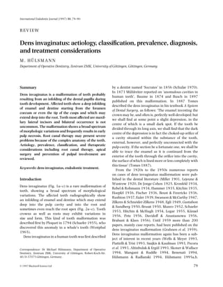
  • 1. International Endodontic Journal (1997) 30, 79–90 REVIEW Dens invaginatus: aetiology, classification, prevalence, diagnosis, and treatment considerations M. HÜLSMANN Department of Operative Dentistry, Zentrum ZMK, University of Göttingen, Göttingen, Germany Summary Dens invaginatus is a malformation of teeth probably resulting from an infolding of the dental papilla during tooth development. Affected teeth show a deep infolding of enamel and dentine starting from the foramen coecum or even the tip of the cusps and which may extend deep into the root. Teeth most affected are maxillary lateral incisors and bilateral occurrence is not uncommon. The malformation shows a broad spectrum of morphologic variations and frequently results in early pulp necrosis. Root canal therapy may present severe problems because of the complex anatomy of the teeth. Aetiology, prevalence, classification, and therapeutic considerations including root canal therapy, apical surgery and prevention of pulpal involvement are reviewed. Keywords: dens invaginatus, endodontic treatment. Introduction Dens invaginatus (Fig. 1a–c) is a rare malformation of teeth, showing a broad spectrum of morphological variations. The affected teeth radiographically show an infolding of enamel and dentine which may extend deep into the pulp cavity and into the root and sometimes even reach the root apex (Fig. 2a–c). Tooth crowns as well as roots may exhibit variations in size and form. This kind of tooth malformation was described first by Ploquet in 1794 (Schaefer 1955), who discovered this anomaly in a whale’s tooth (Westphal 1965). Dens invaginatus in a human tooth was first described Correspondence: Dr Michael Hülsmann, Department of Operative Dentistry, Zentrum ZMK, University of Göttingen, Robert-Koch-Str. 40, D-37075 Göttingen, Germany. © 1997 Blackwell Science Ltd by a dentist named ‘Socrates’ in 1856 (Schulze 1970). In 1873 Mühlreiter reported on ‘anomalous cavities in human teeth’, Baume in 1874 and Busch in 1897 published on this malformation. In 1887 Tomes described the dens invaginatus in his textbook A System of Dental Surgery, as follows: ‘The enamel investing the crown may be, and often is, perfectly well-developed; but we shall find at some point a slight depression, in the centre of which is a small dark spot. If the tooth be divided through its long axis, we shall find that the dark centre of the depression is in fact the choked-up orifice of a cavity situated within the substance of the tooth, external, however, and perfectly unconnected with the pulp-cavity. If the section be a fortunate one, we shall be able to trace the enamel as it is continued from the exterior of the tooth through the orifice into the cavity, the surface of which is lined more or less completely with this tissue’ (Tomes 1887). From the 1920s to the 1950s numerous reports on cases of dens invaginatus malformation were published in the dental literature (Miller 1901, Lejeune & Wustrow 1920, De Jonge Cohen 1925, Kronfeld 1934, Rebel & Rohmann 1934, Hammer 1935, Kitchin 1935, Hoepfel 1936, Fischer 1936, Beust & Freericks 1936, Rushton 1937, Euler 1939, Swanson & McCarthy 1947, Zilkens & Schneider-Zilkens 1948, Egli 1949, Gustafson & Sundberg 1950, Bruszt 1950, Munro 1952, Schaefer 1953, Hitchin & McHugh 1954, Logar 1955, Künzel 1956, Petz 1956, Davidoff & Anastassowa 1956, Brabant & Klees 1956). Until 1959 more than 200 papers, mainly case reports, had been published on the dens invaginatus malformation (Grahnen et al. 1959). Dens invaginatus malformation again has been a subject of interest in recent years (Wells & Meyer 1993, Piattelli & Trisi 1993, Szajkis & Kaufman 1993, Pecora et al. 1993, Altinbulak & Ergül 1993, Skoner & Wallace 1994, Mangani & Ruddle 1994, Benenati 1994, Hülsmann & Radlanski 1994, Hülsmann 1995a,b, 79
  • 2. 80 M. Hülsmann Fig. 1 (a) SEM; (b) microscopic and (c) radiographic appearance of a dental invagination. The invagination is completely filled with debris. At the bottom of the invagination the enamel is missing. Ikeda et al. 1995, Oncag et al. 1995, Nagatani et al. 1995, Khabbaz et al. 1995, Duckmanton 1995, Lindner et al. 1995, Bloch-Zupan et al. 1995, Olmez et al. 1995, Hosey & Bedi 1996). Synonyms for this malformation are: Dens in dente, invaginated odontome, dilated gestant odontome, dilated composite odontome, tooth inclusion, dentoid in dente. Fig. 2 (a) Invagination in the maxillary left lateral incisor. The invagination is limited to the tooth crown. The malformation was detected by chance. (b) Invagination in the maxillary left lateral incisor. The invagination is extending into the root. At the bottom of the invagination the enamel is missing. The invagination was suspected because of dysmorphic, peg-shaped tooth crown anatomy. (c) Severe case of dens invaginatus with open apex, diverging root and bizarre form of the invagination with a separate apical opening. The malformation was detected because of retarded tooth eruption of tooth 12. © 1997 Blackwell Science Ltd, International Endodontic Journal, 30, 79–90
  • 3. Dens invaginatus 81 Aetiology of dens invaginatus The aetiology of dens invaginatus malformation is controversial and remains unclear. Over the last decades several theories have been proposed to explain the aetiology of dental coronal invaginations: • Growth pressure of the dental arch results in buckling of the enamel organ (Euler 1939, Atkinson 1943). • Kronfeld (1934) suggested that the invagination results from a focal failure of growth of the internal enamel epithelium while the surrounding normal epithelium continues to proliferate and engulfs the static area. • Rushton (1937) proposed that the invagination is a result of rapid and aggressive proliferation of a part of the internal enamel epithelium invading the dental papilla. He regarded this a ‘benign neoplasma of limited growth’. • Oehlers (1957a,b) considered that distortion of the enamel organ during tooth development and subsequent protrusion of a part of the enamel organ will lead to the formation of an enamel-lined channel ending at the cingulum or occasionally at the incisal tip. The latter might be associated with irregular crown form. • The ‘twin-theorie’ (Bruszt 1950) suggested a fusion of two tooth-germs. • Infection was considered to be responsible for the malformation by Fischer (1936) and Sprawson (1937). • Gustafson & Sundberg (1950) discussed trauma as a causative factor, but could not sufficiently explain why just maxillary lateral incisors were affected and not central incisors. Most authors, meanwhile, consider dens invaginatus as a deep folding of the foramen coecum during tooth development which in some cases even may result in a second apical foramen (Schulze 1970). On the other hand the invagination also may start from the incisal edge of the tooth. Genetic factors cannot be excluded (Grahnen 1962, Casamassimo et al. 1978, Ireland et al. 1987, Hosey & Bedi 1996). Fig. 3 Classification of invaginated teeth by Oehlers (1957). the confines of the crown not extending beyond the amelocemental junction. Type II: an enamel-lined form which invades the root but remains confined as a blind sac. It may or may not communicate with the dental pulp. Type III: a form which penetrates through the root perforating at the apical area showing a ‘second foramen’ in the apical or in the periodontal area. There is no immediate communication with the pulp. The invagination may be completely lined by enamel, but frequently cementum will be found lining the invagination. Oehlers (1957a,b) also described different crown forms (normal with a deep lingual or palatal pit; conical, barrel-shaped or peg-shaped with an incisal pit) relating to the three groups mentioned above. In addition, Ulmansky & Hermel (1964) and Vincent-Townend (1974) described an ‘incipient dens in dente’, a deep palatal or lingual pit completely lined by enamel with no communication to the pulp. Oehlers (1958) also presented radicular invaginations. Schulze & Brand (1972) proposed a more detailed classification (Fig. 4), also including invaginations starting at the incisal edge or the top of the crown and also including dysmorphic root configurations. Prevalence of dens invaginatus Classification of dens invaginatus The first classification of invaginated teeth was published by Hallet (1953). The most commonly used classification proposed by Oehlers (1957a) is shown in Fig. 3. He described the anomaly occurring in three forms: Type I: an enamel-lined minor form occurring within © 1997 Blackwell Science Ltd, International Endodontic Journal, 30, 79–90 A summary of the results of investigations on the prevalence of invaginated teeth is presented in Table 1. It is difficult to compare findings because of the differences in study design, sample size and composition, and diagnostic criteria. The teeth most affected are maxillary lateral incisors and bilateral occurrence is not uncommon and occurs in 43% of all cases (Grahnen et al. 1959).
  • 4. 82 M. Hülsmann Fig. 4 Classification of dens invaginatus by Schulze & Brand (1972) (from: Zahnärztliche Welt/Reform 81, 569–73, with kind permission). Swanson & McCarthy (1947) were the first to present bilateral dens invaginatus malformation, Conklin (1968) presented a patient with all maxillary central and lateral incisors affected, and Burton et al. (1980) published a case with six teeth involved, the maxillary incisors and also the maxillary canines. Krolls (1969) detected dental invaginations in maxillary central incisors as well as in several maxillary and mandibular bicuspids in one patient. Conklin (1978) found the anomaly in four mandibular incisors in one patient. Double invaginations in one tooth were reported by Rushton (1937), Archer & Silverman (1950), Oehlers (1957a), Grahnen et al. (1959), Ulmansky & Hermel (1964), Schulze & Brand (1972), Brill & Phillips (1974), Bhatt & Dholakia (1975), Conklin (1975), Cole et al. (1978), Gotoh et al. (1979), Mader (1979), Rotstein et al. (1987a), and Vairabhaya (1989). Triple invaginations were described by Hitchin & McHugh (1954) and Mader (1977). In rare cases even primary teeth seem to be affected (Rabinowitch 1952). Cases of dental invaginations in supernumerary teeth have been presented as well as in canines and bicuspids (Archer & Silverman 1950, Schwenzer 1957, Kruse 1962, Klammt 1969, Conklin 1975, Banner 1978, Table 1 Investigations on the prevalence of dens invaginatus Author/Country Year Sample Frequency Mühlreiter (Austria) Atkinson (USA) Boyne (USA) Stephens (unknown) Shafer (USA) Hallet (unknown) 1873 2.8% of teeth 1943 500 extracted lateral maxillary incisors (histological investigation) 500 lateral maxillary incisors 1952 1000 maxillary incisors 0.3% of teeth 1953 1953 1953 150 full-mouth surveys 1000 patients 2542 full-mouth surveys 1953 586 full-mouth surveys Amos (USA) Grahnen et al. (Sweden) Ulmansky & Hermel (Israel) Poyton & Morgan (Canada) Miyoshi et al. ( Japan) Fujiki et al. ( Japan) Thomas (USA) Gotoh et al. ( Japan) Ruprecht et al. (Saudi Arabia) Ruprecht et al. (Saudi Arabia) 1955 1959 1000 full mouth surveys 203 full mouth surveys 3020 right maxillary incisors 8% 3.6% (cited by Hovland & Block 1977) 1.3% of patients (cited by Hovland & Block 1977) 6.6% of maxillary lateral incisors 0.5% of maxillary central incisors (cited by Hovland & Block 1977) 5.1% of patients 6.9% (students of dentistry) 2.7% of patients 1964 500 full mouth surveys 2% of patients 1966 5000 full mouth surveys 1971 1974 extracted maxillary lateral incisors 2126 lateral maxillary incisors 1974 1886 full mouth surveys 0.25% of patients (cited by Hovland & Block 1977) 38.5% of teeth (cited by Gotoh et al. 1979) 4.2% of teeth (cited by Gotoh et al. 1979) 7.74% of patients 1979 766 lateral maxillary incisors 9.66% of teeth 1986 1581 full mouth surveys 1.7% of patients 1987 300 full mouth surveys 10% of patients 10% of teeth © 1997 Blackwell Science Ltd, International Endodontic Journal, 30, 79–90
  • 5. Dens invaginatus Shifman & Tamir 1979, Bottomley & Johnson 1982, Eldeeb 1984, Rotstein et al. 1987a, Morfis 1992, Bramante et al. 1993, Altinbulak & Ergül 1993, Hülsmann & Radlanski 1994, Tavano et al. 1994). 83 pulpal involvement of teeth with coronal invaginations may occur a short time after tooth eruption early diagnosis is mandatory to instigate preventive treatment. Clinical features Histological findings Several reports on microscopic, ultrastructural and microradiographic investigations of teeth with a dens invaginatus malformation show a wide range of findings and thus reproduce the macroscopic variety of this anomaly. The dentine below the invagination may be intact without irregularities (Brabant & Klees 1956, Omnell et al. 1960, Piatelli & Trisi 1993) but also may contain strains of vital connective tissue (Omnell et al. 1960) or even fine canals with communication to the dental pulp (Kronfeld 1934, Fischer 1936, Hoepfel 1936, Gustafson & Sundberg 1950, Hitchin & McHugh 1954, Oehlers 1957a, Rushton 1958). Some authors reported hypomineralized or irregularly structured dentine (Omnell et al. 1960, Vincent-Townend 1974, Beynon 1982). The structure and thickness of the enamel lining the invagination also may vary widely. The enamel was described as irregularly structured by Atkinson (1943), Beynon (1982) and Piatelli & Trisi (1993). Beynon (1982) reported hypomineralized enamel at the base of the invagination whereas Morfis (1992), in a chemical analysis, detected up to eight times more phosphate and calcium compared with the outer enamel, but in his analysis magnesium was missing completely. Bloch-Zupan et al. (1995) found differences in structure and compostion between the external and internal enamel. The internal enamel exhibited atypical and more complex rod shapes and its surface presented the typical honeycomb pattern but no perikymata, which, however, were observed on the outer surface of the tooth. Diagnosis of dens invaginatus In most cases a dens invaginatus is detected by chance on the radiograph. Clinically, an unusual crown morphology (‘dilated’, ‘peg-shaped’, ‘barrel-shaped’) or a deep foramen coecum may be important hints, but affected teeth also may show no clinical signs of the malformation. As maxillary lateral incisors are the teeth most susceptible to coronal invaginations these teeth should be investigated thoroughly clinically and radiographically, at least in all cases with a deep pit at the foramen coecum. If one tooth is affected in a patient the contralateral tooth should also be investigated. As © 1997 Blackwell Science Ltd, International Endodontic Journal, 30, 79–90 The invagination allows entry of irritants into an area which is separated from pulpal tissue by only a thin layer of enamel and dentine and presents a predisposition for the development of dental caries. In some cases the enamel-lining is incomplete (Fig. 1). Channels may also exist between the invagination and the pulp (Kronfeld 1934, Hitchin & McHugh 1954). Therefore, pulp necrosis often occurs rather early, within a few years of eruption, sometimes even before root end closure (Swanson & McCarthy 1947, Ulmansky & Hermel 1964, Stepanik 1968, Ferguson et al. 1980, Morfis & Lentzari 1989, Nik-Hussein 1994, Hülsmann & Radlanski 1994) (Fig. 5a, b). Other reported sequelae of undiagnosed and untreated coronal invaginations are abscess formation (Schaefer 1955, Petz 1956, Kendrick 1971, Greenfield & Cambruzzi 1986, Chen et al. 1990, Whyman & McFayden 1994), retention of neighbouring teeth (Schaefer 1955, Petz 1956, Conklin 1975, Mader 1977), displacement of teeth (Schaefer 1955, Petz 1956), cysts (Rebel & Rohmann 1934, Schwenzer 1957, Conklin 1978, Klammt 1969, Burzynski 1973, Samimy 1977, Murphy & Doku 1977, Augsburger & Brandebura 1978, Greenfield & Cambruzzi 1986), and internal resorption (Shapiro 1970, Hülsmann & Radlanski 1994) (Fig. 6). The dental literature on dens invaginatus malformations contains several case reports presenting invaginated teeth coincident with other dental anomalies, malformations and even dental or medical syndromes (Table 2). Treatment considerations Preventive and restorative treatment Teeth with deep palatal or incisal invaginations or foramina coeca should be treated with fissure sealing before carious destruction can occur. A composite restoration and strict periodic review is recommended (Rotstein et al. 1987b, Hülsmann & Radlanski 1994). If no entrance to the invagination can be detected and no signs of pathosis are visible clinically and radiographically no treatment is indicated, but strict observation is recommended (Hülsmann 1995b, Duckmanton 1995, Hülsmann 1996).
  • 6. 84 M. Hülsmann Fig. 5 (a) Small coronal invagination in tooth 22. (b) Three years later arrest of root development and pulp necrosis, with apical periodontitis, and sinus tract have appeared. (c) Fifteen months later: following apexification therapy with calcium hydroxide and obturation of the root canal the periapical status has improved. Root canal treatment A review of the literature shows that extraction of teeth with invaginations was the preferred therapy until the 1970s (Hülsmann 1995a). Grossman (1974) and Creaven (1975) were the first to describe root canal Table 2 Dental anomalies described in association with dens invaginatus Microdontia Macrodontia Hypodontia Oligodontia Taurodontism Gemination and Fusion Supernumerary teeth Amelogenesis imperfecta Invagination in an odontome Multiple odontomes Coronal agenesis Williams syndrome Casamassimo et al. 1978 Ekman-Westberg & Julin 1974 Hülsmann 1995c Conklin 1978, Ruprecht et al. 1986 Casamassimo et al. 1978, Ruprecht et al.1986, Ireland et al. 1987, Chen et al. 1990 Burzynski 1973, Mader 1979, Mader & Zielke 1982, Ruprecht et al. 1986 Schaefer 1953, 1955, Brabant & Klees 1956, Petz 1956, Rushton 1958, Conklin 1975, Mader 1977, Shifman & Tamir 1979, Beynon 1982, Ruprecht et al. 1986, Morfis 1992 Kerebel et al. 1983 Hitchin & McHugh 1954 Robbins & Keene 1964 Hicks & Flaitz 1985 Oncag et al. 1995 treatment of the invagination only (root invagination treatment) and Tagger (1977) and Hovland & Block (1977) the first to present cases treated with conventional root canal therapy (Fig. 7a,b). Root canal treatment may present several problems because of the irregular shape of the root canal system(s). If there are no radiographic signs of pulp pathosis and no communication between the invagination and the root canal, root canal treatment or, in minor cases, even a composite or amalgam filling of the invagination will be adequate (DeSmit & Demaut 1982). When the invagination has a separate apical or lateral foramen, root canal treatment of the invagination is indicated (Grossman 1974, Creaven 1975, Bolanos et al. 1988, Szajkis & Kaufman 1993, Wells & Meyer 1993, Ikeda et al. 1995). In some cases it may be possible to bur through the invagination to get access to the apical foramen. When these minor forms of invaginations are eliminated root canal therapy in most cases will not present further problems. In certain cases the invagination has to be treated as a separate root canal (Cole et al. 1978, Zillich et al. 1983, Eldeeb 1984, Greenfield & Cambruzzi 1986, Mangani & Ruddle 1994, Khabbaz et al. 1995). When pulp necrosis occurs before root-end closure, apexification procedures with calcium hydroxide may be necessary (Ferguson et al. 1980, Morfis & Lentzari 1989, Vairabhaya 1989, Hülsmann & Radlanski 1994, Nagatani et al. 1995) (Fig. 5a–c). © 1997 Blackwell Science Ltd, International Endodontic Journal, 30, 79–90
  • 7. Dens invaginatus 85 Fig. 6 (a) Pulp necrosis, apical periodontitis, sinus tract, and lateral resorptive perforation in tooth 12 showing a dens invaginatus malformation. (b) Clinical view of the tooth crown of the same tooth before treatment. A deep foramen coecum with caries is visible. (c) After preparation of the access cavity the entrance to the invagination is visible at the palatal aspect (arrow). The main root canal orifice can be seen at the top of the cavity at the buccal aspect. The large and irregular volume of the root canal system makes proper shaping and cleaning difficult. Irrigation, supported by ultrasonic cleaning of the root canal system has been described as an efficient means of disinfection (Cunningham et al. 1982) and has therefore been recommended for cleaning of the complex morphology of the root canal system in teeth with dens invaginatus (Skoner & Wallace 1994). For obturation of such teeth warm gutta-percha techniques including vertical condensation or thermoplastic filling techniques have been recommended (Rotstein et al. 1987b, Hülsmann & Radlanski 1994, Mangani & Ruddle 1994). © 1997 Blackwell Science Ltd, International Endodontic Journal, 30, 79–90 Surgical treatment Surgical treatment should be considered in cases of endodontic failure and in teeth which cannot be treated non-surgically because of anatomical problems or failure to gain access to all parts of the root canal system (Harnisch 1970, Hata & Toda 1987, Teplitsky & Singer 1987, Rotstein et al. 1987b, Kulild & Weller 1989, Suchina et al. 1989, Hülsmann & Radlanski 1994, Benenati 1994, Olmez et al. 1995) (Fig. 8a,b). Cole et al. (1978) and Lindner et al. (1995) even proposed intentional replantation with retrograde surgery in otherwise hopeless cases.
  • 8. 86 M. Hülsmann Fig. 7 (a) Right maxillary lateral incisor in the same patient as in Fig. 5. The invagination was detected by preventive radiographic examination, clinically no invagination nor carious lesion were detected. The invagination has resulted in an internal resorption with perforation. (b) Obturation with injectable gutta-percha, resulting in a slight lateral overfill. Fig. 8 (a) Radiograph of tooth 23 showing a bizarre lateral coronal invagination which seems to be open to the lateral periodontium. The main root canal was root canal treated, but no communication to the invagination could be detected. (b) As no access to the invagination was found coronally the invagination was treated surgically. Probably root canal treatment of the main root canal would not have been necessary. © 1997 Blackwell Science Ltd, International Endodontic Journal, 30, 79–90
  • 9. Dens invaginatus 87 Fig. 9 (a) Severe dens invaginatus malformation with acute apical abscess formation. The tooth was judged inoperable and extracted by a general dental practitioner. (b) The photograph of the extracted tooth shows the invagination extending beyond the apex. Extraction Extraction is indicated only in teeth with severe anatomical irregularities that cannot be treated non-surgically or by apical surgery, and in supernumerary teeth (Figs 9a,b and 10). Additionally Rotstein et al. (1987b) advocated extraction when abnormal crown morphology presents aesthetic or functional problems. References Fig. 10 Severe invagination malformation of tooth 22 resulting in acute apical abscess formation. The tooth was judged inoperable and extracted. © 1997 Blackwell Science Ltd, International Endodontic Journal, 30, 79–90 AL T I N B U L A K H, ER G Ü L N (1993) Multiple dens invaginatus. Oral Surgery, Oral Medicine and Oral Pathology 76, 620–2. AM O S ER (1955) Incidence of the small dens in dente. Journal of the American Dental Association 51, 31–3. ARCHER WH, SI L V E R M A N LM (1950) Double dens in dente in bilateral rudimentary supernumerary central incisors. Oral Surgery, Oral Medicine and Oral Pathology 3, 722. AT K I N S O N SR (1943) The permanent maxillary lateral incisor. American Journal of Orthodontics 29, 685–98. AU G S B U R G E R RA, BR A N D E B U R A J (1978) Bilateral dens invaginatus with associated radicular cysts. Oral Surgery, Oral Medicine and Oral Pathology 46, 260–4. BA N N E R H (1978) Bilateral dens in dente in mandibular premolars. Oral Surgery, Oral Medicine and Oral Pathology 45, 827–8. BA U M E R (1874) Zahnmi␤bildungen. I. Ein Zahn im Zahne. Deutsche Vierteljahresschrift für Zahnheilkunde 14, 25–9. BE N E N A T I FW (1994) Complex treatment of a maxillary lateral incisor with dens invaginatus and associated aberrant morphology. Journal of Endodontics 20, 180–2.
  • 10. 88 M. Hülsmann BE U S T TB, FR E E R I C K S FH (1936) Dens in dente. Journal of Dental Research 15, 158. BE Y N O N AD (1982) Developing dens invaginatus (dens in dente). A quantitative microradiographic study and a reconsideration of the histogenesis of this condition. British Dental Journal 153, 255–60. BH A T T AP, DH O L A K I A HM (1975) Radicular variety of double dens in dente. Oral Surgery, Oral Medicine and Oral Pathology 39, 284–7. BL O C H-ZU P A N A, CO U S A N D I E R L, HE M M E R L E J, OB R Y-MU S S E T AM, MA N I E R E MC (1995) Electron microscopical assessments (FDIabstract FC-11# 123). International Dental Journal 45, 306. BO L A N O S OR, MA R T E L L B, MO R S E DR (1988) A unique approach to the treatment of a tooth with dens invaginatus. Journal of Endodontics 14, 315–7. BO T T O M L E Y WK, JO H N S O N MR (1982) Multiple dens in dente in a dentition. Oral Surgery, Oral Medicine and Oral Pathology 54, 478. BO Y N E PJ (1952) Dens in dente: Report of three cases. Journal of the Americal Dental Association 45, 209–10. BR A B A N T H, KL E E S L (1956) Beitrag zur Kenntnis der ‘dens in dente’ benannten Zahnanomalie. Stoma 9, 12–27. BR A M A N T E CM, DE S O U S A SM, TA V A N O SM (1993) Dens invaginatus in mandibular first premolar. Oral Surgery, Oral Medicine and Oral Pathology 76, 389. BR I L L WA, PH I L L I P S JE (1974) Double Dens invaginatus. Oral Surgery, Oral Medicine and Oral Pathology 37, 139–40. BR U S Z T P (1950) Über die Entstehung des ‘Dens in dente’. Schweizer Monatsschrift für Zahnheilkunde 60, 534–42. BU R T O N DJ, SA F F O S RO, SC H E F F E R RB (1980) Multiple bilateral dens in dente as a factor in the etiology of multiple periapical lesions. Oral Surgery, Oral Medicine and Oral Pathology 49, 496–99. BU R Z Y N S K I NJ (1973) Gemination and dens in dente. Oral Surgery, Oral Medicine and Oral Pathology 36, 760–1. BU S C H N (1897) Über Verschmelzung und Verwachsung der Zähne des Milchgebisses und des bleibenden Gebisses. Deutsche Monatsschrift für Zahnheilkunde 15, 469–86. CA S A M A S S I M O PS, NO W A K AJ, ET T I N G E R RL, SC H L E N K E R DJ (1978) An unusual triad: microdontia, taurodontia, and dens invaginatus. Oral Surgery, Oral Medicine and Oral Pathology 45, 107–12. CH E N RJ, YA N G JF, CH A O TC (1990) Invaginated tooth associated with periodontal abscess. Oral Surgery, Oral Medicine and Oral Pathology 69, 659. CO L E GM, TA I N T O R JF, JA M E S GA (1978) Endodontic therapy of a dilated dens invaginatus. Journal of Endodontics 4, 88–90. CO N K L I N WW (1968) Multiple dens in dente. Oral Surgery, Oral Medicine and Oral Pathology 25, 193. CO N K L I N WW (1975) Double bilateral dens invaginatus in the maxillary incisor region. Oral Surgery, Oral Medicine and Oral Pathology 39, 949–52. CO N K L I N WW (1978) Bilateral dens invaginatus in the mandibular incisor region. Oral Surgery, Oral Medicine and Oral Pathology 45, 905–8. CR E A V E N J (1975) Dens invaginatus-type malformation without pulpal involvement. Journal of Endodontics 1, 79–80. CU N N I N G H A M W, MA R T I N H, PE L L E U G, ST O O P S D (1982) A comparison of antimicrobial effectiveness of endosonic and hand root canal therapy. Oral Surgery, Oral Medicine and Oral Pathology 54, 238–41. DA V I D O F F SM, AN A S T A S S O W A MN (1956) Dentoid in dente. Deutsche Zahn-, Mund- und Kieferheilkunde 23, 454–68. DE JO N G E CO H E N TE (1925) Ein neuer Beitrag zur Morphogenese des ‘dens in dente’. Vierteljahrschrift für Zahnheilkunde 41, 125–30. DE SM I T A, DE M A U T L (1982) Nonsurgical endodontic treatment of invaginated teeth. Journal of Endodontics 8, 506–11. EG L I AR (1949) Beitrag zur Frage des ‘Dens in dente’. Deutsche Zahnärztliche Zeitschrift 4, 432–7. DU C K M A N T O N PM (1995) Maxillary permanent central incisor with abnormal crown size and dens invaginatus: case report. Endodontics and Dental Traumatology 11, 150–2. EK M A N-WE S T B O R G B, JU L I N P (1974) Multiple anomalies in dental morphology: macrodontia, multituberculism, central cusps, and pulp invaginations. Oral Surgery, Oral Medicine and Oral Pathology 38, 217–22. EL D E E B ME (1984) Nonsurgical endodontic therapy of a dens invaginatus. Journal of Endodontics 10, 107–9. EU L E R H (1939) Die Anomalien, Fehlbildungen und Verstümmelungen der menschlichen Zähne. pp. 62–7, München, Germany: Lehmann. FE R G U S O N FS, FR I E D M A N S, FR A Z Z E T T O V (1980) Successful apexification technique in an immature tooth with dens in dente. Oral Surgery, Oral Medicine and Oral Pathology 49, 356–59. FI S C H E R CH (1936) Zur Frage des Dens in dente. Deutsche Zahn-, Mundund Kieferheilkunde 3, 621–34. FU J I K I Y, TA M A K I N, KA W A H A R A K, NA B A E M (1974) Clinical and radiographic observations of dens invaginatus. Journal of Dentomaxillofacial Radiology 3, 343–8. GO T O H T, KA W A H A R A K, IM A I K, KI S H I K, FU J I K I Y (1979) Clinical and radiographic study of dens invaginatus. Oral Surgery, Oral Medicine and Oral Pathology 48, 88–91. GR A H N E N H, LI N D A H L B, OM N E L L K (1959) Dens invaginatus. I. A clinical, roentgenological and genetical study of permanent upper lateral incisors. Odontologisk Revy 10, 115–37. GR A H N E N H (1962) Hereditary factors in relation to dental caries and congenitally missing teeth. In: Witkop CJ, ed. Genetics and Disorder. pp. 194–204, New York, USA: McGraw-Hill. GR E E N F E L D RS, CA M B R U Z Z I JV (1986) Complexities of endodontic treatment of maxillary lateral incisors with anomalous root formation. Oral Surgery, Oral Medicine and Oral Pathology 62, 82–8. GR O S S M A N LI (1974) Endodontic case reports. Dental Clinics of North America 18, 509–28. GU S T A F S O N G, SU N D B E R G S (1950) Dens in dente. British Dental Journal 88, 83–88, 111–122, 144–146. HA L L E T GE (1953) The incidence, nature and clinical significance of palatal invagination in the maxillary incisors teeth. Proceedings of the Royal Society of Medicine 46, 491–9. HA M M E R H (1935) Zwei Fälle eigenartiger Zahnmi␤bildung. Deutsche Zahn-, Mund- und Kieferheilkunde 2, 46–52. HA R N I S C H H (1970) Apicectomy in dens in dente. Quintessence International 1, 21–2. HA T A G, TO D A T (1987) Treatment of dens invaginatus by endodontic therapy, apicocurettage, and retrofilling. Journal of Endodontics 9, 469–72. HI C K S MJ, FL A I T Z CM (1985) Dens invaginatus with partial coronal agenesis: report of a case. Journal of Dentistry for Children 52, 217–9. HI T C H I N AD, MC H U G H WD (1954) Three coronal invaginations in a dilated composite odontome. British Dental Journal 97, 90–2. HO E P F E L W (1936) Der Dens in dente. Deutsche Zahn-, Mund- und Kieferheilkunde 3, 67–80. HO S E Y MT, BE D I R (1996) Multiple dens invaginatus in two brothers. Endodontics and Dental Traumatology 12, 44–7. HO V L A N D EJ, BL O C K RM (1977) Nonrecognition and subsequent endodontic treatment of dens invaginatus. Journal of Endodontics 3, 360–2. HÜ L S M A N N M (1995a) Der Dens invaginatus — Ätiologie, Inzidenz und klinische Besonderheiten. Schweizer Monatsschrift für Zahnmedizin 105, 765–76. HÜ L S M A N N M (1995b) Therapie des Dens invaginatus. Schweizer Monatsschrift für Zahnmedizin 105, 1323–34. HÜ L S M A N N M (1995c) Dens invaginatus: incidence, aetiology, © 1997 Blackwell Science Ltd, International Endodontic Journal, 30, 79–90
  • 11. Dens invaginatus diagnosis, and treatment options. Poster-presentation, 6th ESECongress, Tel Aviv, 1995. HÜ L S M A N N M (1996) Severe dens invaginatus malformation. Report of two cases. Oral Surgery, Oral Medicine and Oral Pathology 82, 456–8. HÜ L S M A N N M, RA D L A N S K I R (1994) Möglichkeiten der konservativen Therapie des Dens invaginatus. Deutsche Zahnärztliche Zeitschrift 49, 804–8. IK E D A H, YO S H I O K A T, SU D A H (1995) Importance of clinical examination and diagnosis. Oral Surgery, Oral Medicine and Oral Pathology 79, 88–91. IR E L A N D JE, BL A C K JP, SC U R E S CC (1987) Short roots, taurodontia and multiple dens invaginatus. Journal of Pedodontics 11, 164–75. KE N D R I C K JK (1971) Periapical abscess from dens in dente. Oral Surgery, Oral Medicine and Oral Pathology 31, 838. KE R E B E L B, KE R E B E L LM, DA C U L S I G, DO U R Y J (1983) Dentinogenesis imperfecta with dens in dente. Oral Surgery, Oral Medicine and Oral Pathology 55, 279–85. KH A B B A Z MG, KO N S T A N T A K I MN, SY K A R A S SN (1995) Dens invaginatus in a mandibular lateral incisor. International Endodontic Journal 28, 303–5. KI T C H I N PC (1935) Dens in dente. Journal of Dental Research 15, 117–21. KL A M M T J (1969) Dens invaginatus und Zyste. Deutsche Zahn-, Mundund Kieferheilkunde 53, 336–47. KR O L L S SO (1969) A dentition with multiple dens in dente. Oral Surgery, Oral Medicine and Oral Pathology 27, 648. KR O N F E L D R (1934) Dens in dente. Journal of Dental Research 14, 49–66. KR U S E H (1962) Dens in dente im Prämolarenbereich des Unterkiefers. Deutsche Zahnärztliche Zeitschrift 17, 1532–5. KÜ N Z E L W (1956) Bilaterales Vorkommen des Dens in dente. Zahnärztliche Praxis 7, 1–2. KU L I L D JC, WE L L E R N (1989) Treatment considerations in dens invaginatus. Journal of Endodontics 15, 381–4. LE J E U N E F, WU S T R O W P (1920) Zwei Fälle einer eigenartigen Zahnmi␤bildung. Deutsche Monatsschrift für Zahnheilkunde 38, 15–22. LI N D N E R C, ME S S E R HH, TY A S MJ (1995) A complex treatment of dens invaginatus. Endodontics and Dental Traumatology 11, 153–5. LO G A R A (1955) Über die Genese des ‘Dens in dente’. Österreichische Zeitschrift für Stomatologie 52, 412–23. MA D E R C (1977) Triple dens in dente. Oral Surgery, Oral Medicine and Oral Pathology 44, 966. MA D E R C (1979) Double dens in dente in a geminated tooth. Oral Surgery, Oral Medicine and Oral Pathology 47, 573. MA D E R CL, ZI E L K E DR (1982) Incomplete dens in dente in a fused tooth. Oral Surgery, Oral Medicine and Oral Pathology 53, 439. MA N G A N I F, RU D D L E CJ (1994) Endodontic treatment of a ‘very particular’ maxillary central incisor. Journal of Endodontics 20, 560–1. MI L L E R WD (1901) Einige seltene Zahnanomalien. Deutsche Monatsschrift für Zahnheilkunde 19, 397–410. MI Y O S H I S, FU J I W A R A J, NA K A T A T, YA M A M O T O K, DE G U C H I K (1971) Dens invaginatus in Japanese incisors. Japanese Journal of Oral Biology 13, 539–44. MO R F I S AS (1992) Chemical analysis of a dens invaginatus by S.E.M. microanalyses. Journal of Clinical Pediatric Dentistry 17, 79–82. MO R F I S AS, LE N T Z A R I A (1989) Dens invaginatus with an open apex: a case report. International Endodontic Journal 22, 190–2. MÜ H L R E I T E R E (1873) Die Natur der anomalen Höhlenbildung im oberen Seitenschneidezahne. Deutsche Vierteljahresschrift für Zahnheilkunde 13, 367–72. MU N R O D (1952) Dens in dente. British Dental Journal 92, 92–3. MU R P H Y JB, DO K U HC (1977) Dens in dente: an unusual sequela. Oral Surgery, Oral Medicine and Oral Pathology 43, 530–1. © 1997 Blackwell Science Ltd, International Endodontic Journal, 30, 79–90 89 NA G A T A N I T, HI R A B A Y A S H I M, OS A D A T (1995) Introduction of rootend closure for traumatized immature tooth with Dens invaginatus (FDI-abstract P’166) International Dental Journal 45, 307. NI K-HU S S E I N NN (1994) Dens invaginatus: Complications and treatment of non-vital infected tooth. Journal of Clinical Pediatric Dentistry 18, 303–6. OE H L E R S FA (1957a) Dens invaginatus. I. Variations of the invagination process and associated anterior crown forms. Oral Surgery, Oral Medicine and Oral Pathology 10, 1204–18. OE H L E R S FA (1957b) Dens invaginatus. II. Associated posterior crown forms and pathogenesis. Oral Surgery, Oral Medicine and Oral Pathology 10, 1302–16. OE H L E R S FA (1958) The radicular variety of dens invaginatus. Oral Surgery, Oral Medicine and Oral Pathology 11, 1251–60. OL M E Z S, UZ A M I S M, E R N (1995) Dens invaginatus of a mandibular central incisor: surgical endodontic treatment. Journal of Clinical Pediatric Dentistry 20, 53–6. OM N E L L KA, SW A N B E C K G, LI N D A H L B (1960) Dens invaginatus. II. A microradiographical, histological and micro X-ray diffraction study. Acta Odontologica Scandinavica 18, 303–30. ON C A G A, GU N B A Y S, PA R L A R A (1995) Williams Syndrome. Journal of Clinical Pediatric Dentistry 19, 301–4. PE C O R A JD, CO N R A D O CA, ZU C O L O T T O WG, SO U S A NE T O MD, SA Q U Y PC (1993) Root canal therapy of an anomalous maxillary central incisor: a case report. Endodontics and Dental Traumatology 9, 260–2. PE T Z R (1956) Beitrag zum ‘Dens in dente’. Deutsche Stomatologie 6, 100–5. PI A T E L L I A, TR I S I P (1993) Dens invaginatus: a histological study of undemineralized material. Endodontics and Dental Traumatology 9, 191–5. PO Y T O N GH, MO R G A N GA (1966) Dens in dente. Dental Radiography and Photography 39, 27–33. RA B I N O W I T C H BZ (1952) Dens in dente: primary tooth. Report of a case. Oral Surgery, Oral Medicine and Oral Pathology 5, 1312–4. RE B E L HH, RO H M A N N C (1934) ‘Dens in dente’. Deutsche Zahnärztliche Wochenschrift 37, 83–5. RO B B I N S IM, KE E N E HJ (1964) Multiple morphologic dental anomalies. Oral Surgery, Oral Medicine and Oral Pathology 17, 683–90. RO T S T E I N I, ST A B H O L Z A, FR I E D M A N S (1987a) Endodontic therapy for dens invaginatus in a maxillary second premolar. Oral Surgery, Oral Medicine and Oral Pathology 63, 237–40. RO T S T E I N I, ST A B H O L Z A, HE L I N G I, FR I E D M A N S (1987b) Clinical considerations in the treatment of dens invaginatus. Endodontics and Dental Traumatology 3, 249–54. RU P R E C H T A, BA T N I J I S, SA S T R Y K, EL-NE W E I H I E (1986) The incidence of dental invagination. Journal of Pedodontics 10, 265–72. RU P R E C H T A, SA S T R Y K, BA T N I J I S, LA M B O U R N E A (1987) The clinical significance of dental invagination. Journal of Pedodontics 11, 176–81. RU S H T O N MA (1937) A collection of dilated composite odontomas. British Dental Journal 63, 65–85. RU S H T O N MA (1958) Invaginated teeth (dens in dente): Contents of the invagination. Oral Surgery, Oral Medicine and Oral Pathology 11, 1378–87. SA M I M Y B (1977) Dens invaginatus: a potential hazard to the pulp. Quintessence International 8, 91–2. SC H A E F E R H (1953) Über das Vorkommen des ‘dens in dente’. Schweizer Monatsschrift für Zahnmedizin 63, 779–87. SC H A E F E R H (1955) Zur Klinik des dens in dente. Deutsche Zahnärztliche Zeitschrift 10, 988–93. SC H U L Z E C (1970) Developmental abnormalities of the teeth and the jaws. In: Gorlin O, Goldman H, eds. Thoma’s Oral Pathology. pp. 96–183, St. Louis, USA: Mosby.
  • 12. 90 M. Hülsmann SC H U L Z E C, BR A N D E (1972) Über den Dens invaginatus (Dens in dente). Zahnärztliche Welt/Reform 81, 569–73, 613–20, 653–60, 699–703. SC H W E N Z E R N (1957) Zur Klinik und Histologie des Dens in dente. Deutsche Zahnärztliche Zeitschrift 12, 1233–8. SH A F E R WG (1953) Dens in dente. New York Dental Journal 19, 220–5. SH A P I R O L (1970) Dens in dente. Oral Surgery, Oral Medicine and Oral Pathology 30, 782. SH I F M A N A, TA M I R A (1979) Dens invaginatus with concrescent supernumerary tooth. Oral Surgery, Oral Medicine and Oral Pathology 47, 391. SK O N E R JR, WA L L A C E JA (1994) Dens invaginatus: another use for the ultrasonics. Journal of Endodontics 20, 138–40. SP R A W S O N EC (1937) Odontomes. British Dental Journal 62, 177–201. ST E P A N I K G (1968) Dens in Dente. Oral Surgery, Oral Medicine and Oral Pathology 26, 332. ST E P H E N S R (1953) The diagnosis, clinical significance and treatment of minor palatal invaginations in maxillary incisors. Proceedings of the Royal Society of Medicine 46, 499–503. SU C H I N A JA, LU D I N G T O N JR, MA D D E N RM (1989) Dens invaginatus of a maxillary lateral incisor: Endodontic treatment. Oral Surgery, Oral Medicine and Oral Pathology 68, 467–71. SW A N S O N WF, MC C A R T H Y FM (1947) Bilateral dens in dente. Journal of Dental Research 26, 167–71. SZ A J K I S S, KA U F M A N AY (1993) Root invagination treatment: a conservative approach in endodontics. Journal of Endodontics 19, 576–8. TA G G E R M (1977) Nonsurgical endodontic therapy of tooth inva- gination. Oral Surgery, Oral Medicine and Oral Pathology 43, 124–9. TA V A N O SMR, SO U S A SMG, BR A M A N T E CM (1994) Dens invaginatus in first mandibular premolar. Endodontics and Dental Traumatology 10, 27–9. TE P L I T S K Y P, SI N G E R D (1987) Radicular anomaly of a maxillary canine; Endodontic treatment. Journal of Endodontics 13, 81–4. TH O M A S JG (1974) A study of dens in dente. Oral Surgery, Oral Medicine and Oral Pathology 38, 653–5. TO M E S J (1887) A System of Dental Surgery, 3rd edn. pp. 652–5. London, UK: J. & A. Churchill. UL M A N S K Y M, HE R M E L J (1964) Double dens in dente in a single tooth. Oral Surgery, Oral Medicine and Oral Pathology 17, 92–7. VA I R A B H A Y A L (1989) Nonsurgical endodontic treatment of a tooth with double dens in dente. Journal of Endodontics 15, 323–5. VI N C E N T-TO W N E N D J (1974) Dens invaginatus. Journal of Dentistry 2, 234–8. WE L L S DW, ME Y E R RD (1993) Vital root canal treatment of a Dens in Dente. Journal of Endodontics 19, 616–7. WE S T P H A L A (1965) Ein kleines Kuriosum um den ersten ‘Dens in dente’. Zahnärztliche Mitteilungen 55, 1066–70. WH Y M A N RA, MA C F A D Y E N EE (1994) Dens in dente associated with infective endocarditis. Oral Surgery, Oral Medicine and Oral Pathology 78, 47–50. ZI L K E N S K, SC H N E I D E R-ZI L K E N S H (1948) Zur Lehre des ‘Dens in dente’. Deutsche Zahnärztliche Zeitschrift 3, 392–402. ZI L L I C H RM, AS H JL, CO R C O R A N JF (1983) Maxillary lateral incisor with two roots and dens formation: A case report. Journal of Endodontics 9, 143–4. © 1997 Blackwell Science Ltd, International Endodontic Journal, 30, 79–90