Corticospinal system
•Als PPTX, PDF herunterladen•
5 gefällt mir•4,067 views

Pyramidal, upper motor, corticospinal system anatomy and physiology

Empfohlen
Weitere ähnliche Inhalte
Was ist angesagt?
Was ist angesagt? (20)
Andere mochten auch
Andere mochten auch (20)
Ähnlich wie Corticospinal system
Ähnlich wie Corticospinal system (20)
Mehr von PS Deb
Mehr von PS Deb (20)
Kürzlich hochgeladen
Kürzlich hochgeladen (20)
Corticospinal system
- 2. ORGANIZATION OF MOTOR NERVOUS SYSTEM
- 3. CONTROL OF VOLUNTARY MOVEMENT Idea Association cortex Premotor + Motor cortex Basal Ganglia Lateral cerebellum Movement Intermediate Cerebellum ExecutionPlanning
- 4. 1906 - SIR CHARLES SCOTT SHERRINGTON 1906- The Integrative Action of the Nervous system that describes the synapse and motor cortex Spinal reflex 1932 Nobel Prize
- 5. LEYTON SSF & SHERRINGTON CS (1917). OBSERVATIONS ON THE EXCITABLE CORTEX OF THE CHIMPANZEE, ORANG-UTAN AND GORILLA. Q J EXP PHYSIOL 11, 135–222. Figure 1. Motor maps of the gorilla cortex A, scale drawing of the left hemisphere of one of Leyton & Sherrington’s experiments on a gorilla (gorilla 1). The numbers and letters encode a wide range of different primary movements evoked by faradic stimulation. Eye movements (372–388) were generally evoked from an area further rostral from the motor cortex. Owing to lack of space, many motor effects were not plotted. B, simplified ‘map’ showing ‘responses grouped diagrammatically’,
- 7. FUNCTIONAL ORGANIZATION OF THE PRIMARY MOTOR CORTEX
- 8. MOTOR CORTEX AFFERENT 1. Adjacent cortex 1. the somatosensory areas of the parietal cortex, 2. theadjacent areas of the frontal cortex anterior to the motor cortex, and 3. the visual and auditory cortices. 2. Opposite cerebral hemisphere. 3. Somatosensory fibers directly from the ventrobasal complex of the thalamus. 4. Tracts from the ventrolateral and ventroanterior nuclei of the thalamus, which in turn receive signals from the cerebellum and basal ganglia 5. Fibers from the intralaminar nuclei of the thalamus (RAS).
- 9. CONVERGENCE OF MOTOR CONTROL ON THE ANTERIOR MOTOR NEURON
- 11. MORE INTRICATE MOTOR MAPS Intracortical micro-stimulation of layer V confirms the spatial motor map of Penfield Stimulation of small regions of the map activated single muscles, suggesting that vertical columns of cells in the motor cortex were responsible for controlling the actions of particular muscles. Microstimulation with simultaneous EMG recording shows that organized movements represented in motor map Individual pyramidal cells terminates on group of muscles in anterior horn cells in mosaic fashion
- 12. LATERAL AND MEDIAL SYSTEM The initiation of skilled voluntary movement in primates Loss of precise movement, retained power movement by lateral corticospinal destruction Anterior corticospinal system destruction produce axial muscle deficits that cause difficulty with balance, walking, and climbing.
- 13. CORTICAL CONTROL OF MOVEMENT 1947 Chang, Ruth could stimulate individual muscle by monkey motor cortex stimulation 1954 Bernhard and Bohm : single shock stimulation produces monosynaptic response in forelimb Landgren, Phillips and Porter (1962). stimulated the surface of the motor cortex while recording intracellularly from motoneurons and demonstrated the existante of a cluster of neurons which projected monosynaptically to motor neurons 1953, Malis, Pribram and Kruger showed that the motor cortex received afferent inputs from the periphery in the absence of the sensory cortex
- 14. CELLULAR ORGANIZATION OF M1 1. The neocortex (most of the cerebral cortex) consists of six layers of histologically and functionally distinct cells 2. Each region of M-1 (and the remainder of the neocortex) is organized as units of interconnected columns of several hundred neurons arranged perpendicular to the cortical surface and including all six cortical layers 3. Stimulation of a given column may activate a single muscle ; more usually stimulation of a column activates several muscles to produce a coordinated movement 4. Note: this is sometimes expressed as "The motor cortex thinks in terms of movements, not muscles“ 5. Groups of columns control groups of alpha motoneurons to determine the force and direction and velocity of movement 6. Descending axons forming the cortical output from M-1 (and from the cortex in general) arise from the pyramidal cells in cortical layer 5 7. Pyramidal cells in layers 2 and 3 send axons to other regions of the ipsilateral cortex (layer 2) and to corresponding areas of the contralateral cortex via the corpus callosum (layer 3) 8. Note: Axial regions of the body are well represented in corpus callosal axons connecting corresponding contralateral regions of the motor cortex but distal regions are less well represented.
- 15. DIRECT CORTICOSPINAL CONTROL OF MOTOR NEURONS IS NECESSARY FOR FINE CONTROL OF THE DIGITS
- 16. CORTICOMOTONEURONAL (CM) CELL IS ACTIVE DEPENDS ON THE MOTOR TASK
- 17. ACTIVITY IN INDIVIDUAL NEURONS OF THE PRIMARY MOTOR CORTEX IS RELATED TO MUSCLE FORCE AND DIRECTION OF MOVEMENT
- 18. DIFFERENT AREAS OF CORTEX ARE ACTIVATED DURING SIMPLE, COMPLEX, AND IMAGINED SEQUENCES OF FINGER MOVEMENTS (XENON PET)
- 19. CELL ACTIVITY IN THE MOTOR CORTEX DEPENDS ON WHETHER A SEQUENCE OF MOVEMENTS IS GUIDED BY VISUAL CUES OR BY PRIOR TRAINING
- 20. A SET-RELATED NEURON IN THE DORSAL PREMOTOR AREA BECOMES ACTIVE WHILE THE MONKEY PREPARES TO MAKE A MOVEMENT TO THE LEFT
- 21. THE VISUOMOTOR TRANSFORMATIONS REQUIRED FOR REACHING AND GRASPING INVOLVE TWO DIFFERENT PATHWAYS
- 22. Individual neurons in the ventral premotor area fire during specific hand actions only
- 23. MOTOR PLANNING
- 24. A. ACTIVITY IN THE NEURON AS THE MONKEY OBSERVES ANOTHER MONKEY MAKE A PRECISION GROUP. B. ACTIVITY IN THE SAME NEURON AS THE MONKEY OBSERVES THE HUMAN EXPERIMENTER MAKE THE PRECISION GRIP. C. ACTIVITY IN THE SAME NEURON AS THE MONKEY ITSELF PERFORMS A PRECISION GRIP. (FROM RIZZOLOTTI ET AL 1996.) Mirror Neurons
- 27. THE SOMATOTOPIC ORGANIZATION OF THE MOTOR CORTEX IS PLASTIC
- 28. AS A MOVEMENT BECOMES MORE PRACTICED, IT IS REPRESENTED MORE EXTENSIVELY IN PRIMARY MOTOR CORTEX
- 29. SUMMARY Primary Motor Cortex: Codes force and direction of movement Spinal motor neuron are directly under control for precise movement. Dorsal Premotor Cortex Movement related neuron encodes sensorimotor transformation for visual and sensory cue Fire before movement Ventral Premotor Cortex Encodes learned motor act fire before movement All cortical neurons are adaptable and plastic
- 30. THANKS
Hinweis der Redaktion
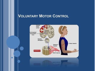
- Commands for voluntary movement originate in cortical association areas. The movements are planned in the cortex as well as in the basal ganglia and the lateral portions of the cerebellar hemispheres, as indicated by increased electrical activity before the movement. The basal ganglia and cerebellum both funnel information to the premotor and motor cortex by way of the thalamus. Motor commands from the motor cortex are relayed in large part via the corticospinal tracts to the spinal cord and the corresponding corticobulbar tracts to motor neurons in the brain stem. However, collaterals from these pathways and a few direct connections from the motor cortex end on brain stem nuclei, which also project to motor neurons in the brain stem and spinal cord. These pathways can also mediate voluntary movement. Movement sets up alterations in sensory input from the special senses and from muscles, tendons, joints, and the skin. This feedback information, which adjusts and smoothes movement, is relayed directly to the motor cortex and to the spinocerebellum. The spinocerebellum projects in turn to the brain stem. The main brain stem pathways that are concerned with posture and coordination are the rubrospinal, reticulospinal, tectospinal, and vestibulospinal tracts and corresponding projections to motor neurons in the brain stem. Ganong 21st
- Charles Scott Sherrington was born on November 27, 1857, at Islington, London. He was the son of James Norton Sherrington, of Caister, Great Yarmouth, who died when Sherrington was a young child. Sherrington's mother later married Dr. Caleb Rose of Ipswich, a good classical scholar and a noted archaeologist, whose interest in the English artists of the Norwich School no doubt gave Sherrington the interest in art that he retained throughout his life.In 1876 Sherrington began medical studies at St. Thomas's Hospital and in 1878 passed the primary examination of the Royal College of Surgeons, and a year later the primary examination for the Fellowship of that College. After a short stay at Edinburgh he went, in 1879, to Cambridge as a noncollegiate student studying physiology under Michael Foster, and in 1880 entered Gonville and Caius College there.In 1881 he attended a medical congress in London at which Sir Michael Foster discussed the work of Sir Charles Bell and others on the experimental study of the functions of nerves that was then being done in England and elsewhere in Europe. At this congress controversy arose about the effects of excisions of parts of the cortex of the brains of dogs and monkeys done by Ferrier and Goltz of Strasbourg. Subsequently, Sherrington worked on this problem in Cambridge with Langley, and with him published, in 1884, a paper on it. In this manner Sherrington was introduced to the neurological work to which he afterwards devoted his life.In 1883 Sherrington became Demonstrator of Anatomy at Cambridge under Professor Sir George Humphrey, and during the winter session of 1883-1884 at St. Thomas's Hospital he demonstrated histology.The years 1884 and 1885 were eventful ones for Sherrington, for during the winter of 1884-1885 he worked with Goltz at Strasbourg, in 1884 he obtained his M.R.C.S., and in 1885 a First Class in the Natural Sciences Tripos at Cambridge with distinction. During this year he published a paper of his own on the subject of Goltz's dogs. In 1885 he also took his M.B. degree at Cambridge and in 1886 his L.R.C.P.In 1885 Sherrington went, as a member of a Committee of the Association for Research in Medicine, to Spain to study an outbreak of cholera, and in 1886 he visited the Venice district also to investigate the same disease, the material then obtained being examined in Berlin under the supervision of Virchow, who later sent Sherrington to Robert Koch for a six weeks' course in technique. Sherrington stayed with Koch to do research in bacteriology for a year, and in 1887 he was appointed Lecturer in Systematic Physiology at St. Thomas's Hospital, London, and also was elected a Fellow of Gonville and Caius College, Cambridge. In 1891 he was appointed in succession to Sir Victor Horsley, Professor and Superintendent of the Brown Institute for Advanced Physiological and Pathological Research in London. In 1895 he became Professor of Physiology at the University of Liverpool.During his earlier years in Cambridge, Sherrington, influenced by W. H. Gaskell and by the Spanish neurologist, Ramón y Cajal, whom he had met during his visit to Spain, took up the study of the spinal cord. By 1891 his mind had turned to the problems of spinal reflexes, which were being much discussed at that time, and Sherrington published several papers on this subject and, during 1892-1894, others on the efferent nerve supply of muscles. Later, from 1893-1897, he studied the distribution of the segmented skin fields, and made the important discovery that about one-third of the nerve fibres in a nerve supplying a muscle are efferent, the remainder being motor.At Liverpool he returned to his earlier study of the problem of the innervation of antagonistic muscles and showed that reflex inhibition played an important part in this. In addition to this, however, he was studying the connection between the brain and the spinal cord by way of the pyramidal tract, and he was at this time visited by the American surgeon Harvey Cushing, then a young man, who stayed with him for eight months.In 1906 he published his well-known book: The Integrative Action of the Nervous System, being his Silliman Lectures held at Yale University the previous year, and in 1913 he was invited to become Waynfleet Professor of Physiology at Oxford, a post for which he had unsuccessfully applied in 1895, and here he remained until his retirement in 1936. Here he wrote, and published in 1919, his classic book entitled Mammalian Physiology: a Course of Practical Exercises, and here he regularly taught the students for whom this book was written.In physique Sherrington was a well-built, but not very tall man with a strong constitution which enabled him to carry out prolonged researches.During the First World War, as Chairman of the Industrial Fatigue Board, he worked for a time in a shell factory at Birmingham, and the daily shift of 13 hours, with a Sunday shift of 9 hours, did not, at the age of 57, tire him. From his early years he was short-sighted, but he often worked without spectacles.The predominant notes of his character as a man were his humility and friendliness and the generosity with which he gave to others his advice and valuable time. An interesting feature of him is that he published, in 1925, a book of verse entitled The Assaying of Brabantius and other Verse, which caused one reviewer to hope that «Miss Sherrington» would publish more verse. He was also sensitive to the music of prose, and this and the poet in him, but also the biologist and philosopher, were evident in his Rede Lecture at Cambridge in 1933 on The Brain and its Mechanism, in which he denied our scientific right to join mental with physiological experience.The philosopher in him ultimately found expression in his great book, Man on his Nature, which was the published title of the Gifford Lectures for 1937-1938, which Sherrington gave. As is well known, this book, published in 1940, centres round the life and views of the 16th century French physician Jean Fernel and round Sherrington's own views. In 1946 Sherrington published another volume entitled The Endeavour of Jean Fernel.Sherrington was elected a Fellow of the Royal Society of London in 1893, where he gave the Croonian Lecture in 1897, and was awarded the Royal Medal in 1905 and the Copley Medal in 1927. In 1922 the Knight Grand Cross of the Order of the British Empire and in 1924 the Order of Merit were conferred upon him. He held honorary doctorates of the Universities of Oxford, London, Sheffield, Birmingham, Manchester, Liverpool, Wales, Edinburgh, Glasgow, Paris, Strasbourg, Louvain, Uppsala, Lyons, Budapest, Athens, Brussels, Berne, Toronto, Montreal, and Harvard.As a boy and a young man Sherrington was a notable athlete both at Queen Elizabeth's School, Ipswich, where he went in 1871, and later at Gonville and Caius College, Cambridge, for which College he rowed and played rugby football; he was also a pioneer of winter sports at Grindelwald.In 1892 Sherrington married Ethel Mary, daughter of John Ely Wright, of Preston Manor, Suffolk. After some years of frail health, during which, however, he remained mentally very alert, he died suddenly of heart failure at Eastbourne in 1952.From Nobel Lectures, Physiology or Medicine 1922-1941, Elsevier Publishing Company, Amsterdam, 1965 This autobiography/biography was written at the time of the award and first published in the book series Les Prix Nobel. It was later edited and republished in Nobel Lectures. To cite this document, always state the source as shown above.
- Figure 1. Motor maps of the gorilla cortexA, scale drawing of the left hemisphere of one of Leyton & Sherrington’s experiments on a gorilla (gorilla 1). The numbers and letters encode a wide range of different primary movements evoked by faradic stimulation. Eye movements (372–388) were generally evoked from an area further rostral from the motor cortex. Owing to lack of space, many motor effects were not plotted. B, simplified ‘map’ showing ‘responses grouped diagrammatically’, as the authors put it.The paper by Leyton & Sherrington (1917) has had a profound and lasting impact for ninety or so years since its publication.It is notable in many different ways. Leyton & Sherrington (1917) provided the first detailed proof that there was indeed localization of function within the cerebral cortex. The durability of their report probably owes most to the fact that Leyton & Sherrington (1917)were the first to establish precisely the true extent of the motor area, and to provide the first detailed ‘motor map’ of the primate motor cortex. In addition, they showed that surgical extirpation of the cortical tissue that, when stimulated, gave rise to movement of a particular body part, resulted in a widespread weakness and loss of use of that same body part.There was, however, substantial recovery in the weeks that followed, recovery that was not lost on lesioning either the adjacenttissue in the same hemisphere or the equivalent cortical area of the opposite hemisphere. Finally, they were able to trace the course of the degenerating corticofugal and corticospinalfibres. They observed widespreaddegeneration in the cervical cord after a lesion of the hand and arm cortical area and noted that after such a lesion in the chimpanzee (p. 185), ‘the whole of the cross-area of ventral horn has scattered through it many degenerating fibres. . .’, which I think is the first report of the direct cortico-motoneuronal projection, a projection whose existence was confirmed physiologically by Bernhard & Bohm (1954), and one that appears to be unique to primates (Porter & Lemon, 1993).The mysterious chronology of the paperThis classic paper was very long in gestation. After the early accounts appeared in 1901 and in 1903, further details of their experiments were given in Sherrington’s Silliman Lectures (published as The Integrative Action of the Nervous System in 1906). However, the full paper was not submitted to the Quarterly Journal of Physiology until October 1916. There is a much-repeated story, probably first promulgated by John Fulton in his 1952 biography, that Sherrington delayed publication until after the death of Victor Horsley, with whom he had a very heated row. It was believed that Sherrington wanted to avoid a repeat of the disagreement in 1894 over primacy of research on pyramidal tract degeneration. Horsley, the famous neurosurgeon and co-inventor the Horsley–Clark stereotaxic method, died in July 1916 from fatal sunstroke in Mesopotamia (where he was stationed during the War).However, this account has been seriously questioned (Vilenskyet al. 2003) and there must have been other reasons for the delay; these might have included the expense and difficulty of obtaining the additional animals needed for the study, and the facts that Leyton left Liverpool not long after the 1903 paper was published, to take up a Chair in Leeds, and that the the work was undoubtedly interrupted by the outbreak of the GreatWar. Why was the study carried out?The decision to investigate the excitable cortex of anthropoid apes probably arose because the findings of motor responses evoked by cortical stimulation in animals such as dogs, rabbits and monkeys were challenged as of being of little relevance to humans (where have we heard that before?).Sherrington considered that experiments in ‘higher apes’, being closer to humans than any other species, might provide the answer. The initial experiment (in a chimpanzee) having provided a positive result, they decided to complete a full series of studies, and this paper summarizes agreat many different experiments in a total of 22 chimpanzees, three orang-utans three gorillas and other animals, all investigated under deep anaesthesia with chloroform and ether. One of them involved an experiment in which the same induction coil was used to stimulate, simultaneously, the cortex of a cat, amonkey and a chimpanzee, and this demonstrated unequivocally that the threshold for evoking motor response was very similar in all three species.Rereading this paper, one is struck by it having all the hallmarks of a really modern piece of neuroscience: a multidisciplinary mixture of state-of-the art electrophysiology, behavioural analysis and detailed neuroanatomy, all linked together to test a strong central hypothesis: ‘the motor cortex can be regarded as a synthetic organ for compounding and recompounding in varied ways movements of varied kinds. . . . ’ The authors considered that the movements they evoked were like words in a basic vocabulary of voluntary movement, words that could be combined in myriad ways to make sentences: purposeful movements involving co-ordinated activation of many muscles in a precise spatio-temporal sequence.There are a great many key points of lasting value in this paper, some of which are discussed below.What can be deduced about brain motor function from electrical stimulation?Sherrington was very clear that the nature of the motor responses was in part due to the type of stimulation used. Leyton & Sherrington (1917) employed ‘faradic’ stimulation (alternating current provided by an induction coil), were in favour of unipolar stimulation (using a large andremote indifferent electrode) and insisted on studying responses evoked by a brief period of stimulation at the lowest current intensities. In this way they reduced the possibility that the responses were due to physical spread of the induced current through the tissue. Stronger and longer lasting stimulation also raises the possibility that the responses are due to physiological spread of synaptic activity generated locally at the stimulation site but permeating through both short- and long-distance connections to recruit even quite remote cortical and subcortical structures.As Phillips & Porter (1977), in their elegant discussion of Leyton & Sherrington (1917) put it: ‘Thus faradic stimulation is disqualified, equally with “galvanic” as tool for evoking natural function but it can be used as ‘a tool for mapping the outputs that are available for selection by the intracortical activities that it cannot itself evoke.’ Previous investigators, including Ferrier and Beevor & Horsley, had used relatively large currents and thus been able to evoke motor responses from the primary sensory as well as from the primary motor cortex.Leyton & Sherrington (1917) refined the approach by carefully controlling current intensity, and demonstrated that the motor area did not extend posterior to the central sulcus. They were careful to point out that even a motor zone could influence sensation, a prescient comment given thatmany of the descending corticospinalfibres are involved in control of afferent input (see Lemon & Griffiths, 2005; Lemon, 2008). The quality of the movements evoked by electrical stimulationLeyton & Sherrington (1917), describing the ‘fractional quality’ of the motor responses evoked, write that ‘the individual movements, elicited by somewhat minutely localised stimulations, are, broadly speaking, fractional, in the sense that each, though co-ordinately executed, forms,so to say, but a unitary part of some more complex act, that would, to attain its purpose, involve combination of that unitary movement with others to make up a useful whole.’ They were also keen to emphasize the complexity of some of the responses, which often comprised a‘1st movement’ (e.g. thumb extension) with accompanying 2nd (e.g. index finger extension), 3rd (wrist extension) or even 4th movements. They also stressed the ‘functional instability of cortical motor points’; the responses were not fixed, but showed properties such as facilitation,reversal and deviation, especially when a given cortical point was retested after the adjacent area had been stimulated. Finally, they recognized that movements of the eyes were likely to be controlled differently from movements of the rest of the body, since the area giving rise to eye movements was located rostral to and distinct from the main motor cortex (see Fig. 1), in what we now call the frontal eye fields.How many different kinds of movements can be evoked and how are they ‘mapped’ across the cortical surface?Leyton & Sherrington (1917) were clearly struck by the rich variety of different movements that could be evoked from the motor area. They reported more than 400 different ‘1st movements’ in a table that takes up 7 pages (would the current editors of Experimental Physiology allowsuch indulgence?). Each movement was carefully inscribed on a scale drawing of the exposed cortex (Fig. 1A). They were careful to note that the map is not fixed in nature but that responses showed instability in time (see above), and that even the boundaries between different body ‘areas’(e.g. between the face and arm area) were labile. Movement of a given body part (e.g. the thumb) could be evoked from an extensive region of motor cortex, many square millimetres in area, and this representation overlapped significantly with that of other, adjacent body parts (e.g.thumb with index finger, wrist with elbow etc.).Similar findings were published 20 years later by Penfield & Boldrey (1937) for the human brain. Somehow the complexity of the experimental findings was digested into a greatly simplified ‘homunculus’ published after another long delay by Penfield & Rasmussen (1952). This cartoon suggested a strictly somatotopic representation within the primary motor area, M1, which bore little relation to the original experimentaldata. However, the homunculus provided an appealing notion of how voluntary motor control might be represented in the cortex, and has since been reproduced in every relevant textbook. It often appears alongside Sherrington’s (1906) map of the gorilla cortex, which bears some resemblance to figure 10 of Leyton & Sherrington (1917), a figure which is not referred to at all in the text (more editorial indulgence?). Thisfigure, in which the responses were grouped diagrammatically (sic), is reproduced here as Fig. 1B. Its simplicity conceals the wealth of data that each of their mapping experiments yielded (Fig. 1A). While the orderly mapping of different areas of the body is well substantiated, evidence for a strict somatotopy within each area (e.g. fixed non-overlapping ordering in the lateral to medial direction of thumb, then index, middle, ring and little finger movement) has been hard to find, and modern investigators (see Lemon, 1988; Schieber&Hibbard, 1993;Rathelot&Strick, 2006) have turned away from this concept to one much closer to the multiple and overlapping representation of movement described by Leyton & Sherrington (1917). This type of representation is well suited for the many and varied combination of ‘fractional’ movements into useful actions or, as they put it: ‘. . .that from movements of locally restricted parts, e.g. movements of a finger or of a limb joint (movements themselves discrete andindividually separable in the motor cortex), the up building of larger combinations varied in character and serviceable for purposes of different and varied kind, prehensile, defensive, locomotor, mimetic, masticatory, deglutitional, orientational etc. is one of the main offices performed by themotor cortex.’ We are now learning how individual motor cortex neurons combine and recombine their actions (Jackson et al. 2003).Why is the cortex involved in motor control?Many investigators have raised the issue of why the basic building blocks of all types of movements are represented at all in the neocortex, one of brain’s ‘higher’ centres; surely these could be managed by subcorticalcentres, including the spinal cord? Leytonand Sherrington recognized, ofcourse, that the spinal cord could generate many types of movements. However, ‘It would seem that in order to preserve the possibility of being interchangeably compounded in a variety of ways, successive or simultaneous, these movements must lie, as more or less discrete and separableelements, within the grasp of the organ whichhas the varied compounding of them.’ Beautifully put! They also recognized that the acquisition of new combinations would make an important contribution to the learning of motor skills: ‘The acquirement of skilled movements, though certainly a process involving far wider areas of the cortex than the excitable zone itself, may be presumed to find in the motor cortex an organ whose synthetic properties are part of the physiological basis which renders that acquirement possible.’ConclusionLeyton and Sherrington’s seminal paper provided a key set of observations that have guided and inspired research ever since. They were clearly impressed by the degree of recovery after large cortical lesions and recognized that some kind of compensatory changes must be at work.They also recognized the importance of finding the neural substrate of recovery. It was only much later that it became clear that plasticity within the cortex is itself an active, use-dependent process, and that this process can be harnessed to improve recovery of function. Ultimately, Leyton and Sherrington’s work opened up an exciting new chapter in the physiology of the cortex, which ultimately is having far-reaching consequences for our understanding of the brain and for the rehabilitation of patients with brain injury.ReferencesBernhard CG & Bohm E (1954). Cortical representation and functional significance of the corticomotoneuronal system. AMA Arch Neurol Psychiatry 72, 473–502. Granit R (1966). Charles Scott Sherrington: An Appraisal. Thomas Nelson and Sons, London. Gr¨unbaum ASF & Sherrington CS (1901).Observations on the physiology of the cerebral cortex of some of the higher apes (preliminary communication). Proc Roy Soc Lond69, 206–209. Gr¨unbaum ASF & Sherrington CS (1903). Observations on the physiology of the cerebral cortex of the anthropoid apes. Proc Roy Soc Lond72, 152–155. Jackson A, Gee VJ, Baker SN & Lemon RN (2003). Synchrony between neurons with similar muscle fields in monkey motor cortex. Neuron 38, 115–125.Lemon RN (1988). The output map of the primate motor cortex. Trends Neurosci11, 501–506.Lemon RN (2008). Descending pathways in motor control. Ann Rev Neurosci; DOI: 10.1146/annurev.neuro.31.060407.125547.Lemon RN & Griffiths J (2005). Comparing the function of the corticospinal system in different species: organizational differences for motor specialization? Muscle Nerve 32, 261–279.Leyton SSF & Sherrington CS (1917). Observations on the excitable cortex of the chimpanzee, orang-utan and gorilla. Q J Exp Physiol11, 135–222.PenfieldW&Boldrey E (1937). Somatic motor and sensory representation in the cerebral cortex of man as studied by electrical stimulation. Brain 60, 389–443.PenfieldW&Rasmussen T (1952). The Cerebral Cortex of Man. Macmillan, New York. Phillips CG & Porter R (1977). Corticospinal Neurones. Their Role in Movement. Academic Press, London. Porter R & Lemon RN(1993). Corticospinal Function and Voluntary Movement. Clarendon Press, Oxford.Rathelot J-A & Strick PL (2006). Muscle representation in the macaque motor cortex: an anatomical perspective. Proc NatlAcadSci USA 103, 8257–8262.Schieber MH & Hibbard LS (1993). How somatotopic is the motor cortex hand area? Science 261, 489–492.Sherrington CS (1906). The Integrative Action of the Nervous System. Cambridge University Press.Stewart MJ (1922). Albert Sidney FrankauLeyton (obituary). J PatholBacteriol25, 109–112.Vilensky JA, Stone JL & Gilman S (2003). Feud and fable: the Sherrington–Horsley polemic and the delayed publication. J HistNeurosci12, 368–375.
- The introduction in the 1960s of intracorticalmicrostimulation (a more refined method of cortical activation) allowed a more detailed understanding of motormaps. Microstimulation entails the delivery of electrical currents an order of magnitude smaller than those used by Sherrington and Penfield. By passing the current through the sharpened tip of a metal microelectrode inserted into the cortex, the upper motor neurons in layer V that project to lower motor neuroncircuitry can be stimulated focally. Although intracortical stimulation generally confirmed Penfield's spatial map in the motor cortex, it also showed that the finer organization of the map is rather different than most neuroscientists imagined. For example, when microstimulation was combined with recordings of muscle electrical activity, even the smallest currents capable of eliciting a response initiated the excitation of several muscles (and the simultaneous inhibition of others), suggesting that organized movements rather than individual muscles are represented in the map (see Box B). Furthermore, within major subdivisions of the map (e.g., arm, forearm, or finger regions), a particular movement could be elicited by stimulation of widely separated sites, indicating that nearby regions are linked by local circuits to organize specific movements. This interpretation has been supported by the observation that the regions responsible for initiating particular movements overlap substantially.What Do Motor Maps Represent?Electrical stimulation studies carried out by the neurosurgeon Wilder Penfield and his colleagues in human patients (and by Clinton Woolsey and his colleagues in experimental animals) clearly demonstrated a systematic map of the body's musculature in the primary motor cortex (see text). The fine structure of this map, however, has been a continuing source of controversy. Is the map in the motor cortex a “piano keyboard” for the control of individual muscles, or is it a map of movements, in which specific sites control multiple muscle groups that contribute to the generation of particular actions? Initial experiments implied that the map in the motor cortex is a fine-scale representation of individual muscles. Thus, stimulation of small regions of the map activated single muscles, suggesting that vertical columns of cells in the motor cortex were responsible for controlling the actions of particular muscles, much as columns in the somatic sensory map are thought to analyze particular types of stimulus information (see Chapter 9).More recent studies using anatomical and physiological techniques, however, have shown that the map in the motor cortex is far more complex than a columnar representation of particular muscles. Individual pyramidal tract axons are now known to terminate on sets of spinal motor neurons that innervate different muscles. This relationship is evident even for neurons in the hand representation of the motor cortex, the region that controls the most discrete, fractionated movements. Furthermore, cortical microstimulation experiments have shown that a single muscle is represented multiple times over a wide region of the motor cortex (about 2–3 mm in primates) in a complex, mosaic fashion. It seems likely that horizontal connections within the motor cortex create ensembles of neurons that coordinate the pattern of firing in the population of ventral horn cells that ultimately generate a given movement.Thus, while the somatotopic maps in the motor cortex generated by early studies are correct in their overall topography, the fine structure of the map is far more intricate. Unraveling these details of motor maps still holds the key to understanding how patterns of activity in the motor cortex generate a given movement.Figure 17.8. The corticospinal tract. Neurons in the motor cortex give rise to axons that travel through the internal capsule and coalesce on the ventral surface of the midbrain, within the cerebral peduncle. These axons continue through the pons and come to lie on the ventral surface of the medulla, giving rise to the pyramids. Most of these pyramidal fibers cross in the caudal part of the medulla to form the lateral corticospinal tract in the spinal cord. Those axons that do not cross (not illustrated) descend on the same side and form the ventral corticospinal tract (see Figure 17.6). The axons that terminate in the reticular formation of the pons and medulla comprise components of the corticobulbar tract. Transmission of Signals from the Motor Cortex to the MusclesMotor signals are transmitted directly from the cortex to the spinal cord through the corticospinal tract and indirectly through multiple accessory pathways that involve the basal ganglia, cerebellum, and various nuclei of the brain stem. In general, the direct pathways are concerned more with discrete and detailed movements, especially of the distal segments of the limbs, particularly the hands and fingers.Corticospinal (Pyramidal) TractThe most important output pathway from the motor cortex is the corticospinal tract, also called the pyramidal tract, shown in Figure 55–4.The corticospinal tract originates about 30 per cent from the primary motor cortex, 30 per cent from the premotor and supplementary motor areas, and 40 per cent from the somatosensory areas posterior to the central sulcus. After leaving the cortex, it passes through the posterior limb of the internal capsule (between the caudate nucleus and the putamen of the basal ganglia) and then downward through the brain stem, forming the pyramids of the medulla. The majority of the pyramidal fibers then cross in the lower medulla to the opposite side and descend into the lateral corticospinal tracts of the cord, finally terminating principally on the interneurons in the intermediate regions of the cord gray matter; a few terminate on sensory relay neurons in the dorsal horn, and a very few terminate directly on the anterior motor neurons that cause muscle contraction.A few of the fibers do not cross to the opposite side in the medulla but pass ipsilaterally down the cord in the ventral corticospinal tracts. Many if not most of these fibers eventually cross to the opposite side of the cord either in the neck or in the upper thoracic region.These fibers may be concerned with control of bilateral postural movements by the supplementary motor cortex.The most impressive fibers in the pyramidal tract are a population of large myelinated fibers with a mean diameter of 16 micrometers. These fibers originate from giant pyramidal cells, called Betz cells, that are found only in the primary motor cortex. The Betz cells are about 60 micrometers in diameter, and their fibers transmit nerve impulses to the spinal cord at a velocity of about 70 m/sec, the most rapid rate of transmission of any signals from the brain to the cord. There are about 34,000 of these large Betz cell fibers in each corticospinal tract. The total number of fibers in eachcorticospinal tract is more than 1 million, so these large fibers represent only 3 per cent of the total. The other 97 per cent are mainly fibers smaller than 4 micrometers in diameter that conduct background tonic signals to the motor areas of the cord.
- Corticospinal system : Role in MovementThe corticospinal and corticobulbar system is the primary pathway for the initiation of skilled voluntary movement. This does not mean that movement—even skilled movement—is impossible without it. Nonmammalian vertebrates have essentially no corticospinal and corticobulbar system, but they move with great agility. Cats and dogs stand, walk, run, and even eat if food is presented to them after complete destruction of this system. Only in primates are relatively marked deficits produced. Careful section of the pyramids producing highly selective destruction of the lateral corticospinal tract in laboratory primates produces prompt and sustained loss of the ability to grasp small objects between two fingers and to make isolated movements of the wrists. However, the animal can still use the hand in a gross fashion and can stand and walk. These deficits are consistent with loss of control of the distal musculature of the limbs, which is concerned with fine skilled movements. On the other hand, lesions of the ventral corticospinal tract produce axial muscle deficits that cause difficulty with balance, walking, and climbing. Guyton
- CEREBRAL CORTICAL CONTROL OF MOVEMENT*HIROSHI ASANUMAThe Rockefeller UniversityNew York, N.Y.Introduction:The purpose of the series of experiments which will be described in this article was to increase our understanding of how the motor cortexcontrols the movement. It has been a continuing controversy whether the motor cortex controls the contraction of single muscles or rathermuscle groups. Hughlings Jackson and his followers, based on the fact that a patient can recover the use of a limb after destruction of thatlimb area previously defined by stimulation, favored the idea of a widespread overlapping of the representation of muscle groups (cf. Ruth1965). This view was strongly supported by Sherrington’s early experiences (Hering and Sherrington, 1897, Sherrington, 1906) in whichstimulation on the surface of the cortex could not produce solitary contraction of a muscle and that the combination of the muscles activatedby cortical stimulation was always in a reciprocal fashion, i. e., “cortical stimulation never produced simultaneous contraction of the trueantagonists” (Sherrington, 1906). The latter evidence led him to think that the motor cortex controls the coordinated contraction of a groupof muscles.The interpretation that the motor cortex “thinks” in terms of movements, not muscles was so persuasive that more than half a century haspassed under the dominance of this theory. In 1947, however, Chang, Ruth and Ward demonstrated evidence that the motor cortex may be able to think in terms of muscles. They stimulated the surface of the monkey cortex, with maximum possible caution, and were able to elicit solitary contraction, although occasionally, of a single muscle. Succeeding experiments by Bernhard and Bohm (1954) demonstrated that a single shock stimulation of the motor cortex activated the fast conducting corticospinal neurons of the pyramidal tract which in turn elicited a monosynaptic response in a forelimb nerve but not in the rest of the nerves. They also showed that the cortical field which produced the monosynaptic response in a certain nerve was more restricted than the fields from which other (polysynaptic) activities in the motoneurons of the same nerve could be evoked. The study about the localization of cortical motor function was further advanced by Landgren, Phillips and Porter (1962). They stimulated the surface of the motor cortex while recording intracellularly from motoneurons and demonstrated the existante of a cluster of neurons which projected monosynaptically to motor neurons and which was confined within a narrow focus of the cortex having a cross section of the order of lmm2. Thus progress in the stimulation experiments of the motor cortex provided evidence that the contraction of individual muscles is represented in small areas of the motor cortex in a mosaic fashion. This conclusion, however, does not resolve the question of “whether the motor cortex thinks in terms of movements or muscles” because as has been pointed out by Bernhard and Bohm (1954) in their stimulation experiments, a given cortical stimulus which facilitated a monosynaptic reflex, at the same time inhibited the monosynaptic reflex of the antagonist in the absence of peripheral influences.In the meantime, another line of investigation of the motor cortex developed depending on the rapid progress of the electrical techniqueswhich enabled recording of the electrical signals associated with the activities of neurons in the central nervous system. In 1953, Malis,Pribram and Kruger showed that the motor cortex received afferent inputs from the periphery in the absence of the sensory cortex. This observation raised the question of the functional significance of these afferent inputs converging onto the motor cortex.The specific purpose of a series of experiments performed in my laboratory has been to further elucidate the details of the localizationof the cortical motor function and then to relate the motor function to the afferent inputs, i. e., to study the input-output relationships. Theterm “motor cortex” is used instead of “motorsensory cortex” because a recent histological study (Hassler and Muhs-Clement, 1964) has shownthat the cat’s pericruciate cortex corresponds to the motor cortex of the primates.
- Cellular Organization 1. The neocortex (most of the cerebral cortex) consists of six layers of histologically and functionally distinct cells2. Each region of M-1 (and the remainder of the neocortex) is organized as units of interconnected columns of several hundred neurons arranged perpendicular to the cortical surface and including all six cortical layers3. Stimulation of a given column may activate a single muscle ; more usually stimulation of a column activates several muscles to produce a coordinated movementNote: this is sometimes expressed as "The motor cortex thinks in terms of movements, not muscles"4. Groups of columns control groups of alpha motoneurons to determine the force and direction and velocity of movement 5. Descending axons forming the cortical output from M-1 (and from the cortex in general) arise from the pyramidal cells in cortical layer 56. Pyramidal cells in layers 2 and 3 send axons to other regions of the ipsilateral cortex (layer 2) and to corresponding areas of the contralateral cortex via the corpus callosum (layer 3)Note: Axial regions of the body are well represented in corpus callosal axons connecting corresponding contralateral regions of the motor cortex but distal regions are less well represented.
- Figure 12-3. Medial (above) and lateral (below) views of the human cerebral cortex, showing the motor cortex (Brodmann's area 4) and other areas concerned with control of voluntary movement, along with the numbers assigned to the regions by Brodmann. (Reproduced, with permission, from Kandel ER, Schwartz JH, Jessell TM [editors]: Principles of Neural Science, 4th ed. McGraw-Hill, 2000.) By means of stimulation experiments in patients undergoing craniotomy under local anesthesia, it has been possible to outline most of the motor projections from the motor cortex. These have been confirmed in unanesthetizedunoperated humans by PET scanning and fMRI (Figure 12-4). The various parts of the body are represented in the precentralgyrus, with the feet at the top of the gyrus and the face at the bottom (Figure 12-5). The facial area is represented bilaterally, but the rest of the representation is unilateral, the cortical motor area controlling the musculature on the opposite side of the body. The cortical representation of each body part is proportionate in size to the skill with which the part is used in fine, voluntary movement. The areas involved in speech and hand movements are especially large in the cortex; use of the pharynx, lips, and tongue to form words and of the fingers and apposable thumbs to manipulate the environment are activities in which humans are especially skilled. The conditions under which the human stimulation studies were performed precluded stimulation of the banks of the sulci and other inaccessible areas. Meticulous study has shown that in monkeys, there is a regular representation of the body, with the axial musculature and the proximal portions of the limbs represented along the anterior edge of the precentralgyrus and the distal part of the limbs along the posterior edge. Another feature of M1 is the presence of considerable overlap in the muscles that are innervated, as well as innervation of synergic muscles separated by considerable distances. There has been debate about whether individual muscles or movements are represented in M1, and the most recent evidence indicates that both are represented. The cells in the cortical motor areas are arranged in columns. The cells in each column receive fairly extensive sensory input from the peripheral area in which they produce movement, providing the basis for feedback control of movement. Some of this input may be direct, and some is relayed from somatic sensory area I in the postcentralgyrus. Cerebral dominance, which is discussed in detail in Chapter 16, also affects the motor cortex in humans. Moving the fingers of the left hand is associated mainly with activation of the right motor cortex and vice versa, as measured by imaging techniques (see Chapter 32). However, moving the fingers of the left hand also activates the left motor cortex, particularly in individuals who are right-handed. This correlates with the fact that lesions of the left motor cortex cause motor dysfunction in the left hand as well as the right hand, whereas lesions of the right motor cortex have little effect on the right hand. Supplementary Motor AreaFor the most part, the supplementary motor area projects to the motor cortex. It appears to be involved primarily in programming motor sequences. Lesions of this area in monkeys produce awkwardness in performing complex activities and difficulty with bimanual coordination. When human subjects count to themselves without speaking, the motor cortex is quiescent, but when they speak the numbers aloud as they count, blood flow increases in the motor cortex and the supplementary motor area. Thus, the supplementary motor area as well as the motor cortex is involved in voluntary movement when the movements being performed are complex and involve planning. Blood flow increases whether or not a planned movement is carried out. The increase occurs whether the movement is performed by the contralateral or the ipsilateral hand. Premotor CortexThe premotor cortex projects to the brain stem areas concerned with postural control and to the motor cortex as well as providing part of the corticospinal and corticobulbar output. Its function is still incompletely understood, but it may be concerned with setting posture at the start of a planned movement and with getting the individual ready to perform. Posterior Parietal CortexIn addition to providing fibers that run in the corticospinal and corticobulbar tracts, the somatic sensory area and related portions of the posterior parietal lobe project to the premotor area. Lesions of the somatic sensory area cause defects in motor performance that are characterized by inability to execute learned sequences of movements such as eating with a knife and fork. Some of the neurons in area 5 (Figure 12-3) are concerned with aiming the hands toward an object and manipulating it, whereas some of the neurons in area 7 are concerned with hand-eye coordination. Role in MovementThe corticospinal and corticobulbar system is the primary pathway for the initiation of skilled voluntary movement. This does not mean that movement—even skilled movement—is impossible without it. Nonmammalian vertebrates have essentially no corticospinal and corticobulbar system, but they move with great agility. Cats and dogs stand, walk, run, and even eat if food is presented to them after complete destruction of this system. Only in primates are relatively marked deficits produced. Careful section of the pyramids producing highly selective destruction of the lateral corticospinal tract in laboratory primates produces prompt and sustained loss of the ability to grasp small objects between two fingers and to make isolated movements of the wrists. However, the animal can still use the hand in a gross fashion and can stand and walk. These deficits are consistent with loss of control of the distal musculature of the limbs, which is concerned with fine skilled movements. On the other hand, lesions of the ventral corticospinal tract produce axial muscle deficits that cause difficulty with balance, walking, and climbing. Ganong 21stPrimary Motor CortexNote that more than one half of the entire primary motor cortex is concerned with controlling the muscles of the hands and the muscles of speech. Point stimulation in these hand and speech motor areas on rare occasion causes contraction of a singlemuscle; most often, stimulation contracts a group of muscles instead. To express this in another way, excitation of a single motor cortex neuron usually excites a specific movement rather than one specific muscle.To do this, it excites a “pattern” of separate muscles, each of which contributes its own direction and strength of muscle movement.Premotor AreaThe premotor area, also shown in Figure 55–1, lies 1 to 3 centimeters anterior to the primary motor cortex, extending inferiorly into the sylvian fissure and superiorly into the longitudinal fissure, where it abuts the supplementary motor area, which has functions similar to those of the premotor area. The topographical organization of the premotor cortex is roughly the same as that of the primary motor cortex, with themouth and face areas located most laterally; as one moves upward, the hand, arm, trunk, and leg areas are encountered.Nerve signals generated in the premotor area cause much more complex “patterns” of movement than the discrete patterns generated in the primary motor cortex. For instance, the pattern may be to position the shoulders and arms so that the hands are properly oriented to perform specific tasks. To achieve these results, the most anterior part of the premotor area first develops a “motor image” of the total musclemovement that is to be performed.Then, in the posterior premotor cortex, this image excites each successive pattern of muscle activity required to achieve the image.This posterior part of the premotor cortex sends its signals either directly to the primary motor cortex to excite specific muscles or, often, by way of the basal ganglia and thalamus back to the primary motor cortex. Thus, the premotor cortex, basal ganglia, thalamus,and primary motor cortex constitute a complex overall system for the control of complex patterns of coordinated muscle activity.Supplementary Motor AreaThe supplementary motor area has yet another topographical organization for the control of motor function. It lies mainly in the longitudinal fissure but extends a few centimeters onto the superior frontal cortex. Contractions elicited by stimulating this area are often bilateral rather than unilateral. For instance, stimulation frequently leads to bilateral grasping movements of both hands simultaneously; thesemovements are perhaps rudiments of the hand functions required for climbing. In general, this area functions in concert with the premotor area to provide body-wide attitudinal movements, fixation movements of the different segments of the body, positional movements of the head and eyes, and so forth, as background for the finer motor control of the arms and hands by the premotor area and primary motor cortex.
- Some Specialized Areas of Motor Control Found in the Human Motor CortexNeurosurgeons have found a few highly specialized motor regions of the human cerebral cortex (shown in Figure 55–3) that control specific motor functions. These regions have been localized either by electrical stimulation or by noting the loss of motor function when destructive lesions occur in specific cortical areas. Some of the more important regions are the following.Broca’s Area and Speech. Figure 55–3 shows a premotorarea labeled “word formation” lying immediately anterior to the primary motor cortex and immediately above the sylvian fissure. This region is called Broca’s area. Damage to it does not prevent a person from vocalizing, but it does make it impossible for the person to speak whole words rather than uncoordinated utterances or an occasional simple word such as “no” or “yes.” A closely associated cortical area also causes appropriate respiratory function, so that respiratory activation of the vocal cords can occur simultaneously with the movements of the mouth and tongueduring speech. Thus, the premotor neuronal activities related to speech are highly complex.“Voluntary” Eye Movement Field. In the premotor areaimmediately above Broca’s area is a locus for controlling voluntary eye movements. Damage to this area prevents a person from voluntarily moving the eyes toward different objects. Instead, the eyes tend to lock involuntarily onto specific objects, an effect controlled by signals from the occipital visual cortex, as explained in Chapter 51. This frontal area also controls eyelid movements such as blinking.Head Rotation Area. Slightly higher in the motor associationarea, electrical stimulation elicits head rotation. This area is closely associated with the eye movement field; it directs the head toward different objects.Area for Hand Skills. In the premotor area immediatelyanterior to the primary motor cortex for the hands and fingers is a region neurosurgeons have identified as important for “hand skills.” That is, when tumors or other lesions cause destruction in this area, hand movements become uncoordinated and nonpurposeful, a condition called motor apraxia.