18. Excitatory and Inhibitory signals Excitatory and inhibitory currents have competitive effects in a single nerve cell. (Adapted from Eckert et al., 1988.)
Overview Perhaps the major reason that neuroscience remains such an exciting field is the wealth of unanswered questions about the fundamental structure and functions of the human brain. To understand this remarkable organ (and the rest of the nervous system), the myriad cell types that constitute the nervous system must be identified, their interconnections traced, and the physiological role of the resulting circuits defined. Adding to these several challenges is the fact that a specialized anatomical vocabulary has arisen to describe the structure of the nervous system, as well as a specialized set of physiological terms to describe its functions. In light of these conceptual and semantic difficulties, comprehending the brain and the rest of the nervous system is greatly facilitated by a general picture of the organization of the nervous system, and by a review of the basic terms and anatomical conventions used in discussing its structure and function. Summary Nerve cells generate electrical signals to convey information over substantial distances and to transmit it to other cells by means of synaptic connections. The action potential—the signal that conveys information along nerve cell axons—ultimately depends on the resting electrical potential across the neuronal membrane. A resting potential occurs because nerve cell membranes are permeable to one or more ion species subject to an electrochemical gradient. More specifically, a negative membrane potential at rest results from a net efflux of K+ across neuronal membranes that are predominantly permeable to K+. In contrast, an action potential occurs when a transient rise in Na+ permeability allows a net flow of Na+ in the opposite direction across the membrane that is now predominantly permeable to Na+. The brief rise in membrane Na+ permeability is followed by a secondary, transient rise in membrane K+ permeability that repolarizes the neuronal membrane and produces a brief undershoot of the action potential. As a result of these processes, the membrane is depolarized in an all-or-none fashion during an action potential. When these active permeability changes subside, the membrane potential returns to its resting level because of the high resting membrane permeability to K+.
The Remarkable Giant Nerve Cells of Squid Many of the initial insights into how ion concentration gradients and changes in membrane permeability produce electrical signals came from experiments performed on the extraordinarily large nerve cells of the squid. The axons of these nerve cells can be up to 1 mm in diameter—100 to 1000 times larger than mammalian axons. Squid axons are large enough to allow experiments that would be impossible on most other nerve cells. For example, it is not difficult to insert simple wire electrodes inside these giant axons and make reliable electrical measurements. The relative ease of this approach yielded the first intracellular recordings of action potentials from nerve cells and, as will be discussed in the next chapter, the first experimental measurements of the ionic currents that produce action potentials. It also is practical to extrude the cytoplasm from giant axons and measure its ionic composition (see Table 2.1 ). In addition, some giant nerve cells form synaptic contacts with other giant nerve cells, producing very large synapses that have been extraordinarily valuable in understanding the fundamental mechanisms of synaptic transmission (see Chapter 5 ). Giant neurons evidently evolved in squid because they enhanced survival. These neurons participate in a simple neural circuit that activates the contraction of the mantle muscle, producing a jet propulsion effect that allows the squid to move away from predators at a remarkably fast speed. As discussed in Chapter 3 , larger axonal diameter allows faster conduction of action potentials. Thus, squid presumably have these huge nerve cells to escape more successfully from their numerous enemies. Today—more than 60 years after their discovery by John Z. Young at University College, London—the giant nerve cells of squid remain useful experimental systems for probing basic neuronal functions. (A) Diagram of a squid, showing the location of its giant nerve cells. Different colors indicate the neuronal components of the escape circuitry. The first- and second-level neurons originate in the brain, while the third-level neurons are in the stellate ganglion and innervate muscle cells of the mantle. (B) Giant synapses within the stellate ganglion. The second-level neuron forms a series of fingerlike processes, each of which makes an extraordinarily large synapse with a single third-level neuron. (C) Structure of a giant axon of a third-level neuron lying within its nerve. The difference in the diameters of a squid giant axon and a mammalian axon are shown below.
Factors That Affect Net Rate of Diffusion By now it is evident that many substances can diffuse through the cell membrane. What is usually important is the net rate of diffusion of a substance in the desired direction. This net rate is determined by several factors. Effect of Concentration Difference on Net Diffusion Through a Membrane. Figure 4–8 A shows a cell membrane with a substance in high concentration on the outside and low concentration on the inside. The rate at which the substance diffuses inward is proportional to the concentration of molecules on the outside, because this concentration determines how many molecules strike the outside of the membrane each second. Conversely, the rate at which molecules diffuse outward is proportional to their concentration inside the membrane. Therefore, the rate of net diffusion into the cell is proportional to the concentration on the outside minus the concentration on the inside, or: Net diffusion μ (Co - Ci) in which Co is concentration outside and Ci is concentration inside. Effect of Membrane Electrical Potential on Diffusion of Ions— The “Nernst Potential.” If an electrical potential is applied across the membrane, as shown in Figure 4–8 B, the electrical charges of the ions cause them to move through the membrane even though no concentration difference exists to cause movement. Thus, in the left panel of Figure 4–8 B, the concentration of negative ions is the same on both sides of the membrane, but a positive charge has been applied to the right side of the membrane and a negative charge to the left, creating an electrical gradient across the membrane. The positive charge attracts the negative ions, whereas the negative charge repels them. Therefore, net diffusion occurs from left to right.After much time, large quantities of negative ions have moved to the right, creating the condition shown in the right panel of Figure 4–8 B, in which a concentration difference of the ions has developed in the direction opposite to the electrical potential difference. The concentration difference now tends to move the ions to the left, while the electrical difference tends to move them to the right. When the concentration difference rises high enough, the two effects balance each other. At normal body temperature (37°C), the electrical difference that will balance a given concentration difference of univalent ions—such as sodium (Na+) ions—can be determined from the following formula, called the Nernst equation: in which EMF is the electromotive force (voltage) between side 1 and side 2 of the membrane, C1 is the concentration on side 1, and C2 is the concentration on side 2. This equation is extremely important in understanding the transmission of nerve impulses and is discussed in much greater detail in Chapter 5. Effect of a Pressure Difference Across the Membrane. At times, considerable pressure difference develops EMF in millivolts 61 log C C between the two sides of a diffusible membrane. This occurs, for instance, at the blood capillary membrane in all tissues of the body. The pressure is about 20 mm Hg greater inside the capillary than outside. Pressure actually means the sum of all the forces of the different molecules striking a unit surface area at a given instant.Therefore, when the pressure is higher on one side of a membrane than on the other, this means that the sum of all the forces of the molecules striking the channels on that side of the membrane is greater than on the other side. In most instances, this is caused by greater numbers of molecules striking the membrane per second on one side than on the other side.The result is that increased amounts of energy are available to cause net movement of molecules from the high-pressure side toward the low-pressure side. This effect is demonstrated in Figure 4–8 C, which shows a piston developing high pressure on one side of a “pore,” thereby causing more molecules to strike the pore on this side and, therefore, more molecules to “diffuse” to the other side.
Resting Potential There is normally a charge difference across the plasma membrane of a neuron. The outside of the membrane has a positive charge. The inside has a negative charge. Resting potential results from differences between sodium and potassium positively charged ions and negatively charged ions in the cytoplasm. Sodium ions are more concentrated outside the membrane, while potassium ions are more concentrated inside the membrane. This imbalance is maintained by the active transport of ions across the membrane known as the sodium potassium pump. This difference or potential is measured in millivolts. The resting potential is usually about -70mv. Two properties of the neuron membrane permit a resting potential: The lipid bilayer bars the free passage of potassium ions and sodium ions. Ions can flow from one side to the other through channels in transport proteins. There are more potassium ions inside and more sodium ions outside the resting neuron membrane. Potassium ions have a tendency to leak out by facilitated diffusion through channel proteins. Most of the sodium channels are "gated" and remain closed most of the time, keeping the concentration outside high. However, small amounts of sodium do leak in and must be pumped out (and potassium pumped in) by the sodium-potassium pump. Electrical potentials are generated across the membranes of neurons—and, indeed, all cells—because (1) there are differences in the concentrations of specific ions across nerve cell membranes, and (2) the membranes are selectively permeable to some of these ions. These two facts depend in turn on two different kinds of proteins in the cell membrane ( Figure 2.2 ). The ion concentration gradients are established by proteins known as active transporters , which, as their name suggests, actively move ions into or out of cells against their concentration gradients. The selective permeability of membranes is due largely to ion channels , proteins that allow only certain kinds of ions to cross the membrane in the direction of their concentration gradients. Thus, channels and transporters basically work against each other, and in so doing they generate the resting membrane potential, action potentials, and the synaptic potentials and receptor potentials that trigger action potentials. The structure and function of these channels and transporters are described in Chapter 4 . To appreciate the role of ion gradients and selective permeability in generating a membrane potential, consider a simple system in which a membrane separates two compartments containing solutions of ions. In such a system, it is possible to determine the composition of the two solutions and, thereby, control the ion gradients across the membrane. For example, take the case of a membrane that is permeable only to potassium ions (K+). If the concentration of K+ on each side of this membrane is equal, then no electrical potential will be measured across it ( Figure 2.3A ). However, if the concentration of K+ is not the same on the two sides, then an electrical potential will be generated. For instance, if the concentration of K+ on one side of the membrane (compartment 1) is 10 times higher than the K+ concentration on the other side (compartment 2), then the electrical potential of compartment 1 will be negative relative to compartment 2 ( Figure 2.3B ). This difference in electrical potential is generated because the potassium ions flow down their concentration gradient and take their electrical charge (one positive charge per ion) with them as they go. Because neuronal membranes contain pumps that accumulate K+ in the cell cytoplasm, and because potassium-permeable channels in the plasma membrane allow a transmembrane flow of K+, an analogous situation exists in living nerve cells. A continual resting efflux of K+ is therefore responsible for the resting membrane potential. In the hypothetical case just described, an equilibrium will quickly be reached. As K+ moves from compartment 1 to compartment 2 (the initial conditions on the left of Figure 2.3B ), a potential is generated that tends to impede further flow of K+. This impediment results from the fact that the potential gradient across the membrane tends to repel the positive potassium ions that would otherwise move across the membrane. Thus, as compartment 2 becomes positive relative to compartment 1, the increasing positivity makes compartment 2 less attractive to the positively charged K+. The net movement (or flux) of K+ will stop at the point (at equilibrium on the right of Figure 2.3B ) where the potential change across the membrane (the relative positivity of compartment 2) exactly offsets the concentration gradient (the 10× excess of K+ in compartment 1). At this electrochemical equilibrium , there is an exact balance between two opposing forces: (1) the concentration gradient that causes K+ to move from compartment 1 to compartment 2, taking along positive charge, and (2) an opposing electrical gradient that increasingly tends to stop K+ from moving across the membrane ( Figure 2.3B ). The number of ions that needs to flow to generate this electrical potential is very small (per cm2 of membrane, approximately 10-12 moles of K+, or 1012 K+ ions). This last fact is significant in two ways. First, it means that the concentrations of permeant ions on each side of the membrane remain essentially constant, even after the flow of ions has generated the potential. Second, the tiny fluxes of ions required to establish the membrane potential do not disrupt chemical electroneutrality because each ion has an oppositely charged counter ion (chloride ions in the example shown in Figure 2.3 ) to maintain the neutrality of the solutions on each side of the membrane. The concentration of K+ remains equal to the concentration of Cl- in the solutions in compartments 1 and 2, meaning that the separation of charge that creates the potential difference is restricted to the immediate vicinity of the membrane.
Figure 2.1. Recording passive and active electrical signals in a nerve cell. (A) Two microelectrodes are inserted into a neuron; one of these measures membrane potential while the other injects current into the neuron. (B) Inserting the voltage-measuring microelectrode into the neuron reveals a negative potential, the resting membrane potential. Injecting current through the current-passing microelectrode alters the neuronal membrane potential. Hyperpolarizing current pulses produce only passive changes in the membrane potential. While small depolarizing currents also elict only passive responses, depolarizations that cause the membrane potential to meet or exceed threshold additionally evoke action potentials. Action potentials are active responses in the sense that they are generated by changes in the permeability of the neuronal membrane. The best way to observe an action potential is to use an intracellular microelectrode to record directly the electrical potential across the neuronal plasma membrane ( Figure 2.1 ). A typical microelectrode is a piece of glass tubing pulled to a very fine point (with an opening of less than 1 μm diameter) and filled with a good electrical conductor, such as a concentrated salt solution. This conductive core can then be connected to a voltmeter, such as an oscilloscope, to record the transmembrane voltage of the nerve cell. When a microelectrode is inserted through the membrane of the neuron, it records a negative potential, indicating that the cell has a means of generating a constant voltage across its membrane when it is at rest. This voltage, called the resting membrane potential , depends on the type of neuron being examined, but it is always a fraction of a volt (typically -40 to -90 mV). Action potentials represent transient changes in the resting membrane potential of neurons. One way to elicit an action potential is to pass electrical current across the membrane of the neuron. In normal circumstances, this current would be generated by the action of neurotransmitters released by other neurons, or by the transduction of an external stimulus at specialized regions of sensory neurons (sensory receptors in the skin, for example; see Unit II). In the laboratory, however, electrical current suitable for initiating an action potential can be readily produced by inserting a second microelectrode into the same neuron and then connecting the electrode to a battery. If the current delivered in this way is such as to make the membrane potential more negative ( hyperpolarization ), nothing very dramatic happens. The membrane potential simply changes in proportion to the magnitude of the injected current. Such hyperpolarizing responses do not require any unique property of neurons and are therefore called passive electrical responses. A much more interesting phenomenon is seen if current of the opposite polarity is delivered, so that the membrane potential of the nerve cell becomes more positive than the resting potential ( depolarization ). In this case, at a certain level of membrane potential called the threshold potential , an action potential occurs (see Figure 2.1B ). The action potential, which is an active response generated by the neuron, appears on an oscilloscope as a brief (about 1 ms) change from negative to positive in the transmembrane potential. Importantly, the amplitude of the action potential is independent of the magnitude of the current used to evoke it; that is, larger currents do not elicit larger action potentials. The action potentials of a given neuron are therefore said to be all-or-none, because they occur fully or not at all. If the amplitude or duration of the stimulus current is increased sufficiently, multiple action potentials occur, as can be seen in the responses to the three different current intensities shown at the right of Figure 2.1B . It follows, therefore, that the intensity of a stimulus is encoded in the frequency of action potentials rather than in their amplitude. This chapter addresses the underlying question of how nerve cells can generate electrical potentials by distributing ions across the neuronal membrane. Chapter 3 explores more specifically the means by which action potentials are produced and how these signals solve the problem of long-distance electrical conduction within nerve cells. Chapter 4 examines the properties of membrane molecules responsible for producing action potentials. Finally, Chapters 5 – 8 consider how electrical signals are transmitted from one nerve cell to another at synaptic contacts.
Figure 2.5. Resting and action potentials entail permeabilities to different ions. (A) Hypothetical situation in which a membrane variably permeable to Na+ (red) and K+ (yellow) separates two compartments that contain both ions. For simplicity, Cl- ions are not shown in the diagram. (B) Schematic representation of the membrane ionic permeabilities associated with resting and action potentials. At rest, neuronal membranes are more permeable to K+ (yellow) than to Na+ (red); accordingly, the resting membrane potential is negative and approaches E K. During an action potential, the membrane becomes very permeable to Na+ (red); thus the membrane potential becomes positive and approaches E Na. The rise in Na+ permeability is transient, however, so that the membrane again becomes primarily permeable to K+ (yellow), causing the potential to return to its negative resting value. When a neuron receives signals, an abrupt, temporary reversal in the polarity is generated (an action potential). The inside becomes more positive. Any membrane that can produce action potentials is said to show membrane excitability. Voltage change causes voltage-gated channels in the membrane to open. As a result of ion flow through these channels, the inside of a neuron briefly becomes more positive than outside. This animation (Audio - Important) describes sodium - potassium pumps. "Graded" means that the signals at the input zone vary in magnitude depending on the intensity and duration of the stimulus. "Local" means the signal does not usually spread beyond the input zone. However, if the stimulation is strong enough, an adjacent trigger zone may respond. When a stimulus reaches a certain minimum, a threshold-gated channels open and sodium rushes in. In an accelerating way, more and more gates open (this is an example of positive feedback). At threshold, the opening of more gates no longer depends on the stimulus but is self-propagating. These two animations (Audio - Important) describe an action potential: action potential 1. action potential 2. Action potentials are all-or-nothing events. All action potentials are the same size. If stimulation is below threshold level, no action potential occurs. If it is above threshold level, the cell is always depolarized to the same level.
Figure 3.12. Action potential conduction requires both active and passive current flow. Depolarization at one point along an axon opens Na+ channels locally (Point 1) and produces an action potential at this point (A) of the axon (time point t =1). The resulting inward current flows passively along the axon (2), depolarizing the adjacent region (Point B) of the axon. At a later time ( t =2), the depolarization of the adjacent membrane has opened Na+ channels at point B, resulting in the initiation of the action potential at this site and additional inward current that again spreads passively to an adjacent point (Point C) farther along the axon (3). At a still later time ( t =3), the action potential has propagated even farther. This cycle continues along the full length of the axon (5). Note that as the action potential spreads, the membrane potential repolarizes due to K+ channel opening and Na+ channel inactivation, leaving a “wake” of refractoriness behind the action potential that prevents its backward propagation (4). Panel to the left of the figure legend shows the changing membrane potential as a function of time at the points indicated.
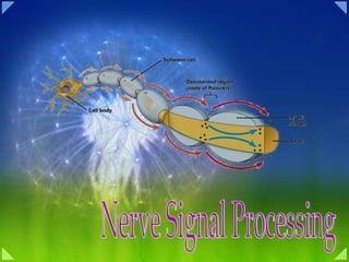
Ramón y Cajal applied Golgi's method to the embryonic nervous systems of many animals, including the human brain. By examining the structure of neurons in almost every region of the nervous system and tracing the contacts they made with one another, Ramón y Cajal was able to describe the differences between classes of nerve cells and to map the precise connections between a good many of them. In this way Ramón y Cajal grasped, in addition to the neuron doctrine, two other principles of neural organization that would prove particularly valuable in studying communication in the nervous system. The first of these has become known as the principle of dynamic polarization. It states that electrical signals within a nerve cell flow only in one direction: from the receiving sites of the neuron (usually the dendrites and cell body) to the trigger region at the axon. From there, the action potential is propagated unidirectionally along the entire length of the axon to the cell's presynaptic terminals. Although neurons vary in shape and function, the operation of most follows this rule of information flow. Later in this chapter we shall describe the physiological basis of this principle.
The Refractory Period The depolarization that produces Na+ channel opening also causes delayed activation of K+ channels and Na+ channel inactivation, leading to repolarization of the membrane potential as the action potential sweeps along the length of an axon (see Figure 3.12 ). In its wake, the action potential leaves the Na+ channels inactivated and K+ channels activated for a brief time. These transitory changes make it harder for the axon to produce subsequent action potentials during this interval, which is called the refractory period . Thus, the refractory period limits the number of action potentials that a given nerve cell can produce per unit time. As might be expected, different types of neurons have different maximum rates of action potential firing due to different types and densities of ion channels. The refractoriness of the membrane in the wake of the action potential explains why action potentials do not propagate back toward the point of their initiation as they travel along an axon.
Mechanism of directly gated synaptic transmission at a neuromuscular junction. (A) Cell bodies of motor neurons. (B and C) Myelinated axons of motor neurons innervate skeletal muscle fibers. As the motor axon reaches a specialized region on the muscle membrane (motor end-plate), it loses its myelin sheath and gives off several fine branches. Presynaptic boutons (swellings) are present at the terminals of these branches. (D) The presynaptic boutons have synaptic vesicles containing acetylcholine
Mechanism by Which an Action Potential Causes Transmitter Release from the Presynaptic Terminals—Role of Calcium Ions The membrane of the presynaptic terminal is called the presynaptic membrane . It contains large numbers of voltage-gated calcium channels . When an action potential depolarizes the presynaptic membrane, these calcium channels open and allow large numbers of calcium ions to flow into the terminal.The quantity of transmitter substance that is then released from the terminal into the synaptic cleft is directly related to the number of calcium ions that enter. The precise mechanism by which the calcium ions cause this release is not known, but it is believed to be the following. When the calcium ions enter the presynaptic terminal, it is believed that they bind with special protein molecules on the inside surface of the presynaptic membrane, called release sites . This binding in turn causes the release sites to open through the membrane, allowing a few transmitter vesicles to release their transmitter into the cleft after each single action potential. For those vesicles that store the neurotransmitter acetylcholine, between 2000 and 10,000 molecules of acetylcholine are present in each vesicle, and there are enough vesicles in the presynaptic terminal to transmit from a few hundred to more than 10,000 action potentials.
Action of the Transmitter Substance on the Postsynaptic Neuron—Function of “Receptor Proteins” The membrane of the postsynaptic neuron contains large numbers of receptor proteins , also shown in Figure 45–6. The molecules of these receptors have two important components: (1) a binding component that protrudes outward from the membrane into the synaptic cleft—here it binds the neurotransmitter coming from the presynaptic terminal—and (2) an ionophore component that passes all the way through the postsynaptic membrane to the interior of the postsynaptic neuron. The ionophore in turn is one of two types: (1) an ion channel that allows passage of specified types of ions through the membrane or (2) a “second messenger” activator that is not an ion channel but instead is a molecule that protrudes into the cell cytoplasm and activates one or more substances inside the postsynaptic neuron. These substances in turn serve as “second messengers” to increase or decrease specific cellular functions.
A. An excitatory input at the base of a dendrite causes inward current to flow through cation-selective channels (Na+ and K+). This current flows outward across the membrane capacitance at the initial segment where it produces a large depolarizing synaptic potential. B. An inhibitory input causes an outward (Cl-) current at the synapse on the cell body and an inward current across the membrane capacitance at other regions of the cell, producing a large hyperpolarization at the initial segment. C. The shunting action of inhibition. When the cell receives both excitatory and inhibitory synaptic current, the channels opened by the inhibitory pathway shunt the excitatory current, thereby reducing the excitatory synaptic potential.
Figure 2-7 Inhibitory interneurons can produce either feed forward or feedback inhibition. A. Feed-forward inhibition is common in monosynaptic reflex systems, such as the knee-jerk reflex (see Figure 2-5). Afferent neurons from extensor muscles excite not only the extensor motor neurons, but also inhibitory neurons that prevent the firing of the motor cells in the opposingflexor muscles. Feedforward inhibition enhances the effect of the active pathway by suppressing the activity of other, opposing, pathways. B. Negative feedback inhibition is a self-regulating mechanism. The effect is to dampen activity within the stimulated pathway and prevent it from exceeding a certain critical maximum. Here the extensor motor neurons act on inhibitory interneurons, which feed back to the extensor motor neurons themselves and thus reduce the probability of firing by these cells. A stretch reflex such as the knee jerk is a simple behavior produced by two classes of neurons connecting at excitatory synapses. But not all important signals in the brain are excitatory. In fact, half of all neurons produce inhibitory signals. Inhibitory neurons release a transmitter that reduces the likelihood of firing. As we have seen, even in the knee-jerk reflex, the sensory neurons make both excitatory connections and connections through inhibitory interneurons. Excitatory connections with the leg's extensor muscles cause these muscles to contract, while connections with certain inhibitory interneurons prevent the antagonist flexor muscles from being called to action. This feature of the circuit is an example of feed-forward inhibition (Figure 2-7A). Feedforward inhibition in the knee-jerk reflex is reciprocal , ensuring that the flexor and extensor pathways always inhibit each other, so only muscles appropriate for the movement, and not those that oppose it, are recruited. Neurons can also have connections that provide feedback inhibition. For example, an active neuron may have excitatory connections withboth a target cell and an inhibitory interneuron that has its own feedbackconnection with the active neuron. In this way signals from the active neuron simultaneously excite the target neuron and the inhibitory interneuron, which thus is able to limit the ability of the active neuron to excite its target (Figure 2-7B). We will encounter many examples of feed-forward and feedback inhibition when we examine more complex behaviors in later chapters.
Criteria That Define a Neurotransmitter Three primary criteria have been used over the years to confirm that a molecule acts as a neurotransmitter at a given chemical synapse. 1. The substance must be present within the presynaptic neuron. Clearly, a chemical cannot be secreted from a presynaptic neuron unless it is present there. Because elaborate biochemical pathways are required to produce neurotransmitters, showing that the enzymes and precursors required to synthesize the substance are present in presynaptic neurons provides additional evidence that the substance is used as a transmitter. Note, however, that since the transmitters glutamate, glycine, and aspartate are also needed for protein synthesis and other metabolic reactions in all neurons, their presence is not sufficient evidence to establish them as neurotransmitters. 2. The substance must be released in response to presynaptic depolarization, and the release must be Ca2+-dependent . Another essential criterion for identifying a neurotransmitter is to demonstrate that it is released from the presynaptic neuron in response to presynaptic electrical activity, and that this release requires Ca2+ influx into the presynaptic terminal. Meeting this criterion is technically challenging, not only because it may be difficult to selectively stimulate the presynaptic neurons, but also because enzymes and transporters efficiently remove the secreted neurotransmitters. 3. Specific receptors for the substance must be present on the postsynaptic cell. A neurotransmitter cannot act on its target unless specific receptors for the transmitter are present in the postsynaptic membrane. One way to demonstrate receptors is to show that application of exogenous transmitter mimics the postsynaptic effect of presynaptic stimulation. A more rigorous demonstration is to show that agonists and antagonists that alter the normal postsynaptic response have the same effect when the substance in question is applied exogenously. High-resolution histological methods can also be used to show that specific receptors are present in the postsynaptic membrane (by detection of radioactively labeled receptor antibodies, for example). Fulfilling these criteria establishes unambiguously that a substance is used as a transmitter at a given synapse. Practical difficulties, however, have prevented these standards from being applied at many types of synapses. It is for this reason that so many substances must be referred to as "putative" neurotransmitters. Demonstrating the identity of a neurotransmitter at a synapse requires showing (1) its presence, (2) its release, and (3) the postsynaptic presence of specific receptors. Chemical Substances That Function as Synaptic Transmitters More than 50 chemical substances have been proved or postulated to function as synaptic transmitters. Many of them are listed in Tables 45–1 and 45–2, which give two groups of synaptic transmitters. One group comprises small-molecule, rapidly acting transmitters . The other is made up of a large number of neuropeptides of much larger molecular size that are usually much more slowly acting. The small-molecule, rapidly acting transmitters are the ones that cause most acute responses of the nervous system, such as transmission of sensory signals to the brain and of motor signals back to the muscles. The neuropeptides, in contrast, usually cause more prolonged actions, such as long-term changes in numbers of neuronal receptors, long-term opening or closure of certain ion channels, and possibly even longterm changes in numbers of synapses or sizes of synapses.
Small-Molecule, Rapidly Acting Transmitters Class I Acetylcholine Class II: The Amines Norepinephrine Epinephrine Dopamine Serotonin Histamine Class III: Amino Acids Gamma-aminobutyric acid (GABA) Glycine Glutamate Aspartate Class IV Nitric oxide (NO) Two Major Categories of Neurotransmitters By the 1950s, the list of neurotransmitters (defined by the criteria described in Box A ) had expanded to include four aminesepinephrine, norepinephrine, dopamine, and serotoninin addition to ACh. Over the following decade, three amino acidsglutamate, g-aminobutyric acid (GABA), and glycinewere also shown to be neurotransmitters. Subsequently, other small molecules were added to the list, and considerable evidence now suggests that histamine, aspartate, and ATP should be included ( Figure 6.3 ). The most recent class of molecules discovered to be transmitters are a large number of polypeptides; since the 1970s, more than 100 such molecules have been shown to meet at least some of the criteria outlined in Box A . For purposes of discussion, it is useful to separate this variety of agents into two broad categories based simply on their size ( Figure 6.3 ). Neuropeptides are relatively large transmitter molecules composed of 3 to 36 amino acids ( Figure 6.4 ). Individual amino acids, such as glutamate and GABA, as well as the transmitters acetylcholine, serotonin, and histamine, are much smaller than neuropeptides and have therefore come to be called small-molecule neurotransmitters . Within the category of small-molecule neurotransmitters, the biogenic amines (dopamine, norepinephrine, epinephrine, serotonin, and histamine) are often discussed separately because of their similar chemical properties and postsynaptic actions. Figure 6.3. Examples of small-molecule and peptide neurotransmitters. Small-molecule transmitters can be subdivided into acetylcholine, the amino acids, purines, and biogenic amines. The catcholamines, so named because they all share the catechol moiety (i.e., a hydroxylated benzene ring), make up a distinctive subgroup within the biogenic amines. Serotonin and histamine contain an indole ring and an imidazole ring, respectively. Size differences between the small-molecule neurotransmitters and the peptide neurotransmitters are indicated by the space-filling models for glycine, norepinephrine, and methionine enkephalin. (Carbon atoms are black, nitrogen atoms blue, and oxygen atoms red.) Figure 6.4. Neuropeptides vary in length, but usually contain between 3 and 36 amino acids. Note that one peptide can include the sequence of other neuroactive peptides. For example, b-endorphin contains both a-endorphin and methionine enkephalin (olive box). Table 45–2 Neuropeptide, Slowly Acting Transmitters or Growth Factors Hypothalamic-releasing hormones Thyrotropin-releasing hormone Luteinizing hormone–releasing hormone Somatostatin (growth hormone inhibitory factor) Pituitary peptides Adrenocorticotropic hormone (ACTH) b-Endorphin a-Melanocyte-stimulating hormone Prolactin Luteinizing hormone Thyrotropin Growth hormone Vasopressin Oxytocin Peptides that act on gut and brain Leucine enkephalin Methionine enkephalin Substance P Gastrin Cholecystokinin Vasoactive intestinal polypeptide (VIP) Nerve growth factor Brain-derived neurotropic factor Neurotensin Insulin Glucagon From other tissues Angiotensin II Bradykinin Carnosine Sleep peptides Calcitonin Neuropeptides The neuropeptides are an entirely different class of transmitters that are synthesized differently and whose actions are usually slow and in other ways quite different from those of the small-molecule transmitters. The neuropeptides are not synthesized in the cytosol of the presynaptic terminals. Instead, they are synthesized as integral parts of large-protein molecules by ribosomes in the neuronal cell body. The protein molecules then enter the spaces inside the endoplasmic reticulum of the cell body and subsequently inside the Golgi apparatus, where two changes occur: First, the neuropeptide-forming protein is enzymatically split into smaller fragments, some of which are either the neuropeptide itself or a precursor of it. Second, the Golgi apparatus packages the neuropeptide into minute transmitter vesicles that are released into the cytoplasm. Then the transmitter vesicles are transported all the way to the tips of the nerve fibers by axonal streaming of the axon cytoplasm, traveling at the slow rate of only a few centimeters per day. Finally, these vesicles release their transmitter at the neuronal terminals in response to action potentials in the same manner as for small-molecule transmitters. However, the vesicle is autolyzed and is not reused. Because of this laborious method of forming the neuropeptides, much smaller quantities of them are usually released than of the small-molecule transmitters. This is partly compensated for by the fact that the neuropeptides are generally a thousand or more times as potent as the small-molecule transmitters. Another important characteristic of the neuropeptides is that they often cause much more prolonged actions. Some of these actions include prolonged closure of calcium channels, prolonged changes in the metabolic machinery of cells, prolonged changes in activation or deactivation of specific genes in the cell nucleus, and/or prolonged alterations in numbers of excitatory or inhibitory receptors. Some of these effects last for days, but others perhaps for months or years. Our knowledge of the functions of the neuropeptides is only beginning to develop.
Small-Molecule, Rapidly Acting Transmitters In most cases, the small-molecule types of transmitters are synthesized in the cytosol of the presynaptic terminal and are absorbed by means of active transport into the many transmitter vesicles in the terminal. Then, each time an action potential reaches the presynaptic terminal, a few vesicles at a time release their transmitter into the synaptic cleft. This usually occurs within a millisecond or less by the mechanism described earlier. The subsequent action of the smallmolecule type of transmitter on the membrane receptors of the postsynaptic neuron usually also occurs within another millisecond or less. Most often the effect is to increase or decrease conductance through ion channels; an example is to increase sodium conductance, which causes excitation, or to increase potassium or chloride conductance, which causes inhibition. Recycling of the Small-Molecule Types of Vesicles. The vesicles that store and release small-molecule transmitters are continually recycled and used over and over again. After they fuse with the synaptic membrane and open to release their transmitter substance, the vesicle membrane at first simply becomes part of the synaptic membrane. However, within seconds to minutes, the vesicle portion of the membrane invaginates back to the inside of the presynaptic terminal and pinches off to form a new vesicle. And the new vesicular membrane still contains appropriate enzyme proteins or transport proteins required for synthesizing and/or concentrating new transmitter substance inside the vesicle. Acetylcholine is a typical small-molecule transmitter that obeys the principles of synthesis and release stated earlier.This transmitter substance is synthesized in the presynaptic terminal from acetyl coenzyme A and choline in the presence of the enzyme choline acetyltransferase .Then it is transported into its specific vesicles. When the vesicles later release the acetylcholine into the synaptic cleft during synaptic neuronal signal transmission, the acetylcholine is rapidly split again to acetate and choline by the enzyme cholinesterase , which is present in the proteoglycan reticulum that fills the space of the synaptic cleft.And then again, inside the presynaptic terminal, the vesicles are recycled; choline is actively transported back into the terminal to be used again for synthesis of new acetylcholine. Acetylcholine is secreted by neurons in many areas of the nervous system but specifically by (1) the terminals of the large pyramidal cells from the motor cortex, (2) several different types of neurons in the basal ganglia, (3) the motor neurons that innervate the skeletal muscles, (4) the preganglionic neurons of the autonomic nervous system, (5) the postganglionic neurons of the parasympathetic nervous system, and (6) some of the postganglionic neurons of the sympathetic nervous system. In most instances, acetylcholine has an excitatory effect; however, it is known to have inhibitory effects at some peripheral parasympathetic nerve endings, such as inhibition of the heart by the vagus nerves. Figure 6.8. Acetylcholine metabolism in cholinergic nerve terminals. The synthesis of acetylcholine from choline and acetyl CoA requires choline acetyltransferase. Acetyl CoA is derived from pyruvate generated by glycolysis, while choline is transported into the terminals via a Na+-dependent transporter. After release, acetylcholine is rapidly metabolized by acetylcholinesterase and choline is transported back into the terminal. Acetylcholine Acetylcholine is the neurotransmitter at neuromuscular junctions, at synapses in the ganglia of the visceral motor system, and at a variety of sites within the central nervous system. Whereas a great deal is known about the function of cholinergic transmission at the neuromuscular junction and at ganglionic synapses, the actions of ACh in the central nervous system are not as well understood. Acetylcholine is synthesized in nerve terminals from acetyl coenzyme A (acetyl CoA, which is synthesized from glucose) and choline, in a reaction catalyzed by choline acetyltransferase (CAT) ( Figure 6.8 ). The presence of CAT in a neuron is thus a strong indication that ACh is used as one of its transmitters. Choline is present in plasma at a concentration of about 10 m M , and is taken up into cholinergic neurons by a high-affinity Na+/choline transporter. About 10,000 molecules of ACh are packaged into each vesicle by a vesicular ACh transporter. In contrast to most other small-molecule neurotransmitters, the postsynaptic action of ACh at many cholinergic synapses (the neuromuscular junction in particular) are not terminated by reuptake but by a powerful hydrolytic enzyme, acetylcholinesterase (AChE). This enzyme is concentrated in the synaptic cleft, ensuring a rapid decrease in ACh concentration after its release from the presynaptic terminal. AChE has a very high catalytic activity (about 5000 molecules of ACh per AChE molecule per second) and hydrolyzes ACh into acetate and choline. As already mentioned, cholinergic nerve terminals typically contain a high-affinity, Na+-choline transporter that takes up the choline produced by ACh hydrolysis. Among the many interesting drugs that interact with cholinergic enzymes are the organophosphates. Compounds such as diphenyl trichloroethane (DTT) and the herbicide 2,4-dichlorophenoxyacetic acid (2,4-D) were originally developed as insecticides. This group also includes some potent chemical warfare agents. One such compound is the nerve gas "Sarin," which was made notorious a few years ago after a group of terrorists released this gas in Tokyo's underground rail system. Organophosphates can be lethal to humans (and insects) because they inhibit AChE, causing ACh to accumulate at cholinergic synapses. This build-up of ACh depolarizes the postsynaptic cell and renders it refractory to subsequent ACh release, causing, among other effects, neuromuscular paralysis
Major cholinergic cell groups. Note two major constellations of cholinergic neurons: cholinergic neurons located in the basal forebrain constellation, including the basal nucleus of Meynert, and cholinergic neurons located in the dorsolateral tegmentum of the pons. There are two main types of cholinergic receptors widely distributed throughout in the brain. These receptors are classified as muscarinic and nicotinic receptors. In certain regions of the brain only the muscarinic subtype is found eg midbrain, medulla, and pons while in other regions eg substantia nigra, locus coeruleus and septum only the nicotinic receptor subtype is found. Both sub-types are located in the corpus striatum, cerebral cortex, hippocampus, thalamus, hypothalamus and cerebellum. Distribution of acetylcholine receptors in the Alzheimer’s disease brain There are two main types of cholinergic receptors widely distributed throughout in the brain; the muscarinic and nicotinic receptors. In certain regions of the brain (eg midbrain, medulla, and pons) only the muscarinic subtype is found while in other regions (eg substantia nigra, locus coeruleus and septum) only the nicotinic receptor subtype is found. Both subtypes are colocalised in the corpus striatum, cerebral cortex, hippocampus, thalamus, hypothalamus and cerebellum. Alzheimer’s disease is associated with a reduction of all cholinergic neurones in the subcortical areas of the brain, leading to reduced availability of acetylcholine.
originate in the median raphe of the brain stem project to many brain and spinal cord areas, especially to the dorsal horns of the spinal cord and to the hypothalamus acts as an inhibitor of pain pathways in the cord, and an inhibitor action in the higher regions of the nervous system believed to help control the mood of the person, perhaps even to cause sleep. Distribution Serotonin-containing neurons have been identified in the midline raphe nuclei of the medulla, pons, and upper brainstem. The serotonin-containing cell groups of the rostral raphe nuclei (e.g., dorsal and median raphe nuclei) send their projections to the dien-cephalon P.120 and telencephalon. The caudal raphe nuclei (e.g., raphe magnus) send their projections to the spinal cord. Physiological and Clinical Considerations Serotonin-containing cells in the raphe regions of the brainstem are believed to play a role in descending pain-control systems. Other serotonin-containing neurons may play a role in mediating affective processes such as aggressive behavior and arousal. Serotonin synthesized in the pineal gland serves as a precursor for the synthesis of melatonin, which is a neurohormone involved in regulating sleep patterns. Serotonin is also believed to play an important role in depression. Serotonin uptake inhibitors, belonging to different chemical families, have been found to be beneficial in the treatment of depression; this effect may be due to an increase in serotonin levels in the brain. For example, fluoxetine (Prozac) selectively blocks reuptake of serotonin and enhances serotonin levels in the brain. It may produce beneficial effects in mental depression via enhancement of transmission through 5-HT 1A receptors. Sumatriptan (Imitrex) is a 5-HT 1D receptor agonist. It is a vasoconstrictor and has proved useful in treating migraine headaches. Designer Drugs of Abuse and Their Relationship With Serotonin Some drugs of abuse mediate their effects through serotonin-containing neurons. For example, Ecstasy has been used as a recreational drug by young adults, especially in large dance parties know as “raves.†Ecstasy includes two drugs, MDMA (3,4, methylene-dioxy-methamphetamine) and MDEA (3,4, methylene-dioxy-ethamphetamine). These drugs, generally called “Adam†and “Eve,†respectively, by drug abusers, have been reported to induce sensory enhancement (a feeling that all is right with this world) and empathogenesis (a feeling of closeness with others and removal of barriers in communication, especially in intimate relationships). These effects are believed to be due to initial release of serotonin by these drugs. Toxic effects of these drugs are dehydration, hyperthermia, tachycardia (increase in heart rate), and sweating. Undesirable side effects following prolonged use of Ecstasy are believed to be caused by the degeneration of the projections of serotonin-containing neurons.
secreted by neurons that originate in the substantia nigra. The termination of these neurons is mainly in the striatal region of the basal ganglia. The effect of dopamine is usually inhibition Parkinsonism is due to degeneration of substantia nigra with reduced dopamine Schizophrenia there is increased dopamine Distribution Dopaminergic neurons are located in the following nuclei (Fig. 8-8). Substantia nigra, which is located bilaterally in the midbrain. Axons of the dopaminergic neurons located in the substantia nigra (pars compacta) ascend rostrally as nigrostriatal projection, providing dopaminergic innervation to neurons located in the corpus striatum (caudate nucleus and putamen). Dopaminergic neurons in the substantia nigra are selectively degenerated in Parkinson's disease. Ventral tegmental area, which is located just medial to the substantia nigra pars compacta. Dopaminergic neurons, located in this area, project as either the mesolimbic or the mesocortical pathway. In the mesolimbic pathway, axons of the dopaminergic neurons ascend in the brainstem and forebrain in the median forebrain bundle to supply limbic structures (amygdala, septal area, and hippocampal formation) and the nucleus accumbens (which is embedded in the ventral straitum). In the mesocortical pathway, axons of the dopaminergic neurons ascend in the median forebrain bundle to provide dopaminergic innervation to the frontal and cingulate cortices. The arcuate nucleus, which is located in the ventrolateral aspect of hypothalamus (not shown in Fig. 8-8). Dopaminergic neurons located in the arcuate nucleus project to the median eminence and release dopamine directly into the hypophyseal portal circulation, which is then carried to the anterior lobe of the pituitary to inhibit the release of prolactin (the hormone primarily responsible for lactation). Physiological and Clinical Considerations Parkinson's Disease This disease is characterized by an expressionless face (clinicians sometimes refer to it as “mask-like faceâ€), slowness of movement (bradykinesia or hypokinetic syndrome), rigidity of extremities and neck, and P.117 tremors in the hands. Patients with Parkinson's disease have a gait that is characterized by short wide-based steps (sometimes referred to as fenestrating gait), stooped posture, and scarcity of normal limb movements. These symptoms are often accompanied by dementia. Dopaminergic neurons located in the substantia nigra are degenerated in this disease, and the release of dopamine in the caudate/putamen is decreased. The mechanism of bradykinesia in Parkinson's disease is discussed in Chapter 20. The main aim of drug therapy for this disease is to replace the deficiency of the transmitter (dopamine) in the basal ganglia (target of nigrostriatal projection). Because dopamine does not cross blood-brain barrier, its immediate metabolic precursor (L-DOPA, or levodopa), which does cross this barrier, is administered orally. L-DOPA is transported into the brain via a large neutral amino acid transporter, and it permeates the striatal tissue, where it is decarboxylated to dopamine. In recent years, a combination of L-DOPA and carbidopa (Sinemet) has been prescribed for this disease. Carbidopa is an inhibitor of dopa-decarboxylase and reduces the decarboxylation of L-DOPA in the peripheral tissues, enabling a greater concentration of this precursor to reach the brain. Carbidopa does not cross the blood-brain barrier. Therefore, conversion of L-DOPA to dopamine does not diminish in the brain. Another potentially useful but controversial approach is to implant fetal midbrain or adrenal medullary tissue into the deteriorating caudate nucleus and putamen. An attractive possibility in the future is transplantation of genetically engineered cells capable of expressing tyrosine hydroxylase that is involved in the synthesis of dopamine. Psychotic Disorders Many adult psychotic disorders, including schizophrenia, are believed to involve increased activity at dopaminergic synapses. Many drugs that are effective in the treatment of these disorders (e.g., phenothiazines, thioxanthines, and butyrophenones) are believed to reduce the dopamine synaptic activity in the limbic forebrain. Further discussion of dopamine and other neurotransmitters in psychiatric disorders is considered in Chapter 28. Cocaine Drug Abuse Cocaine (a local anesthetic drug) blocks the reuptake of dopamine (and norepinephrine) into the nerve terminals. Elevated levels of dopamine in certain brain circuits may be responsible for the euphoric effects of cocaine. This mechanism is not responsible for the local anesthetic effect of cocaine that is mediated via the blockade of neuronal voltage-gated Na + channels. Dopaminergic projections from the ventral tegmental area to the limbic structures, especially the projections to nucleus accumbens, may be involved in emotional reinforcement and motivation associated with cocaine drug addiction. Levels of dopamine in these projections are increased in individuals addicted to cocaine.
secreted by the terminals of many neurons whose cell bodies are located in the brain stem and hypothalamus help control overall activity and mood of the mind, such as increasing the level of wakefulness In most of these areas, norepinephrine probably activates excitatory receptors, but in a few areas, it activates inhibitory receptors instead Norepinephrine is also secreted by most postganglionic neurons of the sympathetic nervous system, where it excites some organs but inhibits others Synthesis and Removal Initial steps in the synthesis of norepinephrine (up to the step of transport and storage of dopamine into the storage vesicles) are described in the section about dopamine. The noradrenergic neurons contain an enzyme, dopamine β-hydroxylase (DBH), which converts dopamine into norepinephrine. It should be noted that the synthesis of norepinephrine takes place in the storage vesicles (Fig. 8-9). The norepinephrine thus formed is ready to be released by exocytosis. Norepinephrine released into the synaptic cleft is removed by the mechanisms described in the dopamine section. Autoinhibition and Negative Feedback Mechanisms Activation of presynaptic adrenergic receptors results in inhibition of the release of norepinephrine. This process is known as autoinhibition and is distinct from negative feedback in which synthesis of the transmitter (norepinephrine in this case) is blocked at its rate-limiting step (i.e., conversion of tyrosine to DOPA by tyrosine hydroxylase). Distribution The major concentration of noradrenergic neurons is in the locus ceruleus (also known as A 6 group of neurons) that is located in the pons. These neurons send projections through the central tegmental tract and the medial forebrain bundle to the thalamus, hypothalamus, limbic forebrain structures (cingulate and parahippocampal gyri, hippocampal formation, and amygdaloid complex), and the cerebral cortex. This group of neurons modulates a variety of physiological functions (e.g., sleep and wakefulness, attention, and feeding behaviors). Physiological and Clinical Considerations Norepinephrine is released as a transmitter from postganglionic sympathetic nerve terminals. Its role in the CNS as a transmitter is not clearly understood. Norepinephrine is believed to play a role in psychiatric disorders such as depression. Drugs used in the treatment of depression (e.g., tricyclic antidepressants like desimipramine) are known to inhibit the reuptake of norepinephrine at the nerve terminals and increase the synaptic levels of P.118 norepinephrine. The exact mechanism by which increased levels of norepinephrine in the CNS mediate the antidepressant action of these drugs is not clearly known.
the prominent glutamatergic pathways are: the cortico-cortical pathways; the pathways between the thalamus and the cortex; and the extrapyramidal pathway o other glutamate projections exist between the cortex, substantia nigra, subthalmic nucleus and pallidum. Glutamate is an excitatory neurotransmitter Physiological and Clinical Considerations Some of the important physiological and clinical considerations relevant to glutamate are as follows. Glutamate has been implicated as a transmitter in a variety of circuits in the brain. For example, excitatory amino acids may be involved in learning and memory processes, as well as motor functions. Glutamate has also been implicated in some chronic neuropathological conditions such as amyotrophic lateral sclerosis (ALS, also known as Lou Gehrig's disease), which is characterized by degeneration of the motor neurons in the anterior horn of the spinal cord, brainstem, and cerebral cortex. Patients suffering from this disease exhibit widespread muscle fasciculations, flaccid weakness, and atrophy of the muscles. The mechanism of the involvement of glutamate in the symptoms of ALS is not clearly known (see also Chapter 9). Prolonged stimulation of neurons by excitatory amino acids results in neuronal death or injury. This effect is known as excitotoxicity. There is predominantly a loss of postsynaptic neurons. Neuronal loss due to excitotoxicity has been implicated in cerebrovascular stroke. Occlusion of arteries supplying the brain results in brain ischemia. Concentrations of excitatory amino acids (glutamate and aspartate) increase in the extracellular spaces in the brain perhaps due to slowing of their uptake by neurons and glia. The P.112 neurons are excited excessively, and a chain of events, not fully identified yet, causes neuronal death. Excessive intracellular Ca 2+ concentrations have been implicated in the neuronal death due to excitotoxicity. Excitotoxic action of glutamate has been implicated in Alzheimer's disease. One of the mechanisms of the loss of neurons concerned with memory in this disease is believed to be overexcitation and, finally, loss of these neurons by glutamate-induced activation of N-methyl-D-aspartic acid (NMDA) receptors (described later in this chapter in the Ionotropic Receptors section). Memantine (Namenda), an NMDA receptor antagonist, is reported to mitigate this effect and slow down the symptoms of the disease.
Distribution GABA is found in high concentrations in the brain and spinal cord but is absent in peripheral nerves or peripheral tissues. Unlike glutamate, GABA is not an essential metabolite, and it is not incorporated into a protein. Physiological and Clinical Considerations Some of the important physiological and clinical considerations regarding this neurotransmitter are as follows. GABA is an inhibitory transmitter in many brain circuits. For example, GABA is used as an inhibitory neurotransmitter by the Purkinje cells in the cerebellum. Alteration of GABAergic circuits has been implicated in neurological and psychiatric disorders like Huntington's chorea, Parkinson's disease, senile dementia, Alzheimer's disease, and schizophrenia. P.114 As mentioned earlier, GAD requires vitamin B 6 as a coenzyme. Therefore, dietary deficiency of vitamin B 6 can lead to diminished GABA synthesis. In a disastrous series of infant deaths, it was noted that vitamin B 6 was omitted in an infant feeding formula. GABA content in the brain of these infants was reduced. Subsequently, there was a loss of synaptic inhibition that caused seizures and death. Since epileptic seizures can be facilitated by lack of neuronal inhibition, increase in the inhibitory transmitter, GABA, is helpful in terminating them. Thus, valproic acid (dipropylacetic acid) is useful as an anticonvulsant because it inhibits GABA transaminase, which is an enzyme that metabolizes GABA, and increases GABA levels in the brain. Barbiturates act as agonists or modulators on postsynaptic GABA receptors and are used to treat epilepsy.
Figure 45–7 “ Second messenger” system by which a transmitter substance from an initial neuron can activate a second neuron by first releasing a “G-protein” into the second neuron’s cytoplasm. Four subsequent possible effects of the G-protein are shown, including 1, opening an ion channel in the membrane of the second neuron; 2, activating an enzyme system in the neuron’s membrane; 3, activating an intracellular enzyme system; and/or 4, causing gene transcription in the second neuron. “ Second Messenger” System in the Postsynaptic Neuron. Many functions of the nervous system—for instance, the process of memory—require prolonged changes in neurons for seconds to months after the initial transmitter substance is gone. The ion channels are not suitable for causing prolonged postsynaptic neuronal changes because these channels close within milliseconds after the transmitter substance is no longer present. However, in many instances, prolonged postsynaptic neuronal excitation or inhibition is achieved by activating a “second messenger” chemical system inside the postsynaptic neuronal cell itself, and then it is the second messenger that causes the prolonged effect. There are several types of second messenger systems. One of the most common types uses a group of proteins called G-proteins . Figure 45–7 shows in the upper left corner a membrane receptor protein. A G-protein is attached to the portion of the receptor that protrudes into the interior of the cell. The G-protein in turn consists of three components: an alpha (a) component that is the activator portion of the G-protein, and beta (b) and gamma (g) components that are attached to the alpha component and also to the inside of the cell membrane adjacent to the receptor protein. On activation by a nerve impulse, the alpha portion of the G-protein separates from the beta and gamma portions and then is free to move within the cytoplasm of the cell. Inside the cytoplasm, the separated alpha component performs one or more of multiple functions, depending on the specific characteristic of each type of neuron. Shown in Figure 45–7 are four changes that can occur. They are as follows: 1. Opening specific ion channels through the postsynaptic cell membrane . Shown in the upper right of the figure is a potassium channel that is opened in response to the G-protein; this channel often stays open for a prolonged time, in contrast to rapid closure of directly activated ion channels that do not use the second messenger system. 2. Activation of cyclic adenosine monophosphate (cAMP) or cyclic guanosine monophosphate (cGMP) in the neuronal cell . Recall that either cyclic AMP or cyclic GMP can activate highly specific metabolic machinery in the neuron and, therefore, can initiate any one of many chemical results, including long-term changes in cell structure itself, which in turn alters long-term excitability of the neuron. 3. Activation of one or more intracellular enzymes . The G-protein can directly activate one or more intracellular enzymes. In turn the enzymes can cause any one of many specific chemical functions in the cell. 4. Activation of gene transcription. This is one of the most important effects of activation of the second messenger systems because gene transcription can cause formation of new proteins within the neuron, thereby changing its metabolic machinery or its structure. Indeed, it is well known that structural changes of appropriately activated neurons do occur, especially in long-term memory processes. It is clear that activation of second messenger systems within the neuron, whether they be of the G-protein type or of other types, is extremely important for changing the long-term response characteristics of different neuronal pathways.We will return to this subject in more detail in Chapter 57 when we discuss memory functions of the nervous system.