2003 role of incretins in glucose homeostasis and diabetes
•Download as PPT, PDF•
10 likes•2,200 views

Dr. Mdasir Bashir Ph.D. scholar IVRI-Izatnagar
![Incretins ,[object Object],[object Object],[object Object],[object Object]](data:image/gif;base64,R0lGODlhAQABAIAAAAAAAP///yH5BAEAAAAALAAAAAABAAEAAAIBRAA7)
Recommended
More Related Content
What's hot
What's hot (20)
Viewers also liked
Viewers also liked (20)
Similar to 2003 role of incretins in glucose homeostasis and diabetes
Similar to 2003 role of incretins in glucose homeostasis and diabetes (20)
More from Dr.Mudasir Bashir
Recently uploaded
Recently uploaded (20)
2003 role of incretins in glucose homeostasis and diabetes
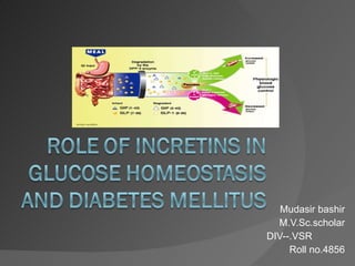
- 1. Mudasir bashir M.V.Sc.scholar DIV--.VSR Roll no.4856
- 24. Incretin therapy in type2DM
- 25. B-cell function and glucagon in T2DM β -Cell mass in Type 2 diabetes -50% -63% ND=non-diabetic; IFG=impaired fasting glucose; T2DM=Type 2 diabetes mellitus Obese Lean
- 28. Action of glucagon Glycogen Glucose Low blood glucose and insulin promotes glucagon release from - cells of pancreas Glucagon stimulates breakdown of glycogen Raises blood glucose
- 30. Postprandial GLP-1 levels are decreased in people with IGT and Type 2 diabetes 2001 Time (min) GLP-1 (pmol/l) * * * * * * * * Meal NGT T2DM IGT * P <0.05 T2DM vs NGT Toft-Nielsen et al. J Clin Endocrinol Metab. 20 15 10 5 0 0 60 120 180 240
- 31. 16 12 8 4 0 2200 0200 0600 1000 1400 1600 Breakfast Lunch Snack Clock time (hours) Glucose (mmol/l) T2DM + GLP-1 Normalisation of diurnal plasma glucose concentrations by continuous IV GLP-1 infusion (1.2 pmol/kg/min) Rachman et al. Diabetologia. 1997 T2DM
- 37. Exenatide and first-phase insulin response in Type 2 diabetes Type 2 diabetes,placebo Type2 diabetes,exenatide Healthy subjects, placebo Fehse et al. J Clin Endocrinol Metab . 2005 Exenatide vs healthy Time (min) Insulin secretion (pmol•kg -1 •min -1 )
- 40. Sitagliptin improves fasting glucose level in monotherapy vs placebo Plasma glucose (mmol/l) Time (weeks) 0 6 12 3 18 24 Placebo Sitagliptin 100 mg 9.0 9.5 8.5 10.0 10.5 8.0 Charbonnel et al. Diabetes Care. 2006; R osenstock et al. Clin Ther. 2006
- 41. Sitagliptin added to metformin [janumet]improves 24-hour glucose profile in Type 2 diabetes Glucose (mmol/l) 8:00 Day 1 13:00 19:00 0:00 Day 2 7:30 6 7 8 9 10 13 11 12 Dose 1 7:30 Dose 2 18:30 Breakfast Lunch Dinner Placebo + metformin Sitagliptin 50 mg bd + metformin Time
Editor's Notes
- Degradation of GLP-1 GLP-1 is a 30 amino acid peptide. This peptide is rapidly degraded by the enzyme DPP-4, present in human serum and universally expressed on endothelial cells. DPP-4 hydrolyses the peptide between alanine in position 2 and the remainder of the amino acid chain. After cleavage, the resulting peptide fragment does not exhibit incretin activity [Gallwitz et al, 1994; Mentlein et al, 1993]. GLP-1 exerts its insulinotropic effects by binding to receptors on β-cells. One approach to improving GLP-1 activity is through the administration of incretin mimetics. These compounds are molecular analogues of GLP-1 which have the ability to activate β-cell receptors but which are structurally different enough to reduce or prevent degradation by DPP-4. A second approach involves the use of incretin enhancers; these prolong the bioactivity of GLP-1 by inhibiting the action of DPP-4 [Drucker 2006; Drucker 2001]. REFERENCES Drucker DJ. Cell Metab . 2006;3:153-165. Drucker DJ. Curr Pharm Des . 2001;7:1399-1412. Gallwitz B, Witt M, Paetzold G, et al. Eur J Biochem . 1994;225:1151-1156. Mentlein R, Gallwitz B, Schmidt WE. Eur J Biochem . 1993;214:829-835.
- Postprandial GLP-1 levels are decreased in people with IGT and Type 2 diabetes This slide illustrates plasma GLP-1 response to meal ingestion in age- and weight-matched individuals with normal glucose tolerance (NGT) compared with individuals with impaired glucose tolerance (IGT) and Type 2 diabetes (n=102). The meal test began 3 days after discontinuation of oral glucose-lowering medication and following an overnight fast. Blood was sampled before the start of the meal and over 4 hours post-meal. Plasma GLP-1 levels rose shortly after meal ingestion in all groups. GLP-1 concentrations were significantly lower in the Type 2 diabetes group compared with the NGT group beginning at 60 minutes and continuing for an additional 90 minutes ( P <0.05). A similar trend was apparent for IGT compared with NGT. These results demonstrate that the meal-stimulated GLP-1 response is depressed in individuals with both Type 2 diabetes and IGT compared with their NGT counterparts. REFERENCE Toft-Nielsen M, Damholt M, Madsbad S, et al. J Clin Endocrinol Metab . 2001;86:3717-3723.
- GLP-1 enhancement Postprandial plasma levels of GLP-1 are depressed in Type 2 diabetes, indicating an impaired GLP-1 response to nutrient ingestion. For this reason, continuous GLP-1 infusion therapy has proven useful in short-term studies but is not a practical long-term therapy. The effort to identify effective forms of GLP-1 administration is challenged by its extremely short half-life (the enzyme DPP-4 degrades GLP-1 rapidly following its release from gut cells) [Drucker 2006; Drucker 2001]. Much research has focused on compounds with molecular structures and incretin activity that are similar to GLP-1, but which have longer half-lives because they are not degraded by DPP-4 or are resistant to DPP-4. These compounds include exenatide and liraglutide, which are injectable treatments. Another approach is to identify compounds that inhibit the activity of DPP-4, thus prolonging the half-life of naturally occurring GLP-1. Two oral agents that act as DPP-4 inhibitors are sitagliptin and vildagliptin [Gallwitz, 2006]. REFERENCES Drucker DJ. Cell Metab . 2006;3:153-165. Drucker DJ. Curr Pharm Des . 2001;7:1399-1412. Gallwitz B. Eur Endocr Dis . June 2006:43-46.
- Exenatide and first-phase insulin response in Type 2 diabetes Exenatide is a synthetic form of the naturally occurring peptide exendin-4, originally discovered in the salivary glands of the Gila monster. This 39-unit peptide shares a similar amino acid sequence with GLP-1, but unlike GLP-1, it is not degraded by the enzyme DPP-4. The half-life of exenatide is about 3 hours (compared with less than 5 minutes for GLP-1) [Fehse et al, 2005; Gallwitz, 2006; Meier et al, 2004]. One goal of this study was to assess both first- and second-phase insulin response in individuals with Type 2 diabetes (n=13) treated with exenatide or saline infusion compared with weight- and age-matched saline-infused (n=12) healthy individuals. All oral glucose-lowering medications were discontinued prior to the test, and an intravenous glucose bolus was administered to all participants [Fehse et al, 2005]. When an intravenous glucose challenge followed saline infusion, individuals with Type 2 diabetes, in comparison with healthy individuals, exhibited a sharply reduced insulin response in the first 10 minutes (first phase) and during the remainder of the study (second phase). In contrast, when the same individuals were infused with exenatide, their first-phase insulin response was higher compared with saline ( P <0.05) and similar to the healthy group. Second-phase insulin response was also increased with exenatide infusion compared with saline infusion in both individuals with Type 2 diabetes and healthy individuals ( P <0.001) [Fehse et al, 2005]. These results corroborate our understanding that an underlying cause of hyperglycaemia in Type 2 diabetes is an inadequate first- and second-phase insulin response resulting from reduced β-cell insulin secretion. They also show the promise of exenatide to compensate for this depressed insulin response. REFERENCES Fehse F, Trautmann M, Holst JJ, et al. J Clin Endocrinol Metab . 2005;90:5991-5997. Gallwitz B. Eur Endocr Dis . June 2006:43-46. Meier JJ, Nauck MA, Kranz D, et al. Diabetes. 2004;53:654-662.
- Sitagliptin improves both fasting and post-meal glucose in monotherapy vs placebo Sitagliptin is an orally administered GLP-1-enhancing agent that is a highly selective inhibitor of the enzyme DPP-4 and has minimal inhibitory effects on other enzymes in the DPP family of enzymes. The first graph on this slide shows the results of a large-scale efficacy trial of sitagliptin monotherapy in individuals with Type 2 diabetes. The design included a 2-week placebo baseline period followed by a 24-week treatment that compared sitagliptin 100 mg once daily with placebo. No other oral glucose-lowering agents were used. Fasting plasma glucose decreased rapidly with sitagliptin compared with placebo (net decrease -1.0 mmol/l [-17.1 mg/dl], P <0.001). Meal tolerance tests were conducted both at baseline and following 24 weeks of sitagliptin treatment. The difference in post-meal glucose response compared with baseline in shown in the second graph. Sitagliptin treatment substantially reduced glucose at 1 and 2 hours post-meal (net decrease -2.6 mmol/l [-46.7 mg/dl], P <0.001). These results indicate that sitagliptin monotherapy improves glucose control in both the fasting and postprandial state. REFERENCE Aschner P, Kipnes MS, Lunceford JK, Sanchez M, Mickel C, Williams-Herman DE, for the Sitagliptin Study 021 Group. Diabetes Care. 2006;29:2632-2637.
- Sitagliptin added to metformin improves 24-hour glucose profile in Type 2 diabetes This study evaluated the efficacy of sitagliptin in combination with metformin in individuals whose hyperglycaemia was inadequately controlled by metformin alone. It has been posited that adding sitagliptin to metformin therapy may enhance glucose control by combining metformin’s insulin-sensitising effect with sitagliptin’s GLP-1 enhancement [Brazg et al, 2007; Gallwitz, 2006]. Study participants were treated with metformin plus placebo or sitagliptin 50 mg twice daily during a 4-week period; at the close of the study, each participant’s 24-hour glucose profile was measured [Brazg et al, 2007]. Sitaglitpin combined with metformin produced substantially lower glucose concentrations in both the postprandial and fasting states compared with metformin montherapy. The decrease in 24-hour mean glucose for the sitagliptin-treated group compared with metformin monotherapy was -1.8 mmol/l (-32.8 mg/dl) ( P <0.001) [Brazg et al, 2007]. These results demonstrate that the addition of sitagliptin to metformin improves glycaemic control compared with metformin monotherapy. REFERENCES Brazg R, Xu L, Dalla Man C, Cobelli C, Thomas K, Stein PP. Diabetes Obes Metab. 2007;9:186-193. Gallwitz B. Eur Endocr Dis . June 2006:43-46.