

Empfohlen
Weitere ähnliche Inhalte
Was ist angesagt?
Was ist angesagt? (20)
Andere mochten auch
Andere mochten auch (18)
Ähnlich wie L4 barrett esophagus 2
Ähnlich wie L4 barrett esophagus 2 (20)
Mehr von Mohammad Manzoor
Mehr von Mohammad Manzoor (20)
Kürzlich hochgeladen
Kürzlich hochgeladen (20)
L4 barrett esophagus 2
- 2. Norman Barrett • Norman Rupert Barrett (1903-1979) was an Australian-born British thoracic surgeon who is primarily remembered for describing Barrett’s oesophagus in 1950.
- 3. Barret esophagus • Barrett's oesophagus, sometimes called Barrett syndrome or • Columnar epithelium lined lower oesophagus (CELLO).
- 4. Barrett Esophagus Barrett esophagus is a complication of chronic GERD ( 5-15%) that is characterized by intestinal metaplasia within the esophageal squamous mucosa. • Barrett esophagus is defined as the replacement of the normal distal stratified squamous mucosa by metaplastic columnar epithelium containing goblet cells.
- 6. Epidemiology • The incidence of Barrett esophagus is rising, and it is estimated to occur in as many as 10% of individuals with symptomatic GERD. • Barrett esophagus is most common in white (Caucasian) males and it typically presents between 40 and 60 years of age. • The greatest concern in Barrett esophagus is that it confers an increased risk of esophageal adenocarcinoma.
- 7. A pre-malignant condition Molecular studies suggest that Barrett epithelium may be more similar to adenocarcinoma than to normal esophageal epithelium, consistent with the view that Barrett esophagus is a pre- malignant condition.
- 8. Epithelial dysplasia Epithelial dysplasia, considered to be a pre-invasive lesion, is detected in 0.2% to 2.0% of persons with Barrett esophagus each year and is associated with prolonged symptoms and increased patient age
- 9. 90%Although the vast majority of esophageal adenocarcinomas are associated with Barrett esophagus, it is important to remember that most (90%) individuals with Barrett esophagus do not develop esophageal tumors.
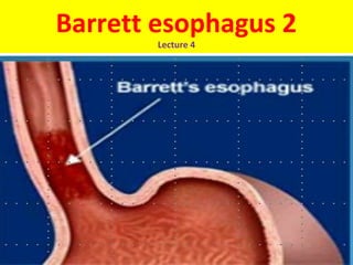
- 10. Red Patches Barrett esophagus can be recognized as one or several tongues or patches of red, velvety mucosa extending upward from the gastroesophageal junction.
- 11. Alternates Metaplastic mucosa alternates with residual smooth, pale squamous (esophageal) mucosa and interfaces with light-brown columnar (gastric) mucosa distally.
- 12. Long segment & Short segment High-resolution endoscopes have increased the sensitivity of Barrett esophagus detection. This has led to subclassification of Barrett esophagus as long segment, in which 3 cm or more of esophagus is involved, or short segment, in which less than 3 cm is involved. It is not yet clear if the risk of dysplasia in short segment disease is less than in long segment Barrett esophagus.
- 13. Long-segment Barrett’s esophagus NEJM 2002; 346: 836
- 14. Short Segment Barrett’s Irregular z-line above hiatal hernia Courtesy of Dr. C. Mel WilcoxCourtesy of Dr. C. Mel Wilcox
- 15. What we want to prevent -- Cancer Arising in Barrett’s Courtesy of Dr. C. Mel WilcoxCourtesy of Dr. C. Mel Wilcox
- 16. Diagnosis• Diagnosis of Barrett esophagus requires both endoscopic evidence of abnormal mucosa above the gastroesophageal junction and histologically documented intestinal metaplasia.
- 17. Goblet cells • Goblet cells, which have distinct mucous vacuoles that stain pale blue by H&E and impart the shape of a wine goblet to the remaining cytoplasm, define intestinal metaplasia and are necessary for diagnosis of Barrett esophagus
- 18. Goblet Cells
- 19. Intestinal metaplasia • The requirement for intestinal metaplasia reflects the fact that this feature correlates with neoplastic risk. • Foveolar mucus cells, which do not have distinct mucous vacuoles are insufficient for diagnosis.
- 20. Role of Endoscopy The requirement for an endoscopic abnormality helps to prevent misdiagnosis if metaplastic goblet cells within the cardia are included in the biopsy.
- 22. Dysplasia • When dysplasia is present, it is classified as low grade or high grade. Increased epithelial proliferation, often with atypical mitoses, nuclear hyperchromasia and stratification, irregularly clumped chromatin, increased nuclear-to-cytoplasmic ratio, and a failure of epithelial cells to mature as they migrate to the esophageal surface are present in both grades of dysplasia.
- 23. Dysplasia cont. • Gland architecture is frequently abnormal and is characterized by budding, irregular shapes, and cellular crowding. • High-grade dysplasia exhibits more severe cytologic and architectural changes. Intramucosal carcinoma is characterized by invasion of neoplastic epithelial cells into the lamina propria.
- 24. Barrett esophagus. A, Normal gastroesophageal junction. B, Barrett esophagus. Note the small islands of paler squamous mucosa within the Barrett mucosa. C, Histologic appearance of the gastroesophageal junction in Barrett esophagus. Note the transition between esophageal squamous mucosa (left) and Barrett metaplasia, with abundant metaplastic goblet cells (right).
- 25. Clinical Features. • Barrett esophagus can only be identified thorough endoscopy and biopsy, which are usually prompted by GERD symptoms (dysphagia, heartburn, regurgitation of sour-tasting gastric contents & attacks of severe chest pain).
- 26. Treatment Surgical resection, or esophagectomy, Photodynamic therapy, Laser ablation, and Endoscopic mucosectomy.
- 27. Future Hope Management of esophageal dysplasia is evolving, and it is hoped that improved molecular understanding of neoplastic progression may allow development of chemopreventive approaches that reduce incidence of esophageal adenocarcinoma.
- 28. Points to Remember 1. In Barrett's esophagus, the tissue lining the esophagus is replaced by tissue that is similar to the lining of the intestine. 2. Barrett's esophagus is associated with gastroesophageal reflux disease (GERD). 3. Improvement in GERD symptoms with acid-reducing drugs may decrease the risk of developing Barrett's esophagus. 4. Barrett's esophagus is diagnosed through an upper gastrointestinal endoscopy and biopsies. 5. People who have Barrett's esophagus should have periodic surveillance endoscopies and biopsies. 6. Endoscopic treatments are used to destroy Barrett's tissue, which will hopefully be replaced with normal esophageal tissue. 7. Removal of most of the esophagus is recommended if a person with Barrett's esophagus is found to have severe dysplasia or cancer and can tolerate a surgical procedure.