Basic CXR Interpretation_Diagnostic Radiography
•
56 gefällt mir•5,281 views

Basic CXR Interpretation_Diagnostic Radiography

Empfohlen
Weitere ähnliche Inhalte
Was ist angesagt?
Was ist angesagt? (20)
Andere mochten auch
Andere mochten auch (20)
Ähnlich wie Basic CXR Interpretation_Diagnostic Radiography
Ähnlich wie Basic CXR Interpretation_Diagnostic Radiography (20)
Mehr von Imhotep Virtual Medical School
Mehr von Imhotep Virtual Medical School (20)
Kürzlich hochgeladen
Kürzlich hochgeladen (20)
Basic CXR Interpretation_Diagnostic Radiography
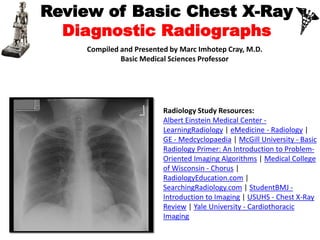
- 1. Review of Basic Chest X-Ray Diagnostic Radiographs Prepared and presented by Marc Imhotep Cray, M.D. BMS/CK Teacher 1
- 2. REVIEW OF NORMAL CHEST ADMINISTRATIVE INITIAL SURVEY Introduction Next 9 slides are modified from using data from: USUHS - Chest X-Ray Review 2
- 3. REVIEW OF NORMAL CHEST A systematic approach to film review and is therefore, designed such that the major areas of the chest should be viewed in the sequential order (see next slide for checklist) 3
- 4. 1.Check patient name, position, technical quality. 2.Soft tissue including breast, chest wall, companion shadow. 3.Review soft tissues and skeletal structures of shoulder girdles and chest wall. 4.Review abdomen for bowel gas, organ size, abnormal calcifications, free air, etc. 5.Review soft tissues and spine of neck. 6.Review spine and rib cage: check alignment, disc space narrowing, lytic or blastic regions, etc. 7.Review mediastinum: A. overall size and shape B. trachea: position C. margins: SVC, ascending aorta, right atrium, left subclavian artery, aortic arch, main pulmonary artery, left ventricle D. lines and stripes: paratracheal, paraspinal, paraesophageal (azygoesophageal), paraaortic E. retrosternal clear space REVIEW OF NORMAL CHEST (2) Sequenced Checklist 4
- 5. REVIEW OF NORMAL CHEST (3) Sequenced Checklist 8. Review hila: A.normal relationships B.size 9. Review lungs and pleura: A.compare lung sizes B.evaluate pulmonary vascular pattern: compare upper to lower lobe, right to left, normal tapering to periphery C.pulmonary parenchyma D.pleural surfaces a.fissures - major and minor - if seen b.compare hemidiaphragms c.follow pleura around rib cage 5
- 6. ADMINISTRATIVE Get in the habit of always checking the following items before anything else. It takes a few seconds and is an important legal safe guard as well. 1.Patient's name. 2.Date exam done (very important if comparing prior exams). 3.Check for position markers - right vs. left, upright. 6
- 7. Other items to check before commencing with clinical review of the film include: 1.Type of film (although this is a chest program, practice noticing if it is a plain film, CT, angio, MRI, etc.) 2.Patients position - supine, upright, lateral, decubitus. 3.Technical quality of exam - learn what are the acceptable limits for the exam. You can't find a subtle pneumothorax if there is patient motion or the film is overexposed. ADMINISTRATIVE (2) 7
- 8. A basic principle to adopt is going from general observations to specific details. Sometimes a change may be so major that the old saying about missing the forest for the trees comes true. For instance, an absent breast shadow on a film of a patient after a mastectomy. After completing your administrative housekeeping, get a general overview of the film before zooming in on tiny detail. Notice the following because it may change the baseline normals you use as reference points, and you may be sensitized to look for specific findings. 1.General Body Size, Shape, and Symmetry 2.Male vs. Female 3.Is this an infant, child, young adult, elderly person? 4.Survey for foreign objects - tubes, IV lines, EKG leads, surgical drains, prosthesis, etc., as well as non-medical objects, bullets, shrapnel, glass, etc. ADMINISTRATIVE (3) 8
- 9. The Chest X-Ray The following radiographic plates are scan ins from: Felson, B., et al.: Principles of Chest Roentgenology. Philadelphia, W.B. Saunders Co., 1973. Fraser, R., et al.: Diagnosis of Diseases of the Chest, 3rd edition. Philadelphia, W.B. Saunders Co., 1988. 9
- 10. Lecture Outline •Densities •Techniques •Anatomy •CXR Interpretation •Common Pathologies 10
- 11. Densities The big two densities are: (1) WHITE - Bone (2) BLACK - Air The others are: (3) DARK GREY- Fat (4) GREY- Soft tissue/water And if anything Man-made is on the film, it is: (5) BRIGHT WHITE - Man-made 11
- 12. Techniques - Projection P-A (relation of x-ray beam to patient) 12
- 13. Techniques - Projection (continued) Lateral 13
- 14. Techniques - Projection (continued) Lateral Decubitus 14
- 15. Rotation 15
- 17. Penetration 17
- 19. Anatomy 19
- 20. Anatomy 20
- 21. Anatomy 21
- 26. Lobes(continued) Left upper lobe with Lingula: 26
- 28. Lobes(continued) Left upper lobe - upper division: 28
- 29. Heart(continued) Right border: Edge of (r) Atrium 2. Left border: (l) Ventricle + Atrium 3. Posterior border: Left Ventricle 4. Anterior border: Right Ventricle 29
- 32. Mediastinum 32
- 33. Hilum Made of: 1. Pulmonary Art.+Veins 2. The Bronchi Left Hilus higher (max 1-2,5 cm) Identical: size, shape, density 33
- 34. Ribs 34
- 35. Lateral CXR 35
- 40. Technical Details • Type • Orientation • Rotation • Inspiration/expiration • Penetration 40
- 41. Lungs: •Lungs • Density • Symmetry • Lesions 41
- 42. Heart Size: 42
- 43. Heart • Size of heart • Size of individual chambers of heart • Size of pulmonary vessels • Evidence of stents, clips, wires and valves • Outline of aorta and IVC and SVC 43
- 44. Mediastinum: • Width • Contour • AP window • Size • Location 44
- 45. Review areas: • Apices • Behind the heart • CP angles • Below the diaphragm • Soft tissues ( breast, surgical emphysema) • Ribs & clavicle • Vertebrae 45
- 46. Identify the lesion → localise the lesion → describe the lesion → give DDx Never stop looking, carry on with your systematic approach!! 46
- 47. Pathology 47 The following radiographic plateare scan ins from: Felson, B., et al.: Principles of Chest Roentgenology. Philadelphia, W.B. Saunders Co., 1973. Fraser, R., et al.: Diagnosis of Diseases of the Chest, 3rd edition. Philadelphia, W.B. Saunders Co., 1988.
- 48. RUL pneumonia 48
- 49. RML pneumonia 49
- 50. RLL pneumonia 50
- 51. LUL pneumonia 51
- 52. LLL pneumonia 52
- 53. • Consolidation on CT 53
- 54. Hilar mass 54
- 55. The Enlarged Hila • Causes: 1. Adenopathies (neoplasia, infection) 2. Primary Tumor 3. Vascular 4. Sarcoidosis 55
- 56. • Multiple Masses 56
- 57. Hilar Lymphadenopathy - BL 57
- 58. 58Hilar Lymphadenopathy - BL
- 61. Heart failure 61
- 62. Pneumothorax 62
- 63. RUL collapse 63
- 64. LLL collapse 64
- 65. Air under the diaphragm 65
- 66. Emphysema 66
- 68. Hiatus hernia 68
- 70. Chest Tube, NG Tube, Pulm. artery cath 70
- 71. 71 Radiology Online Study Resources: Albert Einstein Medical Center – Learning Radiology eMedicine - Radiology GE - Medcyclopaedia McGill University - Basic Radiology Primer: An Introduction to Problem-Oriented Imaging Algorithms Medical College of Wisconsin - Chorus RadiologyEducation.com SearchingRadiology.com StudentBMJ - Introduction to Imaging USUHS - Chest X-Ray Review