Immune System: Immunocytology, Immunophysiology and Intro to Immunopathology
•
17 gefällt mir•1,873 views

The Immune System: Immunocytology, Immunophysiology and Intro to Immunopathology

Empfohlen
Empfohlen
Weitere ähnliche Inhalte
Was ist angesagt?
Was ist angesagt? (20)
Ähnlich wie Immune System: Immunocytology, Immunophysiology and Intro to Immunopathology
Ähnlich wie Immune System: Immunocytology, Immunophysiology and Intro to Immunopathology (20)
Mehr von Imhotep Virtual Medical School
Mehr von Imhotep Virtual Medical School (20)
Kürzlich hochgeladen
Kürzlich hochgeladen (20)
Immune System: Immunocytology, Immunophysiology and Intro to Immunopathology
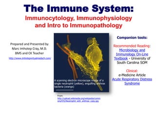
- 1. The Immune System: Immunocytology, Immunophysiology and Intro to Immunopathology Companion tools: Prepared and Presented by Marc Imhotep Cray, M.D. BMS and CK Teacher Recommended Reading: Microbiology and Immunology On-Line Textbook - University of South Carolina SOM http://www.imhotepvirtualmedsch.com/ A scanning electron microscope image of a single neutrophil (yellow), engulfing anthrax bacteria (orange) From: http://upload.wikimedia.org/wikipedia/comm ons/f/f2/Neutrophil_with_anthrax_copy.jpg Clinical: e-Medicine Article Acute Respiratory Distress Syndrome
- 2. Defense Mechanisms • All structures and processes that provide a defense against pathogens. • Defenses: – Innate (nonspecific): • Inherited as part of the structure of each organism. – Adaptive (specific): • Prior exposure (B lymphocytes). N.B.-As much of immunology is also a hematology discussion, for horizontal integration also see — IVMS Hematology-Comprehensive Blood Physiology and Pathology Review IVMS USMLE Step 1 Prep. 2
- 3. Nonspecific Immunity • External: – Skin: • Protective barrier to resist infection. – GI tract: • Gastric juice acidity. – Respiratory tract: • Mucus and cilia. – Urinary tract: • Urine acidity. – Reproductive tract: • Vaginal acidity. IVMS USMLE Step 1 Prep. 3
- 4. Phagocytosis • Distinguish between the kinds of carbohydrates that are produced by mammalian cells and those produced by bacteria. – Bacterial carbohydrates “flag” the cell for phagocytic attack. • 3 major groups of phagocytic cells: – Neutrophils: • 1st to arrive at infection. – Mononuclear phagocyte system: • Macrophages and monocytes. – Organ-specific phagocytes: • Microglia and Kupffer cells. IVMS USMLE Step 1 Prep. 4
- 5. Phagocytosis (continued) • Diapedesis: – Neutrophils and monocytes are able to squeeze through tiny gaps between adjacent endothelial cells. IVMS USMLE Step 1 Prep. 5
- 6. Phagocytosis (continued) • Connective tissue has a resident population of all leukocyte types. – Neutrophils and monocytes are highly mobile. • Recruited to site of infections by chemotaxis: – Movement toward chemical (chemokines) attractants. – If infection has spread, new phagocytes from the blood are attracted to infection. • Phagocytes engulf particles similar to amoeba. IVMS USMLE Step 1 Prep. 6
- 7. Phagocytosis (continued) – Particle becomes surrounded by pseudopods. – Forms vacuole. • Vacuole fuses with lysosomes which digest the particle. – If lysosomes are released into the infected area before vacuole is completely fused: • Contribute to the inflammation. IVMS USMLE Step 1 Prep. 7
- 8. Fever • May be a component of nonspecific defense system. • Cell wall of gram – bacteria contains endotoxin. – Endotoxin stimulate release of cytokines: • Interleukin-1, interleukin-6, and tumor necrosis factor: – Produce fever, increase sleepiness, and decrease plasma iron. IVMS USMLE Step 1 Prep. 8
- 9. Nonspecific Immunity • Body temperature is regulated by hypothalamus (thermoregulatory center). – Thermostat reset by endogenous pyrogens. • Endogenous pyrogens: – Cell wall of gram – bacteria contains endotoxin. – Endotoxin stimulates monocytes and macrophages to release cytokines: • Interleukin-1, interleukin-2, TNF. – Increased activity of neutrophils. – Increased production of interferon. IVMS USMLE Step 1 Prep. 9
- 10. Interferons (Cytokines) • Act as short-acting messengers that protect other cells in the vicinity from viral infection. • a interferon: – Inhibits viral replication, increases NK cells, and induces MHC-I antigens. • b interferon: – Inhibits viral replication, increases NK cells, and induces MHC-I antigens. • g interferon: – Activates macrophages and induces MHC-II antigens. • Immunological defense against infection and cancer. IVMS USMLE Step 1 Prep. 10
- 11. Antigens • Molecules that stimulate the production of specific antibodies. – Combine specifically with antibodies produced. • Foreign to blood and other body fluids. • Immune system can distinguish “self” molecules from nonself antigens. • Large, complex molecules can have different antigenic determinant sites. – Areas of the molecules that stimulate production of, and combine with different antibodies. IVMS USMLE Step 1 Prep. 11
- 12. Haptens • Small organic molecules can become antigens if they bind to proteins. • Become antigenic determinant sites on the proteins. – By binding haptens to proteins in the laboratory, new antigens are created for research or diagnostic purposes. IVMS USMLE Step 1 Prep. 12
- 13. Immunoassays • Tests that use the antigen-antibody complex reaction to produce clumping (agglutination). • Agglutinated particles can be used to assay a variety of antigens. • Generally use antibodies that exhibit specificity for just 1 antigenic determinant site. IVMS USMLE Step 1 Prep. 13
- 14. Lymphocytes and Lymphoid Organs • Derived from stem cells in the bone marrow. – Stem cells produce the specialized blood cells. • Replace themselves by cell division so the stem cell population is not depleted. – Lymphocytes produced by this process seed the thymus, spleen, and lymph nodes. • Produce self-replacing lymphoid colonies in these organs. • Primary lymphoid organs: – Since bone marrow and the thymus produce the T and B lymphocytes. • Both T and B cells function in specific immunity. IVMS USMLE Step 1 Prep. 14
- 15. Lymphocytes (T cells) • Cell mediated immunity: • Lymphocytes that seed the thymus become T cells. – Have surface characteristics and immunological function that differ from other lymphocytes. • Do not secrete antibodies. • Must come in close or direct contact to destroy them. – 65 – 85% of the lymphocytes in blood and most of the lymphocytes in the germinal centers of lymph nodes and spleen. • Attack host cells that have become infected with viruses, fungi, transplanted human cells, and cancerous cells. IVMS USMLE Step 1 Prep. 15
- 16. Thymus • Grows during childhood, gradually regresses after puberty. • Lymphocytes from the fetal liver and spleen; and bone marrow postnatally, seed the thymus: – Become transformed into T cells. • In adulthood, repopulation is accomplished by production in secondary lymphoid organs. IVMS USMLE Step 1 Prep. 16
- 17. Secondary Lymphoid Organs • Lymph nodes, spleen, tonsils, and Peyer’s patches. – Located in areas where antigens could gain entry to the blood or lymph. • Lymphocytes migrate from the primary lymphoid organs to the secondary lymphoid organs. – Spleen filters blood. – Secondary lymph nodes in tonsils and Peyer’s patches filter lymph. IVMS USMLE Step 1 Prep. 17
- 18. B Lymphocytes (B cells) • Humoral immunity: – Most of the lymphocytes that are not T cells are B lymphocytes (B cells). • Processed in the bone marrow. – Function in specific immunity. – B cells combat bacterial and some viral infections. • Secrete antibodies into the blood and lymph. – Provide humoral immunity as blood and lymph are body fluids (humors). – Stimulate production of memory cells: • Important in active immunity. IVMS USMLE Step 1 Prep. 18
- 19. B Lymphocytes (continued) • Others are transformed into plasma cells: – Produce 2000 antibody proteins/sec. when exposed to antigen. • These antigens may be isolated molecules or may be molecules at the surface of an invading foreign cell. – Serves to identify the enemy. • Secrete antibodies that bind to antigens. IVMS USMLE Step 1 Prep. 19
- 20. Local Inflammation • Bacteria enter body (break in skin): – Inflammatory reaction initiated by nonspecific mechanisms of phagocytosis and complement activation. • Complement activation attracts new phagocytes to the area. – After some time, B lymphocytes are stimulated to produce antibodies against specific antigens. – Attachment of antibodies to antigens amplifies nonspecific responses. • Activates complement. – Antibodies promote phagocytic activity of neutrophils, macrophages, and monocytes (opsinization). IVMS USMLE Step 1 Prep. 20
- 21. Local Inflammation (continued) • Leukocytes interact with adhesion molecules in endothelial cell. • Chemotaxis occurs, attracting leukocytes. – Diapedesis occurs. – First to arrive are neutrophils, then monocytes, and T lymphocytes. • Adherence of monocytes to extracellular matrix proteins promotes conversion into macrophages. IVMS USMLE Step 1 Prep. 21
- 22. Local Inflammation (continued) • Mast cells secrete prostaglandins, leukotrienes, histamine, cytokines, and TNF-a. – Increases membrane permeability. – Vasodilation of blood vessels. – Recruit neutrophils. IVMS USMLE Step 1 Prep. 22
- 23. Local Inflammation (continued) • Characteristic effects of inflammation: – Redness and warmth. • Histamine stimulated vasodilation. – Swelling (edema). – Pus. • Accumulation of dead leukocytes. – Pain. – If infection continues, endogenous pyrogens released. IVMS USMLE Step 1 Prep. 23
- 24. Antibodies • Antibody proteins are also known as immunoglobulins. – Found in the gamma globulin class of plasma proteins. • Different antibodies have different structure, as the antibodies have specific actions. IVMS USMLE Step 1 Prep. 24
- 25. Antibodies IVMS USMLE Step 1 Prep. (continued) 25
- 26. Antibody Structure • 100 million trillion antibody molecules that contain 4 polypeptide chains. • 2 long H chains are joined to 2 shorter L chains. – Fab regions are variable. • Provide specificity for a bonding to an antigen. – Fc region of different antibodies are constant. • B cells have antibodies that serve as receptors for antigens. IVMS USMLE Step 1 Prep. 26
- 27. The Complement System • Nonspecific defense system. – The combination of antibodies with antigens does not cause destruction of the antigens or pathogen. • Antibodies serve to identify the targets for immunological attack. – Stimulate opsinization. • Antibody-induced activation of the complement. IVMS USMLE Step 1 Prep. 27
- 28. The Complement System • The complement proteins are designated C-1 to C-9. – These proteins are in an inactive state. • Become activated by the attachment of antibodies to antigens. • Complement proteins can be subdivided into 3 components: – C1: recognization. – C4, C2, C3: activation. – C5-C9: attack (complement fixation). IVMS USMLE Step 1 Prep. 28
- 29. The Complement System (continued) • Classic pathway: • Antibodies of IgG and IgM attach to antigens on invading cell membranes. – Binding to C1 activates the process. • Activated C1 hydrolyzes C4 into C4a and C4b. • C4b binds to the cell membrane and becomes an active enzyme. • C4b splits C2 into C2a and C2b. • C2a attaches to C4b and cleaves C3 into C3a and C3b. – Alternate pathway converges with classic pathway. • Fragment C3b becomes attached to the complex in the cell membrane. • C3b converts C5 to C5a and C5b. IVMS USMLE Step 1 Prep. 29
- 30. Fixation of Complement Proteins IVMS USMLE Step 1 Prep. 30
- 31. Complement Fixation • C5b and C6 through C9 are inserted into bacterial cell membrane, to form a membrane attach complex (MAC). – Create large pores in membrane, causing osmotic influx of H20. • Complement proteins kill the cell. IVMS USMLE Step 1 Prep. 31
- 32. Complement Fragments • Chemotaxis: – C5a acts as a cytokine to attract neutrophils and monocytes to the site. • Attract phagocytes. • Opsinization: – Phagocytes have receptors for C3b. • Form bridges between phagocyte and victim cell. • C3a and C5a stimulate mast cells to secrete histamine: – Increase blood flow and capillary permeability. • Bring in more phagocytes. IVMS USMLE Step 1 Prep. 32
- 33. Killer (Cytotoxic) T Cells • Cell mediated destruction. – Can be identified by the CD8 coreceptor. • Destroy specific cells with antigens on their surface. – Must be in actual contact with their victim cells. • Secrete perforins: – Perforins polymerize in the cell membrane and form cylindrical channels through the membrane. • Osmotic destruction of the cell. IVMS USMLE Step 1 Prep. 33
- 34. Killer (Cytotoxic) T Cells (continued) • Secrete granzymes: – Enter the victim cell activating caspases: • Enzymes involved in apoptosis. • Destruction of victims cell’s DNA. IVMS USMLE Step 1 Prep. 34
- 35. Helper T Cells • Identified by CD4 coreceptor. • Indirectly participate by regulating the response of both T killer and B cells. • B cells must be activated by helper T cells before they produce antibodies. IVMS USMLE Step 1 Prep. 35
- 36. Interaction of Macrophages, Helper T and Killer T cells Insert fig. 15.18 IVMS USMLE Step 1 Prep. 36
- 37. Suppressor T Cells • Indirectly participate in the specific immune response. • Inhibit T cell and B cell activities. • Affect the amount of antibodies secreted. • Moderate immune response. IVMS USMLE Step 1 Prep. 37
- 38. Lymphokines • Interleukin-1: – Secreted by macrophages and other cells. • Activates T cells. • Interleukin-2: – Released by helper T cells. • Activates killer T cells. • Interleukin-3: – Serves as a growth factor. • Activates killer T cells. • Interleukin-4: – Secreted by T cells. • Required for proliferation and clone development of B cells. • G-CSF and GM-CSF: – Promote leukocyte development. IVMS USMLE Step 1 Prep. 38
- 39. Subtypes of Helper T Cells • TH1: – Produces interleukin-2 and gamma interferon. • Promotes cell mediated immunity and activates killer T cells. • Stimulates NO production. – Interleukin-12: • Changes “uncommitted” helper T cells into TH1 cells. • TH2: – Secretes interleukin-4, interleukin-5, and interleukin-10. • Promotes humoral immunity and stimulates B lymphocytes. – Activates mast cells. IVMS USMLE Step 1 Prep. 39
- 40. T Cell Receptor Proteins • Only protein antigens are recognized by most T cells. – T cell receptors cannot bind to free antigens. – In order for T cells to respond to foreign antigens: • Antigen must be presented to T cells on the membrane of antigen presenting cells. – Chief antigen presenting cells are macrophages and dendritic cells. – To interact with correct T cells, dendritic cells migrate to secondary lymphoid organs. • Secrete chemokines. IVMS USMLE Step 1 Prep. 40
- 41. Major Histocompatability Complexes (MHC) • All cells except mature RBCs are genetically marked with histocompatability antigens on the membrane surface. – Also called human leukocyte antigens (HLAs). • The histocompatability antigens are coded for a group of genes called MHC located on chromosome 6. • MHC genes produce 2 classes of MHC molecules: – Class-1. – Class-2. IVMS USMLE Step 1 Prep. 41
- 42. Major Histocompatability Complexes (continued) • MHC class-1: – Produced by all cells but RBCs. – Picks up cytoplasmic peptides and transports to membrane. • Killer T cells (cytotoxic) interact with antigens. – Coreceptor CD8 permits each type of T cell to interact only with a class-1 MHC molecules. IVMS USMLE Step 1 Prep. 42
- 43. Major Histocompatability Complexes (continued) • MHC class-2: – Produced only by antigen-presenting cells and B cells. • Present class-2 MHC together with antigens to helper T cells. • Activates T cells. – Helper T cells react with antigens. • Promotes B cell response. – Appear only on cell membrane when cell is processing antigens. – Coreceptor CD4 interacts with only a specific class of MHC molecule. IVMS USMLE Step 1 Prep. 43
- 44. Coreceptors on Helper and Killer T cells IVMS USMLE Step 1 Prep. 44
- 45. T Cell Response to a Virus • Foreign particle infects body. – Macrophages engulf by phagocytosis. – Partially digested virus particles (foreign antigens) are moved to surface of cell membrane. – Foreign antigens forms a complex with class-2 MHC molecules. – Presented to helper T cells. IVMS USMLE Step 1 Prep. 45
- 46. T Cell Response to a Virus (continued) • Macrophages secrete interleukin-1 and TNF. – Interleukin-1 stimulates cell division and proliferation of T cells. • Activated T cells secrete M-CSF and g- interferon. – Promote the activity of the macrophages. • Killer T cells destroy infected cell if class-1 MHC present. – Helper T cells activate B cells. IVMS USMLE Step 1 Prep. 46
- 47. T Cell Response to a Virus (continued) • Foreign antigens attach to immunoglobulins on B cells. • B cells can present the antigen with class-2 MHC molecules to helper T cells. – Stimulate B cell production, conversion to plasma cells, and antibody production. IVMS USMLE Step 1 Prep. 47
- 48. Destruction of T Lymphocytes • Activated T cells must be destroyed after the infection has cleared. • T cells produce a surface receptor called FAS. – Production of FAS increases during the infection. • After a few days, activated T cells begin to produce FAS ligand. – FAS binds to FAS ligand and triggers apoptosis (cell suicide). • Help maintain immunologically privileged sites. IVMS USMLE Step 1 Prep. 48
- 49. Active Immunity • Primary response: – First exposure to pathogen, immune response insufficient to combat disease. – Latent period of 5-10 days before measurable amounts of specific antibodies appear in blood. • Secondary response: – Subsequent exposure to same antigen. – Antibody production is much more rapid. • Maximum antibody concentration reached in < 2 hrs. – Maintained longer period of time. IVMS USMLE Step 1 Prep. 49
- 50. Primary and Secondary Immune Responses IVMS USMLE Step 1 Prep. 50
- 51. Clonal Selection Theory • Mechanism by which secondary responses are produced. – B lymphocytes inherit the ability to produce a particular antibody. – T lymphocytes inherit the ability to respond to particular antigens. • A given B cell can produce only 1 type of antibody with specificity for 1 antigen. – Inherited specificity reflected in antigen receptor proteins. • Exposure stimulates specific lymphocytes to divide many times until a large population of genetically identical cells (clone) is produced. IVMS USMLE Step 1 Prep. 51
- 52. Clonal Selection Theory (continued) • Some of the cells become plasma cells that secrete primary response. • Others become memory cells that secrete antibodies during the secondary response. • Antigens select lymphocytes that are already able to make antibodies. IVMS USMLE Step 1 Prep. 52
- 53. Active Immunity • Development of a secondary response provides active immunity. • Immunizations induce primary responses. – Inoculating people with pathogens whose viruses have been attenuated or destroyed. IVMS USMLE Step 1 Prep. 53
- 54. Immunological Tolerance • Immunological competence occurs during the 1st month postnatally. – Ability to produce antibodies against non-self antigens while tolerating self-antigens. • Requires continuous exposure to those antigens. • Autoantibodies: – Exposure to self-antigens results in antibody production. • Autoreactive T cells: – Killer T cells attack self-antigens. IVMS USMLE Step 1 Prep. 54
- 55. Passive Immunity • Immune protection produced by the transfer of antibodies to a recipient from a donor. – The donor has been actively immunized. • Person who receives these ready-made antibodies is passively immunized. – Occurs naturally in mother to fetus during pregnancy and mother to infant during nursing. • Immunological competence: – Ability to mount a specific immune response. – Does not develop until 1 month after birth. – Passive immunity disappears when infant is 1 month old. • Infant did not itself produce lymphocytes. IVMS USMLE Step 1 Prep. 55
- 56. Monoclonal Antibodies • Commercially prepared. • Exhibit specificity for one antigenic determinant only. • Results in more sophisticated clinical laboratory tests. – May aid in the diagnosis of cancer. • May result in production of drugs combined with monoclonal antibodies against specific tumor antigens. IVMS USMLE Step 1 Prep. 56
- 57. Tumor Immunology • Tumors are interrelated with the functions of the immune system. – Division of tumor cells is not effectively controlled by normal inhibitory mechanisms. • Tumor cells also dedifferentiate (become similar to less specialized cells of an embryo). – As tumor cells dedifferentiate, they reveal surface antigens that can stimulate the immune destruction of the tumor. IVMS USMLE Step 1 Prep. 57
- 58. Tumor Immunology (continued) • Some of these antigens are proteins produced in embryonic or fetal life. – Are absent at the time immunological competence is established. – Body treats these antigens as foreign. • Release of these antigens provides the basis of laboratory diagnosis of some cancers. IVMS USMLE Step 1 Prep. 58
- 59. Natural Killer (NK) Cells • Lymphocytes that are related to, but distinct from T cells. • Provide first line of cell-mediated defense. – NK cells destroy tumors in a nonspecific fashion. • NK cells attach to cells that lack class-1 MHC antigens. – Release perforins and granzymes. • Do not require prior exposure for sensitization to the tumor antigens. • Stimulated by interferon. IVMS USMLE Step 1 Prep. 59
- 60. Immunotherapy for Cancer • Interleukin-2 activates both killer T and B lymphocytes. – Patients treated with lymphocytes with IL-2 produce lymphokine-activated killer T cells. • Gamma interferon is used to treat particular forms cancer. – Lymphomas, renal carcinomas, melanoma, Kaposi’s sarcoma. • Tumor infiltrating lymphocyte is promising. IVMS USMLE Step 1 Prep. 60
- 61. Effects of Aging and Stress • Cancer risks increase with age. – Aging lymphocytes accumulate genetic errors that decrease effectiveness. – Thymus functions decline. • Stress: – Tumors grow faster. • Increased production of corticosteroids. IVMS USMLE Step 1 Prep. 61
- 62. Diseases Caused by the Immune System • Immune system can be deranged in its ability to tolerate self-antigens while it identifies and attacks foreign antigens. • Diseases caused by the immune system can be grouped into 3 categories: – Autoimmune disease. – Immune complex diseases. – Allergy or hypersensitivity. IVMS USMLE Step 1 Prep. 62
- 63. Autoimmunity • Diseases produced by failure in the immune system to recognize and tolerate self-antigens. – Activates autoreactive T cells and stimulates production of autoantibodies by B cells. • Failure due to: – An antigen that does not normally circulate in the blood may be exposed to the immune system. • Thyroglobulin. – A self-antigen that is otherwise tolerated may be altered by combining with a foreign hapten. • Thrombocytopenia. IVMS USMLE Step 1 Prep. 63
- 64. Autoimmunity (continued) – Antibodies may be produced that are directed against other antibodies. • Rheumatoid arthritis. – Antibodies produced against foreign antigens may cross-react with self-antigens. • Rheumatic fever. – Self-antigens may be presented to the helper T cells together with class-2 MHC molecules. • Type I diabetes. IVMS USMLE Step 1 Prep. 64
- 65. Immune Complex Diseases • Damage produced by inflammatory response. – Antigen-antibody combinations that are free rather than attached to bacterial or other cells: • Activate complement proteins and promotes inflammation. – When large # of immune complexes are continuously formed, inflammation may be prolonged. • Hepatitis B. – Formation of complexes between self-antigens and autoantibodies: • Rheumatoid arthritis. IVMS USMLE Step 1 Prep. 65
- 66. Allergy • Abnormal immune responses to allergens. • Immediate hypersensitivity: – Dendritic cells stimulate TH2 cells to secrete interleukin-4 and interleukin-13: • Stimulate B cells and plasma cells to secrete IgE antibodies. – Do not circulate in the blood. IVMS USMLE Step 1 Prep. 66
- 67. Immediate Hypersensitivity • IgE antibodies attach to mast cells and basophils. – When exposed again to same allergen, the allergen binds to antibodies attached to mast cells and basophils. • Stimulate secretion of histamine, leukotrienes, and prostaglandin D. –IVMS USMLE Step 1 Prep. Produce symptoms. 67
- 68. Delayed Hypersensitivity • Symptoms take longer time to develop (hours to days). – Is a cell mediated T cell response. • Symptoms caused by secretion of lymphokines, not histamine. – Antihistamines provide little benefit. IVMS USMLE Step 1 Prep. 68
- 69. THE END THANK YOU FOR YOUR ATTENTION IVMS USMLE Step 1 Prep. 69
- 70. Resources for further study: Vaccine Research Center Information regarding preventative vaccine research studies Immune System - from the University of Hartford Immunobiology; Fifth Edition – Online version of the textbook by Immunology - BioMed Central (free content) scientific journal The Microbial World - Animal defenses against microbes - Chapter in on-line microbiology textbook Microbiology and Immunology On-Line Textbook - from the University of South Carolina School of Medicine IVMS USMLE Step 1 Prep. 70