Imaging the cv junction.part 1. himadri s das
•Als PPTX, PDF herunterladen•
33 gefällt mir•20,997 views


Empfohlen
Empfohlen
Weitere ähnliche Inhalte
Was ist angesagt?
Was ist angesagt? (20)
Andere mochten auch
Andere mochten auch (20)
Ähnlich wie Imaging the cv junction.part 1. himadri s das
Ähnlich wie Imaging the cv junction.part 1. himadri s das (20)
Mehr von Dr. Himadri Sikhor Das
Mehr von Dr. Himadri Sikhor Das (19)
Kürzlich hochgeladen
Kürzlich hochgeladen (20)
Imaging the cv junction.part 1. himadri s das
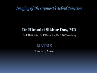
- 1. Imaging of the Cranio-Vertebral Junction Dr Himadri Sikhor Das, MD Dr.P.Hatimota, Dr.P.Hazarika, Dr.C.D.Choudhury. MATRIX Guwahati, Assam
- 2. CV JUNCTION “The C-V junction is a transition site between mobile cranium and relatively rigid spinal column. It is also the site of the medullo spinal junction”. Parts of CV Junction include: The Occiput First Cervical Vertebra (Atlas) Second Cervical Vertebra (Axis) Their articulations and Connecting ligaments
- 3. Embryology of the CV junction AXIS develops from five primary and two secondary centers. Body and arches – II cervical sclerotome Base of Dens – I cervical sclerotome Apical segment of Dens – IV occipital sclerotome.
- 4. Embryology of the CV junction The apical segment is not ossified until 3 years of age. At 12 years it fuses with odontoid to form normal odontoid; failure leads to Os Terminale 1 2 Tip of dens 3 Dens 5 4 Body of dens 6
- 5. Anatomy of the CV junction ATLANTO-AXIAL JOINT: Most active joint in the body, moving approximately 600 times per hour. Normal range of cervical motion is 900 on each side, range of rotation of atlas on axis being 25-530 Rotation of >560 on one side or a R-L diff >80 implies hyper mobility Rotation of <280 implies hypo mobility
- 6. CV Junction
- 7. Anatomy of the CV junction Occipital condyles Lateral mass of atlas Apical Lig Alar Lig Transverse lig Cruciate Ligament vertical band Atlantoaxial joint Tectorial Membrane
- 8. Plain radiographs (standard FFD) Lateral Open mouth Dynamic Flexion & Extension (to r/o atlantoaxial subluxation / occipitalization of atlas) Tomograms –AP and Lateral Vertebral angiogram may be necessary in select cases, especially BI
- 9. Imaging of the CV junction-CT Spiral CT 1-3 mm collimation Pitch =1 Overlapping reconstruction In neutral position With head rotated (Torticollis ) Flexion (? Transverse ligament abnormality ) 2-D axial and multiplanar 3D Dynamic CT (AARF)
- 10. Imaging of the CV junction- MRI Technique Surface / Head coil Contiguous thin sections (2-3mm with 3D acquisitions) Sagittal sections in flexion if C1-C2 Dislocation to be ruled out Sequences SESagittal 3D T1W FSE Sagittal T2W CEMR (Gd )Only if intra/extramedullary lesion is suspected Kinematic MRI – cord compression in AAD, AARF GRE – to delineate bony cortical outline
- 11. X-ray
- 12. X-ray
- 13. X-ray
- 14. Disorders of the CV junction Congenital bony malformations OCCIPUT ANOMALIES Basilar invagination Anomalies of atlas Odontoid abnormality Atlanto-axial instability Others
- 15. Disorders of the CV junction ACQUIRED MALFORMATIONS Trauma Arthritides Infection Degeneration Tumours
- 16. Lines and Angles The important lines are Chamberlain’s line Wackenheim’s clivus canal line Mc Gregor’s line (basal line)
- 17. Lines and Angles The important angles are Basal angle Bull’s angle Height index of Klaus
- 18. Chamberlain’s line Synonym Chamberlain's (Palato-occipital Line) Definition Joins posterior tip of hard palate to posterior tip of Foramen Magnum (opisthion) Tip of dens below this line ±3 mm >7mm or >1/2 of odontoid def basilar Invagination DRAWBACK – posterior rim anatomical variations.
- 19. Mc Gregor’s line Synonym McGregor’s Line Definition Line drawn from posterior tip of Hard palate to lowest part of Occiput Odontoid tip >4.5mm above = Basilar Invagination Should be used when lowest part of occipital bone is not Foramen Magnum.
- 20. Wackenheim’s Line Synonym Wackenheim's (Clivus canal) line Definition Line drawn along clivus into cervical spinal canal Odontoid is ventral and tangential to this line
- 21. McRae’s Line Synonym McRae's (Foramen Magnum) line Definition Joins anterior and posterior edges of Foramen magnum * Tip of odontoid is below this line. ** When sagittal diameter of canal <20mm, neurological symptoms occur – Foramen Magnum Stenosis
- 22. Welcher’s Basal Angle Synonym BASAL ANGLE Definition Angle between two lines drawn from Nasion to tuberculum sella Tuberculum sellae to the basion along plane of the clivus Normal – 1240 - 142 > 1450 = platybasia < 1300 is seen in achondroplasia
- 23. BULL’S ANGLE Line representing prolongation of hard palate and line joining the midpoints of the ant & post arches of C1. Normal : <100 Basilar invagination - >130
- 24. Platybasia – refers only to an abnormally obtuse basal angle, may be asymptomatic, and is not a measure of basilar invagination.
- 25. Basilar impression vs. Basilar invagination • Basilar impression is defined as upward displacement of vertebral column elements into the foramen magnum.
- 26. Specific Anomalies- Basilar Invagination • primary developmental defect implying prolapse of vertebral column into the skull at the base due to softening of the bones at the base of the skull. Causes Hyperparathyroidism Hurler's syndrome Rickets/OM/Scurvy Hajdu-Cheney Syndrome. Paget's disease. Cleidocranial dysostosis Osteogenesis Imperfecta “ As can be seen from the causes almost all of the causes are associated with bone softening conditions due to which the CV junction is invaginated like a sleeve”
- 27. Classification of Basilar Invagination Morphological classification Anterior Paramedian Clivus short Clivus Normal/long Ass. with Platybasia Not ass. with Platybasia Etiological classification (synonyms ) 1.Basilar Coarctation ( congenital-Foramen Magnum syndrome) 2.Basilar Erosion (Inflammatory/Neoplastic/Infective) (RA/Tuberculosis/Nasopharyngeal CA) 3.Basilar Impression (Bone softening conditions) (OI/Paget’s/Osteomalacia)
- 28. BASILAR INVAGINATION : CT Thin sections 1-3 mm Overlapping recon Multiplanar recon : Sag & Coronal
- 31. KINEMATIC MRI IN BI
- 32. ATLANTO-AXIAL SUBLUXATION (AAS) : anterior type Anterior Atlanto-Dental Interval (AADI) : AAS is present when it is >3mm in adults & >5mm in children Measured from posteroinferior margin of ant arch of C1 to the ant surface of odontoid AADI 3-6 mm trans lig. damage AADI >6mm alar lig. damage also AADI >9mm surgical stabilization
- 33. ATLANTO-AXIAL SUBLUXATION (AAS) : anterior type Posterior Atlanto-Dental Interval (PADI) : ** Distance b/w posterior surface of odontoid & anterior margin of post ring of C1 Considered better method as it directly measures the spinal canal Normal : 17-29 mm at C1 PADI <14mm : predicts cord compression
- 34. ATLANTO-AXIAL SUBLUXATION (AAS) : anterior type X-rays in neutral position will miss AAS in 48%. Controlled flexion views always to be done
- 35. ATLANTO-AXIAL SUBLUXATION (AAS) : anterior type
- 36. ATLANTO-AXIAL SUBLUXATION (AAS) : anterior type
- 37. ATLANTO-AXIAL SUBLUXATION (AAS) : anterior type
- 38. AAD
- 39. ATLANTO-AXIAL SUBLUXATION (AAS) : anterior type
- 40. AAD with cord compression
- 41. Pseudosubluxation In children, C2-3 space & sometimes C3-4 space have normal physiologic displacement Line drawn from ant. aspect of spinous process of C1-3 should not be >1mm far from any spines
- 42. RISK FACTORS FOR CORD COMPRESSION IN AAS AADI > 9 mm PADI < 14 mm Basilar Invagination, especially if associated with AAS of any degree Sub axial canal diameter < 14 mm CHI <2
- 43. ATLANTO-AXIAL SUBLUXATION (AAS) : rare types Posterior AAS – rare, associated with deficient odontoid process. Rotatory AAS - (later) Lateral AAS - >2 mm offset of the lateral mass of C1 on C2 on coronal images. It is usually associated with a rotational deformity.
- 44. Specific Anomalies – Occiput anomalies Condylus Tertius (IIIrd occipital condyle) : when proatlas persists or fails to migrate, an ossified remnant is seen at distal end of clivus May form pseudo joint with odontoid or ant arch of C1 and limit mobility of CVJ Increased prevalence of Os Odontoideum seen
- 45. ATLAS ASSIMILATION Represents most cephalic ‘blocked vertebra’ 0.25% of population Usually occurs in association with other anomalies such as BI and Klippel Feil syndrome. Associated with segmentation failures b/w C2-3 : atlanto-axial subluxation in 50%.
- 46. Atlas assimilation with CVJ anomaly
- 48. KLIPPEL-FEIL SYNDROME : classic triad consists of low posterior hairline, short neck and limitation of neck movements.
- 49. KLIPPEL-FEIL SYNDROME Fused vertebrae (usually C2-3 and C5-6 interfaces) Hemivertebrae Atlas occipitalization Spina bifida occulta Scoliosis Urogenital, otological anomalies, Chiari, syndactyly, Sprengel’s etc.
- 50. Atlas rachischisis: posterior >> anterior Both together – ‘split atlas’
- 51. ATLANTO-AXIAL ROTATORY FIXATION Less common cause of Torticollis in children. Fixation usually occurs within normal range of rotation of A-A joint. Fielding types: Type I : fixn without anterior displ of atlas (trans & alar lig intact, MC type) Type II : fixn with ant displ 3-5mm (trans lig deficient) Type III : fixn with ant displ >5mm (trans & alar lig deficient) Type IV : fixn with posterior displacement of atlas (rarest, odontoid is deficient) Type V : AARF with AORF (atlanto-occipital rotatory fixation) combined DIAGNOSIS: X-Ray : asymmetry of lateral masses on open mouth odontoid view. Lateral mass that has rotated forwards appear wider and closer to midline. Asymmetry also on skull lateral.
- 52. ATLANTO-AXIAL ROTATORY FIXATION CT: essential d/t difficulty in obtaining & interpreting X-rays rotated atlas + displacement, fractures, AORF
- 53. ATLANTO-AXIAL ROTATORY FIXATION Dynamic CT: type I AARF vs. other c/o torticollis : Pt. with fixn demonstrate little or no motion of atlas on the axis. Normal pts or with transient torticollis show a reduction or reversal of the rotation. Dynamic MRI also
- 54. ODONTOID ABNORMALITIES Persistent Ossiculum Terminale : Also called Bergman Ossicle. Results from failure of fusion of the terminal ossicle to the rest of odontoid Normally fusion occurs by 12 yrs of age Stable anomaly when isolated with normal height of dens
- 55. Persistent Ossiculum Terminale May mimic type I odontoid # (avulsion of terminal ossicle) : difficult to differentiate at times.