2. Digestion
- The major foods on which the body lives can be
classified as carbohydrates, fats and proteins
- small quantities of vitamins and minerals
- They generally cannot be absorbed in their
natural forms – useless as nutrients without
preliminary digestion
- Hydrolysis of Carbohydrates, fats & proteins
3. Digestion of Carbohydrates
- sucrose, which is the disaccharide known popularly as
cane sugar;
- lactose, which is a disaccharide found in milk;
- starches, which are large polysaccharides present in
almost all nonanimal foods - particularly in potatoes
and the different types of grains
- Other carbohydrates ingested to a slight extent are
amylose, glycogen, alcohol, lactic acid, pyruvic acid,
pectins, dextrins
- Cellulose - no enzymes
4. Digestion of Carbohydrates
- Saliva – ptyalin – alpha amylase – parotid gland
- Hydrolyzes starch into the disaccharide maltose
- the food remains in the mouth only a short time - not
more than 5 per cent of all the starches will be
hydrolyzed
- starch digestion sometimes continues in the body and
fundus of the stomach for as long as 1 hour before the
food becomes mixed with the stomach secretions
- Salivary amylase activity blocked – 40 per cent starches
to maltose
5. Digestion of Carbohydrates
- Pancreatic Alpha amylase – more powerful than
salivary amylase – maltose
- enterocytes lining the villi of the small intestine
contain four enzymes (lactase, sucrase, maltase and
alpha - dextrinase),
- which are capable of splitting the disaccharides
- lactose – glucose + galactose (10 per cent),
- Sucrose - glucose + fructose (10 per cent),
- Maltose - glucose + glucose (80 per cent)
- Final product – water soluble monosaccharides –
absorbed into portal blood
6.
7. Digestion of Proteins
- The dietary proteins are chemically long chains of
amino acids bound together by peptide linkages
- Stomach – pepsin – HCL – digest collagen -
meat
- pepsin only initiates the process of protein
digestion, usually providing only 10 to 20 per cent
of the total protein digestion to convert the protein
to proteoses, peptones and a few polypeptides
- major proteolytic pancreatic enzymes: trypsin,
chymotrypsin, carboxypolypeptidase and
proelastase
8. Digestion of Proteins
- Both trypsin and chymotrypsin split protein
molecules into small polypeptides;
- Carboxypolypeptidase then cleaves individual amino
acids from the carboxyl ends of the polypeptides
- Proelastase, in turn, is converted into elastase, which
then digests elastin fibers that partially hold meats
together.
- Only a small percentage of the proteins are digested all
the way to their constituent amino acids by the
pancreatic juices. Most remain as dipeptides and
tripeptides
9. Digestion of Proteins
- enterocytes that line the villi of the small
intestine, mainly in the duodenum and jejunum
- These cells have a brush border that consists of
hundreds of microvilli projecting from the
surface of each cell – peptidases
- aminopolypeptidase and dipeptidases – larger
polypeptides into tripeptides and dipeptides and a
few into amino acids
- Dipeptides and tripeptides are easily transported
through the microvillar membrane to the interior
of the enterocyte – peptidase – amino acids
10.
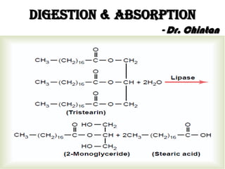
11. Digestion of Fats
- most abundant fats of the diet are the neutral fats,
also known as triglycerides, each molecule of
which is composed of a glycerol nucleus and
three fatty acid side chains
- small quantities of phospholipids, cholesterol and
cholesterol esters
- A small amount of triglycerides is digested in the
stomach by lingual lipase that is secreted by
lingual glands in the mouth and swallowed with
the saliva
- less than 10 per cent and generally unimportant
12. Digestion of Fats
- The first step in fat digestion is physically to break the
fat globules into very small sizes so that the water-
soluble digestive enzymes can act on the globule
surfaces.
- This process is called emulsification of the fat, and it
begins by agitation in the stomach to mix the fat with
the products of stomach digestion.
- Then, most of the emulsification occurs in the
duodenum under the influence of bile - large quantity
of bile salts as well as the phospholipid lecithin.
- Bile salts & lecithin – water & fat soluble – detergent
action
13. Digestion of Fats
- The lipase enzymes are water-soluble
compounds and can attack the fat globules only
on their surfaces
- enzyme for digestion of the triglycerides is
pancreatic lipase
- the enterocytes of the small intestine contain
enteric lipase
- Most of the triglycerides of the diet are split by
pancreatic lipase into free fatty acids and 2-
monoglycerides
14.
15. Digestion of Fats
- Bile salts, when in high enough concentration in
water, have the propensity to form micelles, which
are small spherical, cylindrical globules
- 3 to 6 nanometers in diameter composed of 20 to
40 molecules of bile salt
- Inside fats – outside surface water soluble
- The bile salt micelles also act as a transport
medium to carry the monoglycerides and free
fatty acids - relatively insoluble, to the brush
borders of the intestinal epithelial cells
16.
17. Digestion of Fats
- Bile salts released back into the chyme & again reused
- the enzyme cholesterol ester hydrolase to hydrolyze
the cholesterol ester,
- phospholipaseA2 to hydrolyze the phospholipid
- The bile salt micelles play the same role in “ferrying”
free cholesterol and phospholipid molecule that
they play in “ferrying” monoglycerides and free fatty
acids
- no cholesterol is absorbed without this function of the
micelles
18.
19. Absorption
- The stomach is a poor absorptive area of the GIT because it
lacks the typical villus type of absorptive membrane, and also
because the junctions between the epithelial cells are tight
junctions.
- Only a few highly lipid-soluble substances, such as
alcohol and some drugs like aspirin, can be absorbed in small
quantities
- absorptive surface of the small intestinal mucosa, showing
many folds called valvulae conniventes (or folds of
Kerckring), which increase the surface area of the absorptive
mucosa about 3-fold.
- These folds extend circularly most of the way around the
intestine and are especially well developed in the duodenum
and jejunum
20.
21. Absorption
- millions of small villi
- The villi lie close to one another in the upper small
intestine, but their distribution is less profuse in the
distal small intestine
- enhances the total absorptive area 10-fold
- each intestinal epithelial cell on each villus is
characterized by a brush border consisting of as many
as 1000 microvilli
- This increases the surface area exposed to the intestinal
materials another 20-fold – total 1000-fold
22.
23.
24. Absorption of Water
- Isosmotic Absorption
- Diffusion - usual laws of osmosis
- when the chyme is dilute enough, water is
absorbed through the intestinal mucosa into the
blood of the villi almost entirely by osmosis
- when hyperosmotic solutions are discharged
from the stomach into the duodenum - water
usually will be transferred by osmosis to make the
chyme isosmotic with the plasma
25. Absorption of Ions
- Active Transport of Sodium - Energy - ATP
- the intestines must absorb 25 to 35 grams of
sodium each day
- Whenever significant amounts of intestinal
secretions are lost to the exterior - extreme
diarrhea - the sodium reserves of the body can
sometimes be depleted to lethal levels within
hours
- Chyme - more Na than brush border
26.
27. Absorption of Ions
When a person becomes dehydrated, large
amounts of aldosterone secreted by the cortices
of the adrenal glands
↓
increased activation of the enzyme and
transport mechanisms for all aspects of sodium
absorption by the intestinal epithelium
↓
increased sodium absorption in turn causes
secondary increases in absorption of chloride
ions, water
28. Absorption of Ions
- Absorption of Chloride Ions in the Duodenum
and Jejunum
- chloride ion absorption is rapid and occurs mainly
by diffusion
- absorption of sodium ions through the epithelium
creates electronegativity in the chyme and
electropositivity in the paracellular spaces between
the epithelial cells.
- Chloride ions move along this electrical gradient to
“follow” the sodium ions
29. Absorption of Ions
- Absorption of Bicarbonate Ions in the
Duodenum and Jejunum
- large quantities of bicarbonate ions must be
reabsorbed from the upper small intestine
because large amounts of bicarbonate ions have
been secreted into the duodenum in both
pancreatic secretion and bile
- The bicarbonate ion is absorbed in an indirect
way
30. Absorption of Ions
- When sodium ions are absorbed, moderate
amounts of hydrogen ions are secreted into the
lumen of the gut in exchange for some of the
sodium
- These hydrogen ions in turn combine with the
bicarbonate ions to form carbonic acid
(H2CO3), which then dissociates to form water
and carbon dioxide
- The water remains as part of the chyme in the
intestines, but the carbon dioxide is readily
absorbed into the blood and subsequently expired
through the lungs
31. Absorption of Ions
- Secretion of Bicarbonate Ions in the Ileum and
Large Intestine
- The epithelial cells on the surfaces of the villi in
the ileum as well as on all surfaces of the large
intestine have a special capability of secreting
bicarbonate ions in exchange for absorption of
chloride ions
- it provides alkaline bicarbonate ions that
neutralize acid products formed by bacteria in
the large intestine
32. Absorption of Ions
- New epithelial cells secrete sodium chloride and
water into the intestinal lumen - This secretion in
turn is reabsorbed by the older epithelial cells
- The toxins of cholera and of some other types of
diarrheal bacteria can stimulate the secretion so
greatly that this secretion often becomes much
greater than can be reabsorbed
- Cholera toxin – cAMP – opening of CL channels –
active Na pump – water osmosis
- too much of a good thing can be lethal; Rx -
NaCl
33. Absorption of Ions
- Calcium ions are actively absorbed into the blood
especially from the duodenum – PTH, vitamin D
- Iron ions are also actively absorbed from the small
intestine – body need for Hb
- Potassium, magnesium, phosphate can also be
actively absorbed through the intestinal mucosa
- Monovalent (Na, K, Cl) easily absorbed than
Divalent (Ca)
34.
35. Absorption of Nutrients
- Absorption of Carbohydrates
- form of monosaccharides - small fraction are
absorbed as disaccharides
- Glucose Is Transported by a Sodium Co-
Transport Mechanism
- First is active transport of sodium ions through
the basolateral membranes of the intestinal
epithelial cells into the blood, thereby depleting
sodium inside the epithelial cells
36. Absorption of Glucose
- Second, decrease of Na inside the cells causes
Na from the intestinal lumen to move through
the brush border of the epithelial cells to the
cell interiors by a process of facilitated
diffusion
- Na ion combines with a transport protein, but
the transport protein will not transport the Na
to the interior of the cell until the protein itself
also combines with some other substance such
as glucose
37. Absorption of Glucose
- - the low concentration of sodium inside the cell
literally “drags” sodium to the interior of the cell
and along with it the glucose at the same time
- Once inside the epithelial cell, transport proteins
and enzymes cause facilitated diffusion of the
glucose through the cell’s basolateral membrane
into the paracellular space and from there into the
blood
- Galactose – same as glucose
- Fructose – facilitated diffusion
38.
39. Absorption of Proteins
- in the form of dipeptides, tripeptides & free amino
acids
- sodium co-transport mechanism
- secondary active transport
- Few amino acids via facilitated diffusion
- five types of transport proteins for transporting
amino acids and peptides have been found in the
luminal membranes of intestinal epithelial cells
40.
41. Absorption of Fats
- the micelles perform a “ferrying” function
that is highly important for fat absorption
- In the presence of an abundance of bile
micelles, about 97 per cent of the fat is
absorbed
- in the absence of the bile micelles, only 40 to
50 per cent can be absorbed
42. Absorption of Fats
After entering the epithelial cell, the fatty acids and
monoglycerides are taken up by the cell’s smooth ER
↓
they are mainly used to form new triglycerides that
are subsequently released in the form of
chylomicrons through the base of the epithelial cell,
↓
flow upward through the thoracic lymph duct and
empty into the circulating blood
- short and medium chain fatty acids (more
water-soluble) are absorbed directly into the
portal blood
43.
44. Absorption in Large Intestine
- About 1500 milliliters of chyme normally pass
through the ileocecal valve into the large intestine
each day
- Most of the water and electrolytes in this chyme
are absorbed in the colon, usually leaving less
than 100 milliliters of fluid to be excreted in
the feces
- Proximal half of the colon - absorbing colon
- Distal colon - storage colon
45. Absorption in Large Intestine
- active absorption of sodium - electrical potential
gradient created by absorption of the sodium
causes chloride absorption
- tight junctions between the epithelial cells of the
large intestinal epithelium - prevents significant
amounts of back diffusion of ions
- absorb sodium ions more completely –
aldosterone
- Secretion of HCO3 – absorption of Cl
46. Absorption in Large Intestine
- Absorption of NaCl → water by osmosis
- large intestine can absorb a maximum of 5 to 8
liters of fluid and electrolytes each day – when
more than this – excess as diarrhea
- Lactobacilli - vitamin K, vitamin B12, thiamine,
riboflavin and various gases (carbon dioxide,
hydrogen gas and methane)
- The bacteria-formed vitamin K is especially
important because the amount of this vitamin in
the daily ingested foods is normally insufficient
to maintain adequate blood coagulation
47. Composition of the Feces
- 3/4th water
- 1/4th solid
- 30 per cent dead bacteria,
- 10 to 20 per cent fat,
- 10 to 20 per cent inorganic matter,
- 2 to 3 per cent protein,
- 30 per cent undigested fiber from the food, bile
pigment and sloughed epithelial cells
- Colour due to stercobilin, urobilin