SlideShare ist ein Scribd-Unternehmen logo
1 von 36
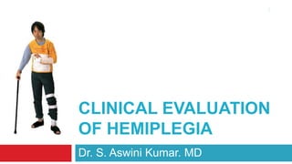
Clinical evaluation of hemiplegia Dr. S. Aswini Kumar. MD 1
Anatomy of Brain Fore brain receiving sensory information from various sensory inputs of body processing the information received and correlating them with prior ones thinking, perceiving, producing and understanding language controlling motor function and autonomic functions Mid brain:  auditory and visual  responses Hind brain balancing equilibrium and co-ordination 2
Physiology of brain 3 Frontal lobe: Provides executive control over much of the brain's higher functions Consciousness, self-awareness, judgment, initiation, motivation  Planning, sequencing, word formation, control over emotional responses Parietal lobe: Perceives, analyzes, and assembles touch information from the body  Integrates visual, auditory, and touch information to formulate complete impression of the world  Left - letters come together to form words and where words are put together in thoughts  Right - recognizing shapes, being aware of one's body in space  Temporal lobe: Hearing, memory acquisition, perception, and categorization of objects Comprehension of language, listening, reading; music   Occipital lobe: Dedicated entirely to vision  In terms of detection, identification, and interpretation of objects.
Handedness & Contra-laterality of brain control 90% of general population-right handed 10% left handed Handedness By birth not by training Test by natural skill; not learned skills Throwing stones, kicking football Determination of hemispherical dominance Right handed –  99% left dominant hemisphere Left handed 70% of left handed – left dominant hemisphere 4
Blood supply of brain 5
The Circle of Willis 6 Named after Thomas Willis (1621–1673) Anterior cerebral artery (left and right) Anterior communicating artery Internal carotid artery (left and right) Posterior cerebral artery (left and right) Posterior communicating artery (left and right) Physiologic significance In event of narrowed or blocked vessel preserve the cerebral perfusion avoid the symptoms of ischemia Considerable anatomic variation exists
The internal capsule 7 Anterior limb:  lenticulostriate branches of middle cerebral artery (superior half) recurrent artery of Heubner off of the anterior cerebral artery (inferior half) Genu:  lenticulostriate branches of middle cerebral artery Posterior limb:  lenticulostriate branches of middle cerebral artery (superior half)  anterior choroidal artery off of the internal carotid artery (inferior half)
Corticospinal tract 8 Originates from pyramidal cells in layer V of the cerebral cortex Axons that travel down through the brain stem and spinal cord - upper motor neurons Long axons to the motor cranial nerve nuclei mainly of the contralateral side of the  midbrain (cortico-mesencephalic tract)  pons (cortico-pontine tract)  medulla oblongata (cortico-bulbar tract) spinal cord (corticospinal tract)
Pathophysiology of ischemic stroke 9
Ischemic penumbra and clinical significance 10 A central area of irreversible infarction  the point of maximum insult called “core” Surrounding area of potentially reversible Called as ischemic penumbra. It has two different segments Inner area of diffusion abnormality Outer area of perfusion abnormality Hypoxia of the cells near the location of the original insult Target for revascularization if given within 60 minutes
History taking in Hemiplegia 11 When did the event start? When was he last found to be in a normal state? What is the total duration of the illness? If multiple, of each episode? What according to the patient or relatives were the initial presenting symptoms? What was the exact mode of onset; was it abrupt, sudden, sub-acute or gradual? When was the maximum deficit noted; was it in the beginning or later? What was the progress of the initial symptoms; static, progressing or regressing? What were the associated symptoms; in CNS as well as CVS, RES and GIT? What investigations he has under gone so far and what are the ones planned? What treatment the patient has received so far and what the ones planned?
History specific for assessing the CNS function 12 Was there any loss of consciousness in the beginning/later; did he recover from it? Is he able to co-operate in interview and the physical examination? What is the emotional state of the patient; memory and intelligence? Is speech affected and if so in what way? Motor, sensory or conductive aphasia? Which of the cranial nerves are affected and what are the symptoms related? What is the degree of motor weakness, wasting, flaccidity or stiffness of muscles? Are all the modalities of sensations normally appreciated or are they abnormal? Is the patient able to stand with/without support; swaying while standing/walking? Any symptoms of increased intra-cranial tension like headache or vomiting?
Precise and complete neurological examination 13 Confirms the presence of a stroke syndrome, distinguishes stroke from stroke mimics Evaluation of level of consciousness and mental status, speech and gait Cranial nerves, motor function, sensory function, superficial, deep tendon reflexes Special reference to Optic fundus - papilledema III – sign of uncalherniation VI – sign of increased ICT Signs of meningeal irritation Signs of head injury
1. Is the patient having neurological problem? 14 Yes or No?  Or is it only hysterical or malingering? Is it a medical condition simulating hemiplegia? Post ictal Todd’s paralysis,  episode of multiple sclerosis? ADEM? If Yes what are the neurological deficits Hemiplegia, UMN Facial weakness, hemianesthesia, homonymous hemianopia Dysphasia in a right hemiplegia and dysarthria in a left hemiplegia Faciobrachialmonoplegia Crossed hemiplegia Cervical cord lesion?
2. Which are the CNS structures involved? 15 Which are the CNS tracts involved? Pyramidal tract Extra-pyramidal tract Cerebellum Brainstem nucleii Cranial nerve II-papilledema, optic atrophy, III palsy, VI palsy VII UMN or LMN It is it an UMN lesion, LMN lesion of neuronal shock state of UMN lesion? Based on weakness, tone, presence of muscle atrophy or fasciculations A prolonged neuronal shock state predicts poor or late recovery
3. Is there UMN or LMN facial paralysis 16 Inspect the face at rest  for voluntary & mimetic movements Examine symmetry of eye blinking and speech motion Ask to raise eyebrows (frontalis) Close eyes (orbicularisoculi) Bell’s phenomenon (Superior rectus) Show teeth (orbicularisoris) Blow out cheeks (buccinator) Scrunch up nose (nasalis)  Retract chin (platysma) ,[object Object]
Upper half of face spared
Lower half affected
No Bell’s phenomenon
Taste not affected
LMN Facial Palsy
Entire half of face affected
Bell’s phenomenon present
Other signs of pontine lesion
Taste affected,[object Object]
5. What is the site of localization of lesion? 18 Cortex Partial deficit, speech involvement, quadrantinopia, cortical sensory, focal seizures Sub-cortical region Denser lesion, Full hemiplegia,  Internal capsule Dense hemiplegia, sparing of speech, absence of speech defects and seizures Thalamic Hemiparalysis, hemianopia, hemisensory loss and emerging hyperpathia Brain stem Crossed hemiplegia - Nuclear type of cranial nerve lesions + contralateralhemiplegia
6. What is the possible pathology of the lesion? 19 Is it an  ischemic infarct embolic infarct hemorrhagic infarct hemorrhagic transformation of an ischemic infarct  hemorrhage Is there evidence of significant or dangerous cerebral edema? Its it a demyelination Acute Disseminated Encephalomyelitis or Episode of MS Is it a space occupying lesion: cerebral abscess cerebral tumor, cerebral secondaries
7. Is it an ischemic stroke? 20 Blood supply to part of the brain is decreased, leading to dysfunction of the brain Cerebral atherosclerosis (producing flow limiting stenosis of a cerebral vessel) Thrombosis (obstruction of a blood vessel by a blood clot forming locally) Embolism (obstruction due to an embolus from elsewhere in the body) Systemic hypoperfusion (general decrease in blood supply, e.g. in shock) Venous thrombosis (infarcts are more likely to undergo hemorrhagic transformation) Clinical features Start suddenly, over seconds to minutes, and in most cases do not progress further Classically detected by the patient in the morning when waking up May or may not be preceded by episodes of transient ischemic attakcs
8. Is it a TIA/evolving stroke/completed stroke? 21 Transient Ischemic Attacks Acute focal non-convulsive neurological dysfunction caused by reversible ischemia recovering in 24h Evolving stroke Deficit occurs in a progressive or step wise fashion culminating in major  deficit In carotid territory within 24 hours and in vertebrobasilar territory within 72 hours Completed stroke The deficit is prolonged and permanent causing demonstrable parenchymal damage Most completed strokes reach the maximum neurological deficits within an hour of onset Reversible Ischemic Neurological deficit The neurological deficit lasts beyond 24 hours but resolves within 3 weeks
9. Is it in carotid artery/vertebrobasilar territory? Contralateral weakness Contralateral numbness Dysphasia Dysarthria Ipsilateral mono-ocular Contralateral homonymous  Combination of above Bilateral or shifting weakness Bilateral/shifting numbness Diplopia Dysarthria Inco-ordination of upper limbs Ataxia/imbalance/disequilibrium Visual loss in both homonymous ields 22 Carotid Vertebral
10. Is it an Internal carotid artery syndrome Often asymptomatic Reason – collateral circulation Ext. carotid ophthalmic anastamosis Superficial/deep cervical Opposite carotid anterior segment Warning symptoms Episodes of confusion Speech dysfunction Amourosisfugax Fleeting paresthesia Neurological deficits Minimal neurological signs Same as that of MCA territory infarct Contralateralhemiplegia Contralateral sensory symptoms Local examination of carotid Feeble carotid pulsation Feeble temporal artery pulsation Cervical bruit over carotid Carotid doppler angiography 23
11. Is it a Middle cerebral artery syndrome? Largest branch and continuation of ICA Most common site of ischemic stroke Clinical picture depends on site of occlusion: Stem, Superior, Inferior  or LS Contralateral weakness Face UL LL Contralateralhemisensory loss Brocas, Wernecke, conduction, global aphasia Contralateral homonymous hemianopia or Qopia Paresis of conjugate gaze to opposite Gerstmann’s syndrome (dominant parietal) 24
12. Is it a Anterior cerebral artery syndrome? 25 Areas supplied by the ACA include: Medial surface of the frontal lobe  Anterior 4/5th of corpus callosum, parietal lobes Anterior 1/2 internal capsule and basal ganglia 1’ of lateral surface of frontal and parietal lobe If stroke occurs prior to ACoA (A1) well tolerated due to collateral circulation If stroke occurs distal to the ACoA (A2) Paralysis of the contralateral foot and leg Sensory loss in the contralateral foot and leg, Gait apraxia
13. Is it a Anterior choroidal artery syndrome? 26 Supply blood to structures which include internal capsule & cruscerebri lateral geniculate body globuspallidus, tail of caudate nucleus Neurological deficits: ContralateralHemiplegia    Contralateral hemihypesthesia Homonymous hemianopia These arise from ischemic damage to the posterior limb of the internal capsule
14. Is it a Posterior cerebral artery syndrome? 27 Thalamic syndrome of Déjerine-Roussy Hemi-sensory loss along with hemiplegia Followed by an agonizing or searing pain  Also termed as thalamic hyperpathia Other features: Persistent pain Aggravated by heat and cold Even by emotions of listening to music Responds poorly to analgesics.  Up regulation of threshold for pain  Once pain threshold is overcome
15. Is it a crossed hemiplegia? 28 Weber Syndrome Ipsilateral III + Contrlateral HP Benedicts Syndrome Ipsilateral III +  Contralateralhemiplegia and tremor Millard Gubler Syndrome Ipsilateral VI + VII + ContralateralHemiplegia Raymond Foville Ipsilateral VI + VII Medial Medullary Syndrome Ipsilateral XII +ContralateralHemiplegia Benedict’s Weber Raymond Foville Millard Gubler

Weitere ähnliche Inhalte

Was ist angesagt?

Upper and lower motor neuron
Upper and lower motor neuronUpper and lower motor neuron
Upper and lower motor neuronMuhammad Saim
 
Approach to a_patient_presenting_with_hemiplegia
Approach to a_patient_presenting_with_hemiplegiaApproach to a_patient_presenting_with_hemiplegia
Approach to a_patient_presenting_with_hemiplegiaalyaqdhan
 
Ardita ( bells palsy ) case presentation
Ardita ( bells palsy ) case presentationArdita ( bells palsy ) case presentation
Ardita ( bells palsy ) case presentationKamal Sharma
 
Approach to a patient with stroke
Approach to a patient with stroke Approach to a patient with stroke
Approach to a patient with stroke Ashwin Haridas
 
Physical examination of rheumatoid arthritis
Physical examination of rheumatoid arthritisPhysical examination of rheumatoid arthritis
Physical examination of rheumatoid arthritisMa Wady
 
HIGHER MENTAL FUNCTIONS ASSESSMENT
HIGHER MENTAL FUNCTIONS ASSESSMENTHIGHER MENTAL FUNCTIONS ASSESSMENT
HIGHER MENTAL FUNCTIONS ASSESSMENTshuchij10
 
Examination of gastrointestinal system by HX
Examination of gastrointestinal system by HXExamination of gastrointestinal system by HX
Examination of gastrointestinal system by HXDr. Rubz
 
neurological examination ppt
neurological examination pptneurological examination ppt
neurological examination pptkabilansilas
 
Clinical examination paraplegia
Clinical examination paraplegiaClinical examination paraplegia
Clinical examination paraplegiaAbino David
 
case study on parkinson disease
case study on parkinson diseasecase study on parkinson disease
case study on parkinson diseaseeducation4227
 
Clinical Examination of RS
Clinical Examination of RSClinical Examination of RS
Clinical Examination of RSPrajwal Rk
 
Higher mental function
Higher mental functionHigher mental function
Higher mental functionPratap Tiwari
 
Clinical examination of cranial nerves
Clinical examination of cranial nervesClinical examination of cranial nerves
Clinical examination of cranial nervesOriba Dan Langoya
 
a case of lower motor neuron facial nerve palsy
a case of lower motor neuron facial nerve palsya case of lower motor neuron facial nerve palsy
a case of lower motor neuron facial nerve palsySamten Dorji
 

Was ist angesagt? (20)

Upper and lower motor neuron
Upper and lower motor neuronUpper and lower motor neuron
Upper and lower motor neuron
 
Approach to a_patient_presenting_with_hemiplegia
Approach to a_patient_presenting_with_hemiplegiaApproach to a_patient_presenting_with_hemiplegia
Approach to a_patient_presenting_with_hemiplegia
 
Ardita ( bells palsy ) case presentation
Ardita ( bells palsy ) case presentationArdita ( bells palsy ) case presentation
Ardita ( bells palsy ) case presentation
 
Knee examination
Knee examinationKnee examination
Knee examination
 
Musculoskeletal Exam
Musculoskeletal ExamMusculoskeletal Exam
Musculoskeletal Exam
 
General examination
General examinationGeneral examination
General examination
 
Approach to a patient with stroke
Approach to a patient with stroke Approach to a patient with stroke
Approach to a patient with stroke
 
Physical examination of rheumatoid arthritis
Physical examination of rheumatoid arthritisPhysical examination of rheumatoid arthritis
Physical examination of rheumatoid arthritis
 
HIGHER MENTAL FUNCTIONS ASSESSMENT
HIGHER MENTAL FUNCTIONS ASSESSMENTHIGHER MENTAL FUNCTIONS ASSESSMENT
HIGHER MENTAL FUNCTIONS ASSESSMENT
 
Examination of gastrointestinal system by HX
Examination of gastrointestinal system by HXExamination of gastrointestinal system by HX
Examination of gastrointestinal system by HX
 
neurological examination ppt
neurological examination pptneurological examination ppt
neurological examination ppt
 
Clinical examination paraplegia
Clinical examination paraplegiaClinical examination paraplegia
Clinical examination paraplegia
 
case study on parkinson disease
case study on parkinson diseasecase study on parkinson disease
case study on parkinson disease
 
Clinical Examination of RS
Clinical Examination of RSClinical Examination of RS
Clinical Examination of RS
 
Tremors
TremorsTremors
Tremors
 
Sciatica
SciaticaSciatica
Sciatica
 
Sensory Examination
Sensory ExaminationSensory Examination
Sensory Examination
 
Higher mental function
Higher mental functionHigher mental function
Higher mental function
 
Clinical examination of cranial nerves
Clinical examination of cranial nervesClinical examination of cranial nerves
Clinical examination of cranial nerves
 
a case of lower motor neuron facial nerve palsy
a case of lower motor neuron facial nerve palsya case of lower motor neuron facial nerve palsy
a case of lower motor neuron facial nerve palsy
 

Ähnlich wie Cns clinical evaluation of hemiplegia slideshare upload

Stroke and cerebrovascular accident
Stroke and cerebrovascular accidentStroke and cerebrovascular accident
Stroke and cerebrovascular accidentDr. Rubz
 
Cerebral tumors 1
Cerebral tumors 1Cerebral tumors 1
Cerebral tumors 1Sabir Ahmed
 
medicine.Coma managment.(dr.muhamad tahir)
medicine.Coma managment.(dr.muhamad tahir)medicine.Coma managment.(dr.muhamad tahir)
medicine.Coma managment.(dr.muhamad tahir)student
 
1 # CNS symptoms history taking.pdf
1 #  CNS symptoms history taking.pdf1 #  CNS symptoms history taking.pdf
1 # CNS symptoms history taking.pdfUgiYou
 
Part 1 function of brain and history taking of a neurological patient
Part 1 function of brain and history taking of a neurological patientPart 1 function of brain and history taking of a neurological patient
Part 1 function of brain and history taking of a neurological patientAtul Saswat
 
Occulomotor nerves
Occulomotor nervesOcculomotor nerves
Occulomotor nervescooravi
 
Nervous system exam part 1
Nervous system exam part 1Nervous system exam part 1
Nervous system exam part 1MUHAMMAD HOSSAIN
 
1 functional anatomy & physiology final
1 functional anatomy & physiology final 1 functional anatomy & physiology final
1 functional anatomy & physiology final eliasmawla
 
Review Of Anatomy And Physiology Of The Nervous
Review Of Anatomy And Physiology Of The NervousReview Of Anatomy And Physiology Of The Nervous
Review Of Anatomy And Physiology Of The Nervousmycomic
 
Dr magdi sasi cns examination 1
Dr magdi sasi  cns  examination 1Dr magdi sasi  cns  examination 1
Dr magdi sasi cns examination 1cardilogy
 
Neuro assessment hdsp final 10.09
Neuro assessment hdsp final 10.09Neuro assessment hdsp final 10.09
Neuro assessment hdsp final 10.09SCramer123
 
Neuro assessment hdsp final 10.09
Neuro assessment hdsp final 10.09Neuro assessment hdsp final 10.09
Neuro assessment hdsp final 10.09SCramer123
 
Approach to a patient with CNS disease
Approach to a patient with CNS diseaseApproach to a patient with CNS disease
Approach to a patient with CNS diseaseAhsan Sajjad
 
NurseReview.Org - Cranial Nerves Nursing Lecture
NurseReview.Org - Cranial Nerves Nursing LectureNurseReview.Org - Cranial Nerves Nursing Lecture
NurseReview.Org - Cranial Nerves Nursing LectureNurse ReviewDotOrg
 
Module 4 cranial nerve
Module 4 cranial nerveModule 4 cranial nerve
Module 4 cranial nerveGlance Ruiz
 
Cranial Nerves.pdf
Cranial Nerves.pdfCranial Nerves.pdf
Cranial Nerves.pdfEnzonBrito
 
neurology (1).pptx
neurology (1).pptxneurology (1).pptx
neurology (1).pptxYomif3
 
Stroke localization by Dr. Md. firoz
Stroke localization by Dr. Md. firozStroke localization by Dr. Md. firoz
Stroke localization by Dr. Md. firozFirozMohammad8
 

Ähnlich wie Cns clinical evaluation of hemiplegia slideshare upload (20)

Stroke and cerebrovascular accident
Stroke and cerebrovascular accidentStroke and cerebrovascular accident
Stroke and cerebrovascular accident
 
Cerebral tumors 1
Cerebral tumors 1Cerebral tumors 1
Cerebral tumors 1
 
medicine.Coma managment.(dr.muhamad tahir)
medicine.Coma managment.(dr.muhamad tahir)medicine.Coma managment.(dr.muhamad tahir)
medicine.Coma managment.(dr.muhamad tahir)
 
1 # CNS symptoms history taking.pdf
1 #  CNS symptoms history taking.pdf1 #  CNS symptoms history taking.pdf
1 # CNS symptoms history taking.pdf
 
Part 1 function of brain and history taking of a neurological patient
Part 1 function of brain and history taking of a neurological patientPart 1 function of brain and history taking of a neurological patient
Part 1 function of brain and history taking of a neurological patient
 
Occulomotor nerves
Occulomotor nervesOcculomotor nerves
Occulomotor nerves
 
Nervous system exam part 1
Nervous system exam part 1Nervous system exam part 1
Nervous system exam part 1
 
1 functional anatomy & physiology final
1 functional anatomy & physiology final 1 functional anatomy & physiology final
1 functional anatomy & physiology final
 
Review Of Anatomy And Physiology Of The Nervous
Review Of Anatomy And Physiology Of The NervousReview Of Anatomy And Physiology Of The Nervous
Review Of Anatomy And Physiology Of The Nervous
 
Dr magdi sasi cns examination 1
Dr magdi sasi  cns  examination 1Dr magdi sasi  cns  examination 1
Dr magdi sasi cns examination 1
 
Neuro assessment hdsp final 10.09
Neuro assessment hdsp final 10.09Neuro assessment hdsp final 10.09
Neuro assessment hdsp final 10.09
 
Neuro assessment hdsp final 10.09
Neuro assessment hdsp final 10.09Neuro assessment hdsp final 10.09
Neuro assessment hdsp final 10.09
 
Facial nerve
Facial nerveFacial nerve
Facial nerve
 
Approach to a patient with CNS disease
Approach to a patient with CNS diseaseApproach to a patient with CNS disease
Approach to a patient with CNS disease
 
NurseReview.Org - Cranial Nerves Nursing Lecture
NurseReview.Org - Cranial Nerves Nursing LectureNurseReview.Org - Cranial Nerves Nursing Lecture
NurseReview.Org - Cranial Nerves Nursing Lecture
 
Module 4 cranial nerve
Module 4 cranial nerveModule 4 cranial nerve
Module 4 cranial nerve
 
Cranial Nerves.pdf
Cranial Nerves.pdfCranial Nerves.pdf
Cranial Nerves.pdf
 
Neuroanatomy lecture
Neuroanatomy lectureNeuroanatomy lecture
Neuroanatomy lecture
 
neurology (1).pptx
neurology (1).pptxneurology (1).pptx
neurology (1).pptx
 
Stroke localization by Dr. Md. firoz
Stroke localization by Dr. Md. firozStroke localization by Dr. Md. firoz
Stroke localization by Dr. Md. firoz
 

Mehr von Prof. Dr. Aswinikumar Surendran

Mehr von Prof. Dr. Aswinikumar Surendran (20)

His | history module | 002
His | history module | 002His | history module | 002
His | history module | 002
 
Protocol for fever
Protocol for feverProtocol for fever
Protocol for fever
 
Radiology for Undergraduate Part 1
Radiology for Undergraduate Part 1Radiology for Undergraduate Part 1
Radiology for Undergraduate Part 1
 
Fever Of Unknown Origin
Fever Of Unknown OriginFever Of Unknown Origin
Fever Of Unknown Origin
 
Cvs Simple Approach To Chd
Cvs   Simple Approach To ChdCvs   Simple Approach To Chd
Cvs Simple Approach To Chd
 
Life Style Diseases
Life Style DiseasesLife Style Diseases
Life Style Diseases
 
AV Nodal Blocks
AV Nodal BlocksAV Nodal Blocks
AV Nodal Blocks
 
Cardiovascular Risk in Diabetes
Cardiovascular Risk in DiabetesCardiovascular Risk in Diabetes
Cardiovascular Risk in Diabetes
 
Medical Emergencies
Medical EmergenciesMedical Emergencies
Medical Emergencies
 
Principles of Ophthalmoscopy
Principles of OphthalmoscopyPrinciples of Ophthalmoscopy
Principles of Ophthalmoscopy
 
Respiratory System Diagnosis
Respiratory System DiagnosisRespiratory System Diagnosis
Respiratory System Diagnosis
 
History Taking
History TakingHistory Taking
History Taking
 
Carotid Artery Stroke
Carotid Artery StrokeCarotid Artery Stroke
Carotid Artery Stroke
 
Acute Left Ventricular Failure
Acute Left Ventricular FailureAcute Left Ventricular Failure
Acute Left Ventricular Failure
 
Tetralogy Of Fallot
Tetralogy Of FallotTetralogy Of Fallot
Tetralogy Of Fallot
 
Aortic Regurgitation
Aortic RegurgitationAortic Regurgitation
Aortic Regurgitation
 
Aortic Stenosis
Aortic StenosisAortic Stenosis
Aortic Stenosis
 
Acute Rheumatic Fever
Acute Rheumatic FeverAcute Rheumatic Fever
Acute Rheumatic Fever
 
Pleural Effusion
Pleural EffusionPleural Effusion
Pleural Effusion
 
Hypothyroidism
HypothyroidismHypothyroidism
Hypothyroidism
 

Cns clinical evaluation of hemiplegia slideshare upload

  • 1. Clinical evaluation of hemiplegia Dr. S. Aswini Kumar. MD 1
  • 2. Anatomy of Brain Fore brain receiving sensory information from various sensory inputs of body processing the information received and correlating them with prior ones thinking, perceiving, producing and understanding language controlling motor function and autonomic functions Mid brain: auditory and visual responses Hind brain balancing equilibrium and co-ordination 2
  • 3. Physiology of brain 3 Frontal lobe: Provides executive control over much of the brain's higher functions Consciousness, self-awareness, judgment, initiation, motivation  Planning, sequencing, word formation, control over emotional responses Parietal lobe: Perceives, analyzes, and assembles touch information from the body  Integrates visual, auditory, and touch information to formulate complete impression of the world Left - letters come together to form words and where words are put together in thoughts Right - recognizing shapes, being aware of one's body in space Temporal lobe: Hearing, memory acquisition, perception, and categorization of objects Comprehension of language, listening, reading; music   Occipital lobe: Dedicated entirely to vision In terms of detection, identification, and interpretation of objects.
  • 4. Handedness & Contra-laterality of brain control 90% of general population-right handed 10% left handed Handedness By birth not by training Test by natural skill; not learned skills Throwing stones, kicking football Determination of hemispherical dominance Right handed – 99% left dominant hemisphere Left handed 70% of left handed – left dominant hemisphere 4
  • 5. Blood supply of brain 5
  • 6. The Circle of Willis 6 Named after Thomas Willis (1621–1673) Anterior cerebral artery (left and right) Anterior communicating artery Internal carotid artery (left and right) Posterior cerebral artery (left and right) Posterior communicating artery (left and right) Physiologic significance In event of narrowed or blocked vessel preserve the cerebral perfusion avoid the symptoms of ischemia Considerable anatomic variation exists
  • 7. The internal capsule 7 Anterior limb:  lenticulostriate branches of middle cerebral artery (superior half) recurrent artery of Heubner off of the anterior cerebral artery (inferior half) Genu:  lenticulostriate branches of middle cerebral artery Posterior limb:  lenticulostriate branches of middle cerebral artery (superior half) anterior choroidal artery off of the internal carotid artery (inferior half)
  • 8. Corticospinal tract 8 Originates from pyramidal cells in layer V of the cerebral cortex Axons that travel down through the brain stem and spinal cord - upper motor neurons Long axons to the motor cranial nerve nuclei mainly of the contralateral side of the  midbrain (cortico-mesencephalic tract)  pons (cortico-pontine tract)  medulla oblongata (cortico-bulbar tract) spinal cord (corticospinal tract)
  • 10. Ischemic penumbra and clinical significance 10 A central area of irreversible infarction the point of maximum insult called “core” Surrounding area of potentially reversible Called as ischemic penumbra. It has two different segments Inner area of diffusion abnormality Outer area of perfusion abnormality Hypoxia of the cells near the location of the original insult Target for revascularization if given within 60 minutes
  • 11. History taking in Hemiplegia 11 When did the event start? When was he last found to be in a normal state? What is the total duration of the illness? If multiple, of each episode? What according to the patient or relatives were the initial presenting symptoms? What was the exact mode of onset; was it abrupt, sudden, sub-acute or gradual? When was the maximum deficit noted; was it in the beginning or later? What was the progress of the initial symptoms; static, progressing or regressing? What were the associated symptoms; in CNS as well as CVS, RES and GIT? What investigations he has under gone so far and what are the ones planned? What treatment the patient has received so far and what the ones planned?
  • 12. History specific for assessing the CNS function 12 Was there any loss of consciousness in the beginning/later; did he recover from it? Is he able to co-operate in interview and the physical examination? What is the emotional state of the patient; memory and intelligence? Is speech affected and if so in what way? Motor, sensory or conductive aphasia? Which of the cranial nerves are affected and what are the symptoms related? What is the degree of motor weakness, wasting, flaccidity or stiffness of muscles? Are all the modalities of sensations normally appreciated or are they abnormal? Is the patient able to stand with/without support; swaying while standing/walking? Any symptoms of increased intra-cranial tension like headache or vomiting?
  • 13. Precise and complete neurological examination 13 Confirms the presence of a stroke syndrome, distinguishes stroke from stroke mimics Evaluation of level of consciousness and mental status, speech and gait Cranial nerves, motor function, sensory function, superficial, deep tendon reflexes Special reference to Optic fundus - papilledema III – sign of uncalherniation VI – sign of increased ICT Signs of meningeal irritation Signs of head injury
  • 14. 1. Is the patient having neurological problem? 14 Yes or No? Or is it only hysterical or malingering? Is it a medical condition simulating hemiplegia? Post ictal Todd’s paralysis, episode of multiple sclerosis? ADEM? If Yes what are the neurological deficits Hemiplegia, UMN Facial weakness, hemianesthesia, homonymous hemianopia Dysphasia in a right hemiplegia and dysarthria in a left hemiplegia Faciobrachialmonoplegia Crossed hemiplegia Cervical cord lesion?
  • 15. 2. Which are the CNS structures involved? 15 Which are the CNS tracts involved? Pyramidal tract Extra-pyramidal tract Cerebellum Brainstem nucleii Cranial nerve II-papilledema, optic atrophy, III palsy, VI palsy VII UMN or LMN It is it an UMN lesion, LMN lesion of neuronal shock state of UMN lesion? Based on weakness, tone, presence of muscle atrophy or fasciculations A prolonged neuronal shock state predicts poor or late recovery
  • 16.
  • 17. Upper half of face spared
  • 22. Entire half of face affected
  • 24. Other signs of pontine lesion
  • 25.
  • 26. 5. What is the site of localization of lesion? 18 Cortex Partial deficit, speech involvement, quadrantinopia, cortical sensory, focal seizures Sub-cortical region Denser lesion, Full hemiplegia, Internal capsule Dense hemiplegia, sparing of speech, absence of speech defects and seizures Thalamic Hemiparalysis, hemianopia, hemisensory loss and emerging hyperpathia Brain stem Crossed hemiplegia - Nuclear type of cranial nerve lesions + contralateralhemiplegia
  • 27. 6. What is the possible pathology of the lesion? 19 Is it an ischemic infarct embolic infarct hemorrhagic infarct hemorrhagic transformation of an ischemic infarct hemorrhage Is there evidence of significant or dangerous cerebral edema? Its it a demyelination Acute Disseminated Encephalomyelitis or Episode of MS Is it a space occupying lesion: cerebral abscess cerebral tumor, cerebral secondaries
  • 28. 7. Is it an ischemic stroke? 20 Blood supply to part of the brain is decreased, leading to dysfunction of the brain Cerebral atherosclerosis (producing flow limiting stenosis of a cerebral vessel) Thrombosis (obstruction of a blood vessel by a blood clot forming locally) Embolism (obstruction due to an embolus from elsewhere in the body) Systemic hypoperfusion (general decrease in blood supply, e.g. in shock) Venous thrombosis (infarcts are more likely to undergo hemorrhagic transformation) Clinical features Start suddenly, over seconds to minutes, and in most cases do not progress further Classically detected by the patient in the morning when waking up May or may not be preceded by episodes of transient ischemic attakcs
  • 29. 8. Is it a TIA/evolving stroke/completed stroke? 21 Transient Ischemic Attacks Acute focal non-convulsive neurological dysfunction caused by reversible ischemia recovering in 24h Evolving stroke Deficit occurs in a progressive or step wise fashion culminating in major deficit In carotid territory within 24 hours and in vertebrobasilar territory within 72 hours Completed stroke The deficit is prolonged and permanent causing demonstrable parenchymal damage Most completed strokes reach the maximum neurological deficits within an hour of onset Reversible Ischemic Neurological deficit The neurological deficit lasts beyond 24 hours but resolves within 3 weeks
  • 30. 9. Is it in carotid artery/vertebrobasilar territory? Contralateral weakness Contralateral numbness Dysphasia Dysarthria Ipsilateral mono-ocular Contralateral homonymous Combination of above Bilateral or shifting weakness Bilateral/shifting numbness Diplopia Dysarthria Inco-ordination of upper limbs Ataxia/imbalance/disequilibrium Visual loss in both homonymous ields 22 Carotid Vertebral
  • 31. 10. Is it an Internal carotid artery syndrome Often asymptomatic Reason – collateral circulation Ext. carotid ophthalmic anastamosis Superficial/deep cervical Opposite carotid anterior segment Warning symptoms Episodes of confusion Speech dysfunction Amourosisfugax Fleeting paresthesia Neurological deficits Minimal neurological signs Same as that of MCA territory infarct Contralateralhemiplegia Contralateral sensory symptoms Local examination of carotid Feeble carotid pulsation Feeble temporal artery pulsation Cervical bruit over carotid Carotid doppler angiography 23
  • 32. 11. Is it a Middle cerebral artery syndrome? Largest branch and continuation of ICA Most common site of ischemic stroke Clinical picture depends on site of occlusion: Stem, Superior, Inferior or LS Contralateral weakness Face UL LL Contralateralhemisensory loss Brocas, Wernecke, conduction, global aphasia Contralateral homonymous hemianopia or Qopia Paresis of conjugate gaze to opposite Gerstmann’s syndrome (dominant parietal) 24
  • 33. 12. Is it a Anterior cerebral artery syndrome? 25 Areas supplied by the ACA include: Medial surface of the frontal lobe  Anterior 4/5th of corpus callosum, parietal lobes Anterior 1/2 internal capsule and basal ganglia 1’ of lateral surface of frontal and parietal lobe If stroke occurs prior to ACoA (A1) well tolerated due to collateral circulation If stroke occurs distal to the ACoA (A2) Paralysis of the contralateral foot and leg Sensory loss in the contralateral foot and leg, Gait apraxia
  • 34. 13. Is it a Anterior choroidal artery syndrome? 26 Supply blood to structures which include internal capsule & cruscerebri lateral geniculate body globuspallidus, tail of caudate nucleus Neurological deficits: ContralateralHemiplegia   Contralateral hemihypesthesia Homonymous hemianopia These arise from ischemic damage to the posterior limb of the internal capsule
  • 35. 14. Is it a Posterior cerebral artery syndrome? 27 Thalamic syndrome of Déjerine-Roussy Hemi-sensory loss along with hemiplegia Followed by an agonizing or searing pain Also termed as thalamic hyperpathia Other features: Persistent pain Aggravated by heat and cold Even by emotions of listening to music Responds poorly to analgesics. Up regulation of threshold for pain Once pain threshold is overcome
  • 36. 15. Is it a crossed hemiplegia? 28 Weber Syndrome Ipsilateral III + Contrlateral HP Benedicts Syndrome Ipsilateral III + Contralateralhemiplegia and tremor Millard Gubler Syndrome Ipsilateral VI + VII + ContralateralHemiplegia Raymond Foville Ipsilateral VI + VII Medial Medullary Syndrome Ipsilateral XII +ContralateralHemiplegia Benedict’s Weber Raymond Foville Millard Gubler
  • 37. 16. Is it cerebral embolism? 29 1. Cardiac sources – various sites Aortic root Native aortic valve Prosthetic aortic valve Left ventricular chamber Native mitral valve Prosthetic mitral valve Left atrial chamber Pulmonary veins 2. Non-cardiac source
  • 38. 17. Is it a cardiac source of cerebral embolism? 30 Aortic root aneurysm with thrombus Aortic valve Endocarditis acute/subacute Aortic Prosthetic valve Tissue/mechanical Left ventricular mural thrombus Left ventricular aneurysm with thrombus Mitral valve stenosis-rheumatic in origin Mitral valve endocarditis Mitral valve prolapse Atrial fibrillation
  • 39. 18. Is it a non-cardiac source of embolism? 31 Pulmonary venous thrombosis Suppurative lung – abscess, bronchiectasis Bronchogenic carcinoma – secondaries Air embolism Fat embolism Amniotic fluid embolism Paradoxical embolism – PFO Tetralogy of Fallot, Eisenmenger syndrome Carotid artery – cerebral artery embolism
  • 40. 19. Is it a Hemorrhagic stroke? 32 Severe essential hypertension 55-75 years of age Smooth onset over minutes or hours steady progress in spite of treatment Features of increased ICT Types: Epidural/ Subdural Intra-parenchymal Intra-ventricular Sub-arachnoid Thalamic hemorrhage
  • 41.
  • 42. 21. Is it a stroke mimic? Post ictal Todd’s paralysis Transient and follows aseizure Brain infections Fever, headache and papilledema Brain tumors Progressive headache, papilledema Demyelinating Disease (ADEM, MS) Recurrent episodes, distant lesions Hemi-parkinsonism Rigidity rather than spasticity Hypertensive Encephalopathy Accelerated hypertension, convulsion Subdural hematoma Waxing-waning neurological deficit Conversion disorder Stress situation underlying 34
  • 43. Summary Basic Sciences Applied anatomy Functional components Handedness Hemispherical dominance Blood supply Circle of Willis Internal Capsule Corticospinal tract Pathophysiology Neurological Assessment TIA, RIND, Evolved/completed stroke Carotid/vertebrobasilar territory Localization: Cortical/internal capsule Arterial territory: MCA, ACA, AChA Thalamic/crossed hemiplegia Type: Ischemic, embolic, hemorrhage Cardiac/non-cardiac source Location of hemorrhage Young stroke/cerebral palsy 35