Call Girls Shimla Just Call 8617370543 Top Class Call Girl Service Available
Upper Respiratory Infections
1. INFECTIOUS DISEASE
CHAPTER TWO
UPPER RESPIRATORY
TRACT INFECTIONS AND
OTHER INFECTIONS OF
THE HEAD AND NECK
Upper respiratory tract infection
(URI) causes at least of one-half of
all symptomatic illness in the
community, exacting huge tolls
that can be measured as morbidity,
absenteeism from school and
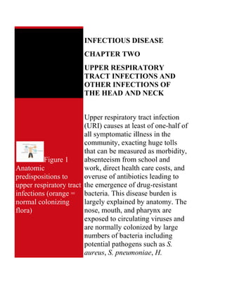
Figure 1
work, direct health care costs, and
Anatomic
overuse of antibiotics leading to
predispositions to
upper respiratory tract the emergence of drug-resistant
infections (orange = bacteria. This disease burden is
largely explained by anatomy. The
normal colonizing
nose, mouth, and pharynx are
flora)
exposed to circulating viruses and
are normally colonized by large
numbers of bacteria including
potential pathogens such as S.
aureus, S. pneumoniae, H.
4. Pathogenesis of paramyxovirus, figure 5) can
symptoms attributable cause colds, but more often cause
to the common cold lower respiratory tract infection or
systemic symptoms.
Adenoviruses, of which there are
at least 47 antigenic types, cause
5% to 10% of colds. Although
echoviruses and coxsackieviruses
have been associated with colds,
they more commonly cause an
undifferentiated flu-like illness or
distinctive syndromes such as
aseptic meningitis or pharyngitis.
Rhinoviruses and parainfluenza
viruses cause outbreaks of cold
symptoms during the fall and late
spring; RSV, adenoviruses, and
coronaviruses cause outbreaks
during the winter and early spring.
Viruses that remain to be
discovered probably cause a
substantial fraction of colds.
Rhinoviruses are transmitted most
efficiently by direct contact. Hand
contact with the eyes and nose is
very common in everyday life.
Rhinoviruses remain viable on
5. skin and also on objects (fomites)
for at least 2 hours. Rhinoviruses
can be recovered from hands in
40% to 90% of persons with colds
and from up to 15% of objects
near persons with colds. However,
brief exposure such as a
handshake or even being around
an infected person for 36 hours
causes transmission in less than
10% of subjects and in one study
transmission was only 38%
between spouses. Rhinoviruses
can also be transmitted by
aerosolization, for example, by
being in a crowded room where
people are sneezing. Kissing does
not seem to be a common mode of
transmission, probably because
only about 10% of persons with
colds have demonstrable virus in
their saliva. Studies carried out in
Antarctica dispel the popular idea
that cold weather increases
susceptibility to rhinovirus
infection.
The nasal epithelium of persons
6. with colds is remarkably intact
even when studied with the
electron microscope. Symptoms
are best explained by physiologic
responses:
release of chemical mediators
of inflammation;
sensitization and irritation of
airway receptors, with
stimulation of the
parasympathetic nervous
system.
After an incubation period of 24 to
72 hours, most patients develop a
sore or scratchy throat which is
followed by nasal obstruction,
rhinorrhea, and sneezing (figure
6). A green or yellow nasal
discharge should not be construed
as evidence of secondary bacterial
infection (neutrophils cause
yellow-green discoloration
because of their natural
myeloperoxidase activity). By the
second and third day of the illness,
rhinitis with nasal congestion
7. replaces sore throat as the major
complaint. By the fourth and fifth
day, nasal symptoms have usually
decreased but in about 30% of
cases are replaced by cough or
quot;chest coldquot;.
Acute Bacterial Sinusitis
Few common problems in primary
care are as confusing as sinusitis.
Acute bacterial sinusitis is vastly
over diagnosed, but chronic
sinusitis can be frustrating and
disabling. The paranasal sinuses
are accessible to direct
examination only by sophisticated
instruments. Adequate specimens
for cultures can be obtained only
by invasive procedures. Low-
grade sinusitis is an intrinsic
feature of the common cold.
Clinicians must distinguish
between self-limited viral
rhinosinusitis and acute bacterial
sinusitis, which usually calls for
8. antibiotic therapy.
Sinusitis is usually caused by
obstruction of the ostia, as from
edema, damage to ciliated
epithelial cells, and/or increased
volume or viscosity of the mucous
secretions. The pathogenesis of
acute sinusitis can be discussed in
3 complementary ways: anatomy,
physiology, and microbiology.
Anatomically, the outflow tract of
the maxillary sinus sits in an
awkward position high on the
medial wall of the sinus cavity and
is connected to the nasal cavity by
a narrow tubular passage known as
the infundibulum. Gravitational
drainage of the maxillary sinus is
therefore tenuous and easily
disrupted. A small area between
the middle and inferior nasal
turbinates where drainage from the
maxillary, ethmoid, and frontal
sinuses converges is known as the
ostiomeatal complex. Anatomic or
physiologic compromise of the
9. ostiomeatal complex predisposes
not only to maxillary sinusitis but
also to infection of multiple
sinuses (pansinusitis). Causes of
mechanical obstruction of sinus
drainage include deviated nasal
septum, polyps, foreign bodies,
tumors, concha bullosa (enlarged
middle turbinates from
pneumatization, present in 10% of
the population), ethmoid bullae,
choanal atresia, and―most
commonly―mucosal swelling.
Mucosal swelling is usually due to
viral infection or allergic
inflammation, but can also be
caused by systemic disorders (such
as cystic fibrosis and the
dyskinetic cilia syndromes) or
injury brought about by trauma,
swimming or diving, or overuse of
topical medication (rhinitis
medicamentosa) or cocaine.
Physiologically, the sinuses are
kept sterile mainly by a
mucociliary blanket that changes 2
to 3 times per hour, a rate
10. sufficient to prevent mucus from
accumulating in the sinuses.
Sinusitis results when mucociliary
drainage of the paranasal sinuses
fails because of mucosal edema,
dysfunction of the ciliated
epithelial cells, or both. Viral
rhinitis and allergic rhinitis disrupt
the epithelium as do cigarette
smoking and intranasal cocaine
use. Swimming predisposes to
sinusitis for reasons that are
somewhat unclear, although
chlorine is known to irritate the
mucosa. Other factors that may
help to preserve the normal
sterility of the paranasal sinuses
include cellular and humoral
immunity and nitric oxide.
Microbiologically, acute sinusitis
typically starts with viral infection
that paves the path for pathogenic
bacteria. The major pathogens are
Streptococcus pneumoniae and
non-typeable strains of
Haemophilus influenzae. Other
bacteria associated with
11. community-acquired sinusitis
include group A streptococci, S.
aureus, Moraxella catarrhalis, and
viridans streptococci. Whether
Chlamydia pneumoniae commonly
causes sinusitis is currently being
studied. Patients who have had
nasogastric tubes in place are
vulnerable to sinusitis caused by
aerobic gram-negative rods such
as Pseudomonas aeruginosa and
Klebsiella pneumoniae. Anaerobic
bacteria are associated with acute
sinusitis mainly in the setting of
dental disease. Anaerobic bacteria
possibly play a role in chronic
sinusitis (discussed separately,
below).
Sinusitis usually begins with
symptoms of the common cold
such as runny nose, nasal
obstruction, sore throat, cough,
and the sensation of “pressure” or
“tightness” in the face. The
symptoms differ somewhat
between children and adults.
Children with sinusitis can have
12. either of two presentations. The
more common presentation
consists of persistent cold
symptoms that is, symptoms
lasting more than 10 days.
Children with persistent sinusitis
seldom complain of headache or
facial pain. Parents of young
children often report malodorous
breath. The less common
presentation consists of severe
cold symptoms―that is, cold
symptoms that are accompanied
by high fever (> 39° C) and
purulent nasal discharge. Some of
these children experience
headaches usually located behind
or around the eye, occasionally
with periorbital edema. Adults
with sinusitis, compared with
children, tend to have more
prominent facial pain, sometimes
with local tenderness, swelling,
and erythema. Pain patterns and
other findings vary according to
which sinuses are involved:
Maxillary sinusitis
13. (the most common
location): Pain is
over the cheekbones
or above the
maxillary 2nd molar
teeth.
Ethmoid sinusitis
(common): Pain is
between the orbits
and the nasal bridge.
Frontal sinusitis
(uncommon): Pain is
over the frontal
bones.
Sphenoid sinusitis
(least common, but
dangerous): Pain can
be frontal, temporal,
orbital, or occipital;
facial pain due to Vth
nerve involvement is
characteristic; pain is
occasionally
perceived over the
vertex of the skull.
Low-grade fever is often present.
Physical examination may reveal
14. tenderness on percussion of the
maxillary or frontal sinuses, and
pinching the bridge of the nose
may bring out tenderness if the
ethmoid sinuses are involved.
However, the sensitivity and
specificity of these findings are
unknown, and the clinical findings
are often subtle.
Acute bacterial sinusitis is often
self-limited, but the frequency
with which this condition resolves
spontaneously is unknown. Direct
sinus puncture for accurate
diagnosis has not been carried out
in placebo-controlled clinical
trials. Serious complications occur
often enough to justify close
follow-up, especially in the
clinically more severe cases. The
most common complication is
progression to chronic sinusitis
(see below). Cavernous sinus
thrombosis can result from
ethmoid, frontal, or sphenoid
sinusitis. Ethmoid sinusitis can
also cause orbital cellulitis. Frontal
15. sinusitis can also cause
osteomyelitis of the frontal bone
with swelling and edema of the
forehead (Pott’s puffy tumor) and
subdural empyema. Sphenoid
sinusitis can be a medical
emergency.
Chronic Sinusitis
When acute sinusitis fails to
resolve and becomes chronic,
cultures may reveal a variety of
opportunistic pathogens including
anaerobic bacteria. Some
authorities feel that the problem is
no longer mainly “infectious” but
rather reflects permanent mucosal
injury. It has been estimated that
chronic sinusitis causes morbidity,
measured as absenteeism from
school, work, or social activities,
of the same magnitude as heart
disease and arthritis.
The prevailing view holds chronic
16. sinusitis to be a disorder of
abnormal anatomy and physiology
of the paranasal sinuses with one
or more causes:
previous acute sinusitis
nasal polyposis (as in the triad
of asthma, allergies, and
aspirin sensitivity)
previous sinus surgery
cystic fibrosis
These processes lead to anatomic
changes including obstruction of
the infundibula and ostia, mucosal
edema and scarring, bone
hypertrophy, polypoid
degeneration, and/or mucosal
fibrosis, rendering the mucociliary
clearance mechanism defunct.
Numerous microorganisms can be
isolated from patients with chronic
sinusitis, but correlation between
culture results and the disease
process is often poor. Mixtures of
aerobic and anaerobic bacteria are
common. The general conclusion
at this time is that in most patients,
17. no single microorganism can be
assigned a pathogenic role. In
some patients, however,
Pseudomonas aeruginosa or
Staphylococcus aureus seems to
be clearly pathogenic, and there
are data suggesting roles for
Haemophilus influenzae and
Moraxella catarrhalis (in
children). Patients with chronic
sinusitis often have exacerbations
analogous to the acute
exacerbations of chronic
obstructive lung disease. In these
instances, especially in children,
Streptococcus pneumoniae and
Haemophilus influenzae may be
important. Numerous bacteria
including gram-negative rods have
been isolated with patients with
post-operative sinusitis. Some
investigators believe that many
patients with chronic sinusitis have
allergic fungal sinusitis (discussed
further below), the disease
manifestations being caused by an
immune response to extramucosal
18. fungi.
The typical history consists of
nasal drainage, obstruction, and
postnasal drip lasting for at least
several months, often against a
background of chronic “sinus
trouble.” Patients often complain
of headache or “sinus pain,”
nocturnal cough, and bad breath.
Loss of smell may also be present.
Fever is unusual. A history of
inhalant allergy is 4.5 times more
common in patients with chronic
sinusitis than in persons without
chronic sinusitis.
Although physical examination
can reveal findings such as nasal
septal deviation or mucosal
changes, imaging studies are
necessary to make a correct
diagnosis. The coronal CT scan
represents the current gold
standard, but axial CT scans are
often useful especially in children.
Sinus endoscopy an also provide
invaluable information.
19. Chronic sinusitis may have a
relapsing or remitting course, but,
untreated, patients seldom become
entirely free of symptoms related
to the paranasal sinuses.
Complications include remodeling
of the facial bones, osteomyelitis,
and―occasionally―invasive
disease of the CNS caused by
bacteria or fungi.
Fungal Sinusitis
This chest Fungal sinusitis is relatively
radiograph shows uncommon, but should be
probable aspergillosis considered in patients with chronic
with an aspergilloma, or sinusitis because of its potentially
fungus ball in the upper serious complications. Aspergillus
lobe of the right lung. species are the most common
Lung diseases that causes of fungal sinusitis, and
damage a lung can fungi of the order Mucorales are
cause cavities that can the most dangerous (rhinocerebral
leave a person more mucormycosis). Various widely
susceptible to distributed pigmented fungi that
developing an are collectively known as
aspergilloma, or fungus dematiaceous molds can cause a
20. ball. The fungus can variety of syndromes that include
then begin secreting life-threatening disease; examples
toxic and allergic of these organisms include
products, which may Alternaria, Bipolaris,
make the person feel ill. Cladosporium, Curvularia, and
CDC/M. Renz Exserohilum. Numerous other
fungi sometimes cause sinusitis.
Five syndromes are currently
recognized.
This patient presented
Simple colonization of
with a case of a
the paranasal sinuses by fungi
periorbital fungal
may be relatively common,
infection known as
although the incidence is
mucormycosis, or
unknown.
phycomycosis.
Sinus mycetoma (fungus
Mucormycosis is a
ball) presents most often as a
dangerous fungal
mass in the maxillary sinus.
infection usually
Underlying disease is usually
occurring in the
not present although some
immunocompromised
patients have nasal polyps and
patient, affecting the
chronic bacterial sinusitis.
regions of the eye,
Patients usually seek medical
nose, and through its
attention for nasal obstruction,
growth and
facial pain, symptoms of
destruction of the
chronic sinusitis, or fetid
periorbital tissues, it
breath (cacosmia). Seizures
will eventually invade
21. the brain cavity. CDC have been reported as a
presenting manifestation.
Allergic fungal sinusitis
usually presents as intractable
sinusitis with nasal polyposis
Pseudallescheria
in patients with atopy. Some
boydii is pathogenic
patients also have allergic
in humans, especially
bronchopulmonary
those who are
aspergillosis. Children with
immunocompromised,
allergic sinusitis may develop
causing infections in
hypertelorism or proptosis
almost all body
when the frontal or ethmoid
regions, and which
sinuses are involved. As
are classified under
mentioned above, some
the broad heading of
“Pseudallescheriasis”. investigators believe that
allergic fungal sinusitis is
CDC
present in a majority of
patients who carry the
diagnosis of quot;chronic
rhinosinusitisquot;.
Acute (fulminant)
invasive fungal sinusitis is
essentially synonymous with
rhinocerebral mucormycosis,
which usually occurs in
patients with diabetes mellitus
or severe immunosuppression
22. from other causes. The
sinuses have been called quot;way
stations to the brainquot; in this
medical emergency, which
classically presents as a
painless black eschar on the
palate or a nasal turbinate
followed by epistaxis,
headache, changes in mental
status, and focal neurologic
symptoms and signs (such as
diplopia). Other fungi
including Aspergillus species,
Fusarium species, and
Pseudallescheria boydii can
cause an identical syndrome,
typically in patients who are
severely immunosuppressed
from disease (including
AIDS) or chemotherapy for
cancer or organ
transplantation.
Chronic invasive fungal
sinusitis can occur not only in
immunocompromised patients
but also in patients who are
immunologically normal.
23. Most of these latter patients
have chronic sinusitis and
nasal polyposis. Causative
organisms include the
dematiaceous molds noted
above; the condition is known
as phaeohyphomycosis. Dense
masses of fungal elements
resembling mycetoma are
found, but there is also
invasion into the mucosa and
then into bone. Patients
present with headache and
localizing symptoms such as
decreased vision and loss of
eye movement (orbital apex
syndrome) or behavioral
changes (mycetoma of the
frontal lobe).
Otitis Externa
Otitis externa, a spectrum of
conditions caused by infection,
Erysipelas is a allergy, or primary skin disease,
dermatologic affects up to 10% of all people
condition, which during their lifetimes. Necrotizing
24. involves the (malignant) otitis externa is a
inoculation of the skin medical emergency seen usually in
and subcutaneous patients with diabetes mellitus or a
tissue with compromised immune system or
streptococcal bacteria in patients who have had prior
causing edema and irradiation of the head.
bright red erythema of
Otitis externa begins with
the affected areas. It is
breakdown in the cerumen barrier.
sometimes referred to
Cerumen, although commonly
as cellulitis. CDC
considered a nuisance, protects
against infection by (1) creating an
acidic environment hostile to
bacterial and fungal growth; (2)
promoting a dry environment
through its hydrophobic
properties; and (3) trapping debris
by its sticky nature. Excessive
cleaning or scratching of the ear
canal promotes breakdown of the
cerumen barrier. Swimming
notoriously predisposes to otitis
externa, the main effect being
promotion of a more alkaline pH,
which in turn promotes bacterial
growth. However, increased
moisture of any origin leads to
maceration of the skin and
25. breakdown. Mechanical trauma
from devices such as ear plugs
(used for hearing conservation in
many industries), headphones,
hearing aids, and diving caps also
predisposes to otitis externa.
Pseudomonas aeruginosa and S.
aureus are common causes of
otitis externa. Group A
streptococci and various gram-
negative rods sometimes cause this
condition, and anaerobic bacteria
are involved in up to 25% of cases.
About 10% of cases are caused by
various fungi, with Aspergillus
species being the most common
followed by Candida species.
Acute localized otitis
externa presents as a single
or several pustules or
furuncles located in the ear
canal, usually due to S.
aureus. Initial symptoms
include itching, pain,
swelling, redness, and
sometime decreased
26. hearing. A small furuncle
that would be
inconspicuous on most
parts of the skin becomes
intensely painful in the
confines of the narrow ear
canal.
Acute diffuse otitis
externa (swimmer’s ear)
typically presents with pain
(otalgia), itching,
discharge, and hearing loss.
The pain can become
intolerable. There is often a
history of recent exposure
to water. Mobile redwood
hot tub systems were
associated with a severe
hemorrhagic form of the
disease. Pseudomonas
aeruginosa is the most
common pathogen. Other
patients give a history of
ear instrumentation,
excessive cleaning,
previous infection, otitis
media, tinnitus, or vertigo.
27. Examination reveals the ear
canal to be diffusely red
and edematous.
Erysipelas involving the
concha and canal is caused
by group A streptococci,
the pathogenesis being
similar to that of erysipelas
elsewhere. Patients present
with a diffusely red and
painful ear. Examination
may reveal hemorrhagic
bullae on the walls of the
canal and on the tympanic
membrane, and there may
be tender regional
lymphadenopathy.
Chronic otitis externa
presents with mild
discomfort and flaking of
the skin of long duration,
often with a history Otitis
media can be graded in
severity from mild (minor
pain and pruritus, minimal
edema of the ear canal) to
28. severe (severe pain and
pruritus, complete
occlusion of the ear canal,
auricular and periauricular
erythema, and, frequently,
fever and
lymphadenopathy).
Malignant otitis externa
is a medical emergency
usually encountered in
patients with diabetes
mellitus. A history of
chronic otitis media with
drainage is often obtained.
Pseudomonas aeruginosa
is the usual causative
organism. Complications
include osteomyelitis of the
temporal bone (a defining
feature of the disease) and
extension to the base of the
skull, with cranial nerve
palsies, meningitis, and
other serious consequences.
Diagnosis of otitis externa is
nearly always made by the history
29. and physical examination.
Pressure on the tragus or pulling
the auricle superiorly causes pain;
the latter maneuver is a valuable
diagnostic aid. Examination of the
ear canal shows erythema and
edema and, in severe cases, partial
or complete occlusion of the canal.
To exclude otitis media, one
should demonstrate with
pneumatic insufflation that the
tympanic membrane is mobile.
However, the tympanic membrane
is often partially or totally
obscured by edema in the ear
canal.
Although otitis externa is usually
considered a self-limited
condition, serious complications
can occur. Perforation of the
tympanic membrane can be caused
by extension of the disease process
or by misguided attempts by
patients or health care providers to
relieve the condition through
mechanical manipulation. Other
complications include stenosis of
30. the ear canal, auricular cellulitis,
or chondritis.
Acute Otitis Media
The importance of otitis media in
primary care cannot be
overemphasized, especially in
pediatrics practice, where it
accounts for about 25% of all
office visits, 50% of office visits
for illness, and 40% of antibiotic
prescriptions. By age 5, between
75% and 95% of children have had
at least one episode of otitis
media. This disease is responsible
for some 25 million office visits
each year, with annual health care
costs estimated to be between 3
and 5 billion dollars. Although
80% of patients with otitis media
are less than 15 years of age, more
than one-fourth of all oral
antibiotic prescriptions in the
United States are written for this
condition. Heavy prescribing of β-
31. lactam antibiotics for otitis media
is thought to be responsible in
large measure for the decreasing
drug susceptibility of S.
pneumoniae strains.
Acute otitis media is often
preceded by viral URI that causes
edema and obstruction of the
eustachian tube, causing an ex
vacuo serous transudate into the
middle ear and paving the way for
pathogenic bacteria. Children are
predisposed because their
eustachian tubes are shorter,
wider, and straighter compared
with those of adults. Appreciation
of otitis media requires knowledge
of the relationship of the
eustachian tube to the
nasopharynx, the middle ear, and
the mastoid air cells. The
eustachian tube normally serves to
regulate pressure in the middle ear,
to protect against nasopharyngeal
sound pressure and secretions, and
to afford a pathway for the
drainage of secretions produced
32. within the middle ear into the
nasopharynx. The latter function
requires an intact mucociliary
system. Factors predisposing to
eustachian tube dysfunction and
otitis media include allergy, cleft
palate, ciliary dysmotility,
immunodeficiency, exposure to
tobacco smoke, exposure to
frequent upper respiratory tract
infections (notoriously, in day care
centers), early age of first
infection, and race. Native
Americans are markedly
predisposed to otitis media for
reasons that are unclear. Adults
can be predisposed to otitis media
on account of diabetes mellitus,
cancer, immune deficiencies, and
injection drug use. The adenoids
have long been implicated in the
pathogenesis of otitis media, for
better or worse, since
inflammation of the adenoids
causes inflammatory
obstruction of the adjacent
eustachian tube orifices;
33. colonization of the adenoids
by pathogenic bacteria
promotes invasion of the
middle ear by these same
bacteria;
yet the adenoids provide local
immunity in the form of
secretory IgA secretion.
The microbiology in otitis media
is similar in adults and children
when tympanocentesis is carried
out. In about 40% of cases, culture
of middle ear fluid fails to show a
bacterial pathogen. This might
reflect a viral etiology or sterile
inflammation. Respiratory
syncytial virus has been found
relatively commonly, with a
special tendency to cause the
disease in children. Influenza
viruses and parainfluenza viruses
also predispose to otitis media.
Streptococcus pneumoniae causes
about 30% to 40% of cases, and
the prevalence of strains with
reduced susceptibility to penicillin
is increasing. H. influenzae causes
34. between 20% and 30% of cases,
and M. catarrhalis between 10%
and 15% of cases, especially in
children. Group A streptococci
cause less than 5% of cases but
can cause up to 10% during the
winter months.
Acute otitis media in children
usually presents with rapid onset
of otalgia, fever, and/or irritability.
Otalgia in young infants is
manifest by pulling on the ear.
Young children can also have
anorexia, loose stools, and
vomiting. Otalgia tends to be the
major symptom in adults. A
minority of patients experience
spontaneous perforation of the
tympanic membrane.
The tympanic membrane is
abnormal, often bulging, with loss
of the usual landmarks. Erythema
of the tympanic membrane alone
is not diagnostic; it can be caused,
for example, by crying. Purulent
fluid is sometimes seen behind the
35. tympanic membrane. The key
procedures are pneumatic
otoscopy and, when indicated,
tympanocentesis. Pneumatic
otoscopy, which is done by gently
squeezing and then releasing a
rubber bulb attached to the
otoscope, provides information
about the mobility of the tympanic
membrane. Tympanocentesis
provides fluid for culture, which is
becoming more important due to
the emergence of drug-resistant
bacteria. Cultures of the
nasopharynx correlate poorly with
cultures of fluid obtained by
tympanocentesis, and are therefore
of limited usefulness.
Acute otitis media must be
distinguished from otitis media
with effusion (serous otitis media).
The latter consists of an
asymptomatic or
hyposymptomatic middle ear
effusion, which can be acute (less
than 3 weeks), subacute (3 weeks
to 3 months), or chronic. Although
36. hearing loss is frequently present
in both acute otitis media and otitis
media with effusion, patients with
otitis media and effusion lack
systemic signs and symptoms such
as otalgia and fever.
The natural history of untreated
otitis media continues to prompt
debate whether most cases should
be treated with antibiotics. A
meta-analysis of the literature
based on data obtained from 5400
children in 33 studies indicates
that 81% percent of children have
spontaneous resolution. However,
it is difficult to predict on clinical
grounds whether an individual
patient s disease will resolve
spontaneously, and the current
consensus opinion in the United
States is that all patients should be
treated.
Chronic suppurative otitis
37. media and mastoiditis
Chronic suppurative otitis media is
a complication of acute otitis
media, usually occurring when
there is a defect in the tympanic
membrane, such as a quot;centralquot;
perforation or a tympanostomy
tube. It is accompanied by
purulent discharge (otorrhea).
Mastoiditis is invariably present.
The associated bacteria seem to
vary depending on whether an
infected cholesteatoma is present.
Cholesteatoma is often associated
with anaerobic bacteria and “skin
flora” microorganisms, and the
otorrhea often has a foul odor.
When cholesteatoma is not
present, gram-negative rods
including Pseudomonas
aeruginosa and E. coli are often
found.
In the pre-antibiotic era,
mastoiditis was often a dramatic
and severe illness with
38. retroauricular inflammation and
serious intracranial complications.
Today, mastoiditis is more
typically an indolent, low-grade,
often painless infection of the
temporal bone that tends to be
clinically silent (quot;masked
mastoiditisquot;) unless a complication
such as brain abscess develops.
Patients at high risk of
complications include newborn
infants, persons with diabetes
mellitus, the elderly, and the
immunocompromised.
Spontaneous resolution is rare, if it
occurs at all. Local complications
of chronic suppurative otitis media
and mastoiditis include bone
destruction, subperiosteal abscess,
facial paralysis, labyrinthitis, and
petrositis. Intracranial
complications include brain
abscess, subdural abscess, epidural
abscess, septic thrombosis of the
lateral sinus, meningitis, and
hydrocephalus. Patients with
chronic suppurative otitis media
39. and/or mastoiditis should be
referred to an otolaryngologist, as
effective treatment usually
requires surgical intervention.
Acute suppurative parotitis
Acute suppurative parotitis, an
uncommon condition, usually
results from decreased salivary
flow, allowing retrograde ascent of
bacteria from Stenson’s duct. It
can also result from ductal
obstruction from mucus or
fibrinous plugs, tumors, or foreign
Figure
bodies). The clinical signs and
Staphylococcal
symptoms include an acutely
parotitis, face of
swollen, indurated check with
elderly woman.
fever and pain. Pus can be
CDC
expressed from Stenson’s duct.
More than 80% of cases are
caused by Staphylococcus aureus.
Most patients are elderly,
debilitated, and dehydrated.
Mortality is high.
40. Acute pharyngitis
Acute pharyngitis (sore throat) is
one of the most common problems
encountered in clinical practice.
Viruses cause most cases as part of
the common cold. However, about
15% of cases, and up to 50% of
cases in children during some
periods, are caused by group A β-
hemolytic streptococci (S.
pyogenes). Although usually self-
limited, streptococcal pharyngitis
demands respect as a cause of
acute rheumatic fever and―less
commonly―major suppurative
complications, acute
glomerulonephritis, and even the
streptococcal toxic-shock
syndrome. The clinician’s task is
to determine in a cost-effective
manner which patients need
treatment and which do not.
Acute pharyngitis has many
known etiologies, and pathogens
remain to be discovered for an
41. estimated 30% of cases. Viral
infections cause sore throat, it is
thought, by generating bradykinin
and lysyl bradykinin, which
stimulate nerve endings. Group A
streptococci and certain other
pathogens including some of the
respiratory viruses cause pain by
invading the mucosa.
Group A streptococci are carried
in the human nasopharynx and
transmitted from person to person
usually by direct contact with
saliva or nasal secretions.
Acquisition is greatest in school-
aged children, suggesting the
gradual development of immunity
over time. Children also serve as a
reservoir for spread among family
members. Asymptomatic
pharyngeal carriage of group A
streptococci is relatively common,
and the factors that cause some
persons to develop acute
pharyngitis and other
complications are poorly
understood. Group C and group G
42. streptococci cause a pharyngitis
syndrome clinically
indistinguishable from that caused
by group A streptococci,
sometimes recognized as
outbreaks related to a common
food source. Group C streptococci
(S. dysgalactiae subspecies
equisimilis) appear to be a
frequent cause of pharyngitis in
college-aged students.
Pharyngitis due to group A
streptococci occurs most
frequently in children between 5
and 15 years of age, usually during
the winter and early spring. In its
severe form the disease starts
abruptly with fever, sore throat,
and odynophagia. Chills,
headache, and abdominal pain are
sometimes present. Examination
reveals diffuse erythema of the
pharynx and tonsils accompanied
by a patchy, purulent tonsillar and
pharyngeal exudate, hypertrophy
of the lymphoid nodules in the
posterior pharyngeal mucosa, and
43. tender cervical lymphadenopathy.
Occasional strains of S. pyogenes
elaborate the erythrogenic toxin of
scarlet fever, resulting in a striking
rash and quot;red strawberry tonguequot;
with enlargement of the papillae.
Rhinorrhea and cough are usually
not present, but may occur.
However, these dramatic
manifestations are absent in many,
perhaps most cases of
streptococcal pharyngitis. Because
the features of group A
streptococcal pharyngitis blend
imperceptibly with those of other
causes of sore throat, numerous
students of the disease have
concluded that the diagnosis must
be secured by laboratory methods
prior to definitive treatment (see
below). Other syndromes include
the following:
The common cold is
often accompanied
by sore throat, which
is frequently the first
symptom but is
44. usually not the main
complaint when
patients seek medical
care. Rhinorrhea,
postnasal drainage,
and cough are
usually more
prominent symptoms
than sore throat.
Fever is seldom
prominent and severe
sore throat with
odynophagia are
uncommon.
Influenza sometimes
presents with sore
throat as the chief
complaint, but is
usually accompanied
by other symptoms
suggesting influenza
such as myalgia,
headache, and cough.
In pharyngitis due to
the common cold or
to influenza, purulent
45. pharyngeal or
tonsillar exudates
and tender cervical
lymphadenopathy are
not present. Tracheal
tenderness may be
present in influenza,
indicating diffuse
viral infection of the
respiratory mucosa.
Adenoviral
pharyngitis, on the
other hand, often
presents with sore
throat as the chief
complaint. Fever,
chills, headache,
malaise, and
myalgias can be
prominent. About
one-third to one-half
of patients with
adenoviral
pharyngitis also have
follicular
conjunctivitis; this
46. syndrome is known
as
pharyngoconjunctiva
l fever. Patients with
adenoviral
pharyngitis often
have pharyngeal
exudate, so the
disease can mimic
streptococcal
pharyngitis.
Epidemics of
pharyngoconjunctiva
l fever occur during
the summer months
in civilian
populations and
during the winter
months in military
recruits.
Infectious
mononucleosis due
to the Epstein-Barr
virus causes
exudative tonsillitis
or pharyngitis in
47. about one-half of
cases. Tonsillar and
pharyngeal exudates
can be prominent. If
examined with
Wright’s stain, the
exudates of
mononucleosis
consist mainly of
mononuclear cells in
contrast to the
exudates of
streptococcal
pharyngitis, which
consist mainly of
polymorphonuclear
neutrophils. Tender
cervical
lymphadenopathy is
often prominent in
the posterior
triangles of the neck
(spinal accessory
chain of nodes),
contrasting with the
prominent anterior
nodal enlargements
48. (jugulodigastric
chain) typical of
bacterial pharyngitis.
Patients with
mononucleosis
usually have
headache, fatigue,
and other features of
the disease such as
palpable
splenomegaly (about
one-half of cases).
Peripheral blood
smear typically
shows lymphocytosis
with atypical
lymphocytes.
Cytomegalovirus
mononucleosis can
also cause sore
throat, but
pharyngeal exudate
is rare.
Primary infection
with HIV, known as
the acute retroviral
49. syndrome,
sometimes presents
with fever and
pharyngitis.
Pharyngeal erythema
can be marked but
exudates do not seem
to occur. This illness
usually occurs within
3 to 6 weeks of the
initial infection,
during a phase of
initial viral
multiplication and
prior to the
appearance of HIV
antibodies. Fever,
lethargy, arthralgia,
and myalgia are
usually prominent
and many patients
have a nonpruritic
maculopapular rash.
Lymphadenopathy,
which can be in the
anterior and/or
posterior triangles,
50. appears about one
week after the onset
of pharyngitis.
Herpes simplex virus
infection can cause
pharyngitis, which
can sometimes
resemble viral or
streptococcal
pharyngitis. Vesicles
and shallow ulcers
on the palate suggest
the herpetic etiology.
These can be
extensive and
confluent, causing
severe oral pain (see
“Mouth Ulcers,”
below). Herpangina
is an uncommon
syndrome caused by
coxsackieviruses and
is seen mainly in
children. It is
characterized by
small (1 to 2 mm)
51. vesicles on the soft
palate, uvula, and
anterior tonsillar
pillars, which rupture
to form small white
ulcers. Fever, sore
throat, and dysphagia
can be severe and
occasional patients
experience anorexia
and abdominal pain
suggesting acute
appendicitis.
Chlamydia
pneumoniae can
cause pharyngitis
with or without
infection of the lower
respiratory tract. No
distinctive features
have been described.
There is some
epidemiologic
evidence that
Mycoplasma
pneumoniae causes
52. pharyngitis, usually
mild and again
without distinctive
features.
Diphtheria should be
mentioned, as it still
causes occasional
cases of pharyngitis
in the United States
in patients who have
not been vaccinated.
Classical diphtheria
(Corynebacterium
diphtheriae) has a
slow onset followed
by marked systemic
toxicity. Sore throat
is usually not severe
despite the finding of
a gray
quot;pseudomembranequot;
adherent to the
tonsillar and
pharyngeal mucosa.
Arcanobacterium
hemolyticum
53. (formerly known as
Corynebacterium
hemolyticum) has
been increasingly
recognized as a cause
of exudative
pharyngitis in
adolescents and
young adults,
associated with a
diffuse and
sometimes pruritic
maculopapular rash
on the trunk and
extremities. Rarely,
this disease can
mimic diphtheria.
Corynebacterium
ulcerans is a rare
cause of pharyngitis
associated with the
ingestion of raw
milk.
Anaerobic
pharyngitis
(Vincent’s angina)
presents with a
54. purulent exudate and
often a foul odor to
the breath. This
uncommon infection
is caused by a
mixture of anaerobic
bacteria and
spirochetes, with
group A streptococci
and S. aureus
sometimes playing a
role. This infection
sometimes
progresses to
peritonsillar abscess
(quinsy) or to septic
thrombophlebitis of
the internal jugular
vein (Lemierre
syndrome).
Yersinia
enterocolitica can
cause exudative
pharyngitis with a
fulminant course
associated with high
55. mortality. Fever,
tender cervical
lymphadenopathy,
and abdominal pain
with or without
diarrhea are
prominent features.
This illness can
occur in outbreak
form due to ingestion
of contaminated food
or beverages.
Tularemia can
present as
pharyngitis in the
typhoidal form of the
disease due to
inhalation of the
organisms, which
can occur while
skinning an infected
rabbit.
Kawasaki
syndrome, a
systemic vasculitis
affecting mainly
56. infants and young
children, can
present with fever
and sore throat.
Diffuse
oropharyngeal
erythema without
exudate is found
during the acute
febrile phase of the
illness. Other
features include
bilateral,
nonpurulent
conjunctivitis,
erythema with
fissuring, cracking,
and bleeding of the
lips, strawberry
tongue, edema of
the hands and feet
with erythema of
the palms and
soles, and an
erythematous rash.
Miscellaneous causes of sore
throat include juvenile rheumatoid
57. arthritis, systemic lupus
erythematosus, bullous
pemphigoid, Behçet’s disease,
paraquat ingestion, and drug
reactions.
Acute Laryngitis
Acute laryngitis is extremely
common, usually occurring as part
of upper respiratory tract infection.
Treatment is symptomatic, but
prolonged hoarseness mandates
the search for other etiologies.
Acute laryngitis is most often
caused by respiratory viruses, but
vocal abuse or gastroesophageal
reflux must also be considered.
Parainfluenza viruses are the usual
cause in patients between ages 5
and 15. Hoarseness complicates up
to 29% of rhinovirus infections,
35% of influenza virus and
adenovirus infections, and 63% of
coronavirus infections according
to various studies. Hoarseness can
also complicate acute
58. streptococcal pharyngitis. H.
influenzae and Moraxella
catarrhalis are often isolated, but
their pathogenic roles are unclear.
Rarely, fungi such as Candida
species, Cryptococcus neoformans
and Coccidioides immitis can
cause laryngitis. Uncommonly,
laryngitis can also be caused by
tuberculosis and blastomycosis.
Acute laryngitis presents as
hoarseness. Speech and/or
swallowing may be painful. The
voice is hoarse, harsh, broken, or
nearly absent. There are often
concomitant symptoms of
common cold including sore
throat. Diagnosis of acute
laryngitis is made on clinical
grounds, typically by the history.
Direct or indirect laryngoscopy
shows erythema and edema of the
vocal folds, sometimes with
submucosal bruising or micro-
hemorrhages (if the patient
continues major voice use or has a
bad cough). Viral cultures and
59. special studies are seldom
indicated.
Odontogenic Infections
Odontogenic infections, the most
common infections of the oral
cavity, begin in and around the
teeth and can spread to cause life-
threatening local or systemic
complications.
The normal oral cavity contains
dense masses of bacteria, of which
more than 80% or more are
anaerobic. Viridans streptococci
(such as S. mitis, S. sanguis, S.
salivarius, and S. mutans)
preferentially colonize one or
another anatomic site.
Streptococcus mutans is the major
cause of dental caries (cavities).
As is well known, poor oral
hygiene facilitates development of
dental plaque, composed mainly of
anaerobic bacteria. Diet high in
60. simple sugars and carbohydrates
predisposes to plaque. Periodontal
disease, on the other hand, is
unrelated to diet but is associated
with poor oral hygiene, increasing
age, and with various congenital
immunodeficiency diseases and
juvenile diabetes mellitus.
Gingivitis and periodontitis are
caused mainly by anaerobic
bacteria. Once established,
suppuration arising in and around
the teeth can spread along fascial
planes, invade bone, or enter the
bloodstream. Deeper infections
requiring surgical drainage, such
as periapical abscesses and deep
fascial space infections, are
usually associated with numerous
anaerobic bacterial species.
Odontogenic infections present
as one of several syndromes:
Periapical abscess and
acute alveolar abscess
represent infection of the
tooth pulp. In the early
61. stages, the tooth is sensitive
to touch and also to both
hot and cold. In the later
stages, the tooth is
extremely painful to heat,
the pain being relieved by
cold.
Gingivitis is often first
manifest by friability and
bleeding of the gums. A
more severe form, known
as acute necrotizing
gingivitis (Vincent’s angina
or trench mouth), presents
with sudden onset of severe
pain and necrosis of the
gingiva, typically with a
grayish pseudomembrane,
halitosis, and systemic
symptoms including fever,
malaise, and generalized
lymphadenopathy.
Periodontitis, broadly
defined as infections of the
periodontium (the
62. supporting structures of the
teeth, which include
alveolar bone, cementum,
and the periodontal
ligament as well as the
gingiva) begins gradually,
usually in early adulthood,
and is associated with
plaque below the gingival
margin. Patients sometimes
complain of itchy
sensations of the gums in
between the teeth, bad taste
in the mouth, vague jaw
pains, and sensitivity to
both hot and cold. Pus can
sometimes be expressed by
pressure on the gingival
margin. Periodontal abscess
can present as a red,
fluctuant swelling of the
gingival margin. Pus can
readily be expressed after
probing.
Pericoronitis consists of
acute localized pain over a
63. partially erupted or
impacted wisdom tooth.
The gums are red and
swollen, and a small
amount of pus can usually
be expressed by pressing
on the gum flap overlying
the partially erupted tooth.
Trismus caused by
irritation of the masseter or
medial pterygoid muscle is
often marked.
Diagnosis of dentoalveolar
infections, gingivitis, and
periodontal infections is based
upon symptoms, examination, and,
when indicated, dental x-rays.
Dental x-rays are especially
valuable for visualizing periapical
abscess and acute alveolar abscess.
Mouth Ulcers
Careful examination of the oral
cavity often provides evidence of
64. important systemic and local
disease. More commonly, primary
care clinicians evaluate patients
with self-limited diseases such as
stomatitis due to the herpes
simplex viruses and aphthous
stomatitis (canker sores). Here we
will briefly review these latter
conditions and their differential
diagnosis.
Herpes simplex viruses 1 and 2
cause fever blisters and, less
commonly, lesions elsewhere in
the mouth including the palate.
The cause of aphthous stomatitis is
unknown. Current opinion favors
an immunopathogenesis involving
T-cell immunity, possibly a
delayed-type hypersensitivity
reaction to an antigen residing
within the epithelium. Some cases
of aphthous stomatitis have been
attributed to drugs. Stress,
smoking, hormonal factors, and
food allergy have also been
invoked, but the evidence is
unconvincing.
65. Herpes simplex stomatitis begins
with small, 1 to 2 mm vesicles that
rupture, leading to ulceration.
Confluent lesions can give rise to
large areas of ulceration. Lesions
typically occur on keratinized or
attached oral mucosal surfaces
such as the lips, the gingiva, the
lateral surfaces of the tongue, or
the palate. Aphthous stomatitis
begins with ulcers of various sizes,
usually from several mm to 1 cm
or more in diameter. Lesions
typically occur on non-keratinized,
unattached mucosal surfaces. Both
types of lesions are painful.
Return to the Infectious Disease
Section of Microbiology and
Immunology On-line
This page copyright 2008, The Board
of Trustees of the University of South
Carolina