
Empfohlen
Empfohlen
Weitere ähnliche Inhalte
Was ist angesagt?
Was ist angesagt? (20)
Andere mochten auch
Andere mochten auch (20)
Ähnlich wie Facial spaces of periodontal interest.
Ähnlich wie Facial spaces of periodontal interest. (20)
Mehr von Dandu Prasad Reddy
Mehr von Dandu Prasad Reddy (14)
Kürzlich hochgeladen
Kürzlich hochgeladen (20)
Facial spaces of periodontal interest.
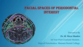
- 1. FACIAL SPACES OF PERIODONTAL INTEREST Presented By Dr. M. Shiva Shanker Ist Year Post Graduate Student , Dept of Periodontics, Mamata Dental College.
- 2. Introduction Classification of spaces Formation of spaces Maxillofacial odontogenic infections Individual spaces Communications Complications Case report conclusion CONTENTS
- 3. Connective tissue - naturally forms a padding between and around the various structures in the neck, and also elsewhere in the body. - tends to be somewhat more dense where it is arranged immediately about organs. When the fascia and spaces of the head and neck of normal bodies are investigated, the looser Connective tissue intervening between organs is torn and pressed against these organs, there by exaggerating any fascial layer already present. The interrelationships of these spaces have been regarded as special importance in the spread of infection. Accurate knowledge of the anatomy of these spaces and prompt drainage of them when they become infected is necessary. INTRODUCTION
- 4. Fascia & spaces above the hyoid bone : Fascia & spaces below the hyoid bone : Superficial layer of fascia Prevertebral Superficial layer of fascia Buccopharyngeal Pretracheal Space of the body of mandible Prevertebral Sub-maxillary gland space. Carotid sheath Masticator space. Retrovisceral space. Space of parotid gland. Retropharyngeal space Danger space or space of Grodinsky and Holyoke. Lateral pharyngeal space. CLASSIFICATION
- 5. Depending upon its relationship with Hyoid bone. Infrahyoid spaces ` (Below the hyoid bone) Suprahyoid spaces (above the hyoid bone) This is further divided into 2 categories. Blind or intrafascial spaces. Pretracheal space Retrovisceral space Space of the body of mandible. Carotid space Space of submaxillary gland. Space 4 or danger space. Masticatory spaces. Space of parotid gland. Peripharyngeal spaces. Retropharyngeal space. Lateral pharyngeal space. Submandibular space. CLASSIFICATION ….
- 6. Depending Primary upon the involvement spaces Secondary spaces CLASSIFICATION ….
- 7. FASCIA The fascia is divided into two major divisions Superficial layer Deep cervical fascia Superficial fascia This arises from the vertebral spinous processes and the ligamentum nuchae and completely encircles the neck to attach again to these. It invests the platysma in the neck, the muscles of facial expression, and the epicranial muscles in the scalp.
- 8. Deep cervical fascia 1. Anterior layer Investing fascia (over the neck) Parotideomassetric Temporal 2. Middle layer Sternohyoid-omohyoid division Sternothyroid-thyrohyoid division Visceral division Buccopharyngeal Pretracheal retropharyngeal
- 9. Deep cervical fascia...... Posterior layer Alar division Prevertebral division The DCF of the neck is contiguous with the mediastenal structures in the thorax through the thoracic inlet. As it emerges from the neck superior to the hyoid bone.
- 10. FASCIAL SPACES
- 11. Space of Burns Fascia anterior to strap muscles between the two sternocleidomastoid muscles splits into two layers just above the sternum Suprsternal space( SOB)
- 12. Pretracheal fascia. The pretracheal fascia passes behind the infrahyoid or strap muscles, infront of the trachea and thyroid gland. The pretracheal layer with the carotid sheath laterally and the prevertebral layer posteriorly, forms the visceral compartment containing the trachea, esophagus and associated structures.
- 13. FORMATION OF SPACES Vestibular space: Medial: mandible or maxilla and overlying periosteum Lateral: vestibular mucosa Superior: buccinator muscle Inferior: buccinator muscle Anterior: intrinsic lip muscles Posterior: lateral pharyngeal and massetric space
- 14. SUB MANDIBULAR SPACE Medial: mylohyoid, hyoglossus, styloglossus muscles Lateral: skin and platysma muscle Superior: mandible, mylohyoid and masseter muscle Mandible Post digastric Stylohyoid muscle Inferior: hyoid bone Anterior: anterior digastric muscle, submental space Posterior: posterior belly of digastric muscle and stylohyoid muscle Ant digastric Mylohyoid
- 15. SUB MENTAL SPACE Mandible Ant digastric Inferior border of the mandible Mylohyoid muscle Hyoid bone
- 16. SUBLINGUAL SPACE Floor of mouth mucosa Mandible Geniohyoid Mylohyoid Hyoid bone
- 17. BUCCAL SPACE Zygomatic arch Skin Buccinator muscle and attachment to maxillary and mandibular alveolus Mandibular and Sub Mandibular space
- 18. BUCCAL SPACE Zygomatic major muscle Depressor anguli oris
- 19. CANINE SPACE Maxilla Nasal bone Levator labii superioris Skin Buccal space
- 20. LATERAL PHARYNGEAL SPACE Sub lingual, sub mandibular spaces Pterygomandibular raphae Hyoid bone Parotid capsule Base of the skull
- 21. RETROPHARYNGEAL SPACE Lateral pharyngeal space Base of the skull Vertebrae C6-T4
- 22. MASSETRIC SPACE Superficial part of masseter Deep part of masseter Mucosa of retromolar triangle of mandible Parotidomassetric fascia
- 23. PTERYGOMANDIBULAR SPACE Parotid gland & its fascia Superior constrictor of pharynx Mandibular ramus Lateral pterygoid muscle Pterygomandibular raphae Buccinator
- 24. SUPERFICIAL TEMPORAL SPACE Temporal fascia Temporalis
- 25. DEEP TEMPORAL SPACE Temporalis Fascia of deep surface of temporalis
- 26. PREVERTEBRAL SPACE Medial: postural neck muscles Lateral: postural neck muscles Superior: base of skull Inferior: coccyx Anterior: alar fascia and retropharyngeal space Posterior: vertebral bodies
- 27. INFRA TEMPORAL SPACE Medial: lateral pterygoid muscle Lateral: temporalis and lateral pterygoid plate tendon and coronoid process Superior: Inferior: Anterior: maxillary tuberosity Posterior: lateral muscle, temporalis muscle, and condyle
- 28. MAXILLOFACIAL ODONTOGENIC INFECTIONS Spread of infection
- 29. STAGES OF INFECTION Characteristic Duration Pain Inoculation 0-3 days Mild-moderate Cellulitis 3-7 days Severe and generalized Size Location Palpation Small Diffuse Soft,doughy, mildly tender Large Diffuse Hard, exquisitely tender Abscess >5 days Moderate-severe localized Small Circumscribed Fluctuant, tender Appearance Normal color Reddened Peripherally reddened Skin quality Normal Thickened Centrally undermined and shiny Surface temperature Slightly heated Hot Moderately heated Loss of function Tissue fluid Minimal or none Edema Levels of malaise Severity Percutaneous bacteria Mild Mild Aerobic Severe Moderately severe Serosanguineous, flecks of Pus pus Severe Moderately severe Severe Moderately severe Mixed Anaerobic and
- 30. MANAGEMENT OF SPACE INFECTIONS Management of infections, mild or severe, always has five general goals: Medical support of the patient Administration of proper antibiotics Surgical removal of the source of infection as early as possible Surgical drainage of the infection Constant reevaluation of the resolution of the infection.
- 31. INDIVIDUAL SPACES Vestibular space Etiology Signs and symptoms Treatment and applied aspects
- 32. CLINICAL PICTURE SHOWING VESTIBULAR SPACE INFECTION
- 33. SUBMANDIBULAR SPACE Facial vessels Hypoglossal nerve Submandibular gland Marginal mandibular branch of VII CONTENTS
- 35. CLINICAL PICTURE OF SUBMANDIBULAR SPACE
- 36. SUBMENTAL SPACE Etiology Signs and symptoms Treatment and aspects Spread of infection applied
- 37. CLINICAL PICTURE OF A SUBMENTAL SPACE
- 38. SUBLINGUAL SPACE Lingual nerves and vessels Submandibular ganglion Submandibular gland CONTENTS
- 39. Etiology Signs and symptoms Treatment and aspects Spread of infection applied
- 40. CLINICAL PICTURE OF A SUBLINGUAL SPACE
- 41. BUCCAL SPACE Parotid duct Buccal branch of VII Facial vessels Buccal fat pad CONTENTS
- 42. Etiology Signs and symptoms Treatment and aspects Spread of infection applied
- 43. CLINICAL PICTURE OF BUCCAL SPACE
- 44. CANINE SPACE Infraorbital nerves and vessels Levator anguli oris CONTENTS
- 46. CLINICAL PICTURE OF A CANINE SPACE
- 47. LATERAL PHARYNGEAL SPACE Etiology Signs and symptoms Treatment and aspects B. Lateral Pharyngeal Space applied
- 48. RETROPHARYNGEAL SPACE Etiology Signs and symptoms Treatment and aspects applied
- 49. PREVERTEBRAL SPACE Etiology Signs and symptoms Treatment and aspects applied
- 51. MASSETRIC SPACE Treatment and applied aspects: Spread of infection
- 52. CLINICAL PICTURE SHOWING MASSETRIC SPACE
- 53. PTERYGOMANDIBULAR SPACE Treatment and aspects: A. Pterygomandibular Space applied
- 54. TEMPORAL SPACE Treatment and applied aspects:
- 55. INFRATEMPORAL SPACE Etiology Signs and symptoms Treatment and aspects applied
- 56. PRETRACHEAL SPACE The pretracheal space is encased by the middle layer of the DCF and contains the thyroid gland, trachea, and esophagus. Infections in this space usually result from thyroiditis or perforation of the anterior cervical esophagus. This space is rarely involved as a result of odontogenic infection
- 57. COMMUNICATIONS
- 58. COMPLICATIONS OF HEAD AND NECK INFECTIONS Extension of infection from local to regional spaces Orbital complications (blindness, ophthalmoplegia) Jugular vein thrombosis Septicemia Metastatic abscesses Airway compromise Aspiration Carotid artery rupture Mediastinitis Osteomyelitis Cutaneous fistula Cranial nerve deficits Cavernous sinus thrombosis Necrotizing fasciitis Maxillary sinusitis, oroantral fistula Septic shock
- 59. Aminoglycosides Gentamicin IM/IV Cephalosporins Cefaclor Cefadoxil cephalexin 3mg/kg/day in equal doses 8 hourly 250-500 mg 8 hourly 500mg-1g 12-24 hrs 250-500mg 6hrs Penicillins Amoxicillin Cloxacyclin 1g initially than 250-500mg 6hrs 250-500mg 6hrly Macrolides Erythromycin Azithromycin 250-500mg 6hrly 10mg/kg upto 500mg initially followed by 5mg/kg upto 250 mg qd- 5 days Tetracyclins Doxcycline 100mg q 24hrs or 50mg q 12 hrs 250mg q 24 hrs ANTIBIOTIC ADMINISTRATION REGIMEN oxytetracycline Clindamycin 150mg q 6hrs FOR ORAL INFECTIONS Metronidazole Vancomycin 500mg q 6hrs IV 1g infused over 1hr before procedure
- 60. For practical purposes, it makes little difference to the surgeon in an uninfected neck whether a facial sheath is present or not. Of some practical importance however are the relationships of the loose connective tissue areas, the fascial spaces, of the head and neck, both because of the structures that transverse or abut against them, and because they may become infected. The interrelationships of these spaces have been regarded as special importance in the spread of infections and many surgeons have urged accurate knowledge of the anatomy of these spaces and a prompt drainage of them when they become infected CONCLUSION
- 61. REFERENCES 1. Grays anatomy-Muscles and fascia of the head, 38th edition, (789-807) 2. Oral and Maxillofacial surgery, Fonseca, vol-5 (77-118) 3. Oral and Maxillofacial surgery, Laskin, Vol-2 (219-252) 4. Oral and Maxillofacial infections, Topazian, 4th edition (158-214) 5. Atlas of minor oral surgery, Hary dym (154) 6. Text book of Clinical periodontology, Newman, Takei, Carranza, 10th edition 7. Anatomical considerations in periodontal periodontology 1971, vol 42, number 10. surgery, journal of 8. Two cases of masticator space abscess initially diagnosed as TMJD, Clarke, Kobe J. Med. Sci., Vol. 54, No. 3, pp. E163-E168, 2008
- 62. THANK YOU………….