How to Extract a Tooth as Atraumatically as Possible (Rosenbach 2015)
•
2 gefällt mir•341 views

Technique article on how to remove teeth as minimally invasively as possible

Empfohlen
Empfohlen
Weitere ähnliche Inhalte
Was ist angesagt?
Was ist angesagt? (20)
Ähnlich wie How to Extract a Tooth as Atraumatically as Possible (Rosenbach 2015)
Ähnlich wie How to Extract a Tooth as Atraumatically as Possible (Rosenbach 2015) (20)
Kürzlich hochgeladen
Kürzlich hochgeladen (20)
How to Extract a Tooth as Atraumatically as Possible (Rosenbach 2015)
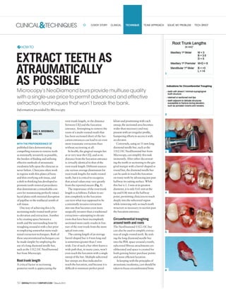
- 1. 72 | DENTALPRODUCTSREPORT.COM | March2015 CLINICAL&TECHNIQUES 1 2 3 4 5 6 COVER STORY CLINICAL TECHNIQUE TEAM APPROACH SOLVE MY PROBLEM TECH BRIEF WITH THE PREPONDERANCE OF published data demonstrating compelling reasons to remove teeth as minimally invasively as possible, the burden of finding and utilizing effective methods of atraumatic exodontia falls upon the clinician as never before. Clinicians often work in regions with thin plates of bone and thin overlying soft tissue, and a shift in thinking has developed to promote tooth removal procedures that demonstrate a remarkable con- cern for maintaining perfectly intact facial plates with minimal disruption of papillae or the midfacial zenith of soft tissue. One way of achieving this is by sectioning multi-rooted teeth prior to elevation and extraction. Another is by creating space between a tooth and the surrounding bone by troughing around it with a bur prior to employing somewhat more tradi- tional extraction techniques. Both of these unconventional techniques can be made simpler by employing the use of a long diamond needle bur, such as the 1312.11C NeoDiamond bur from Microcopy. Root trunk length A critical factor in sectioning posterior teeth is appreciating the root trunk length, or the distance between CEJ and the furcation entrance. Attempting to remove the roots of a multi-rooted tooth that has been sectioned short of the fur- cation entrances can lead to an even more traumatic extraction than without sectioning at all. In health, the gingival margin lies at or very near the CEJ, and so its distance from the furcation entrance is virtually identical to that of the root trunk length. Different sources cite various average dimensions for root trunk lengths for multi-rooted teeth, but it is critical to recognize that actual values may vary greatly from the reported means (Fig. 1). The importance of the root trunk length is as follows: Failure to sec- tion completely to the furcation can turn what was supposed to be a minimally invasive extraction into one that becomes even more surgically invasive than a traditional extraction—attempting to elevate roots that have been incompletely sectioned more easily results in frac- ture of the root trunk from the more apical root cone. The cutting length of an average barrel-shaped bur is 4 mm long and is sometimes greater than 1 mm wide. Use of such a bur often leaves a wide path that, in many cases, won’t even reach the furcation with a single sweep of the bur. Multiple subcrestal bur sweeps are thus indicated to reach the furcation, and because it is difficult to maintain perfect paral- lelism and positioning with each sweep, the sectioned area becomes wider than necessary and may present with an irregular profile, hampering efforts to access it with an elevator. Conversely, using an 11 mm long diamond needle bur, such as the 1312.11C NeoDiamond bur from Microcopy, can simplify this task immensely. After either decoronat- ing the tooth or sectioning to the gin- gival margin with a barrel-shaped or round bur, the diamond needle bur can be sunk to reach the furcation on many teeth by advancing just past halfway its cutting surface. While this bur is 1. 2 mm at its greatest diameter, it is only 0.61 mm at the tip and 0.86 mm at the halfway point, permitting clinicians to reach deeply into the subcrestal region while removing only as much tooth structure as necessary to section past the furcation entrance. Circumferential troughing around teeth and roots The NeoDiamond 1312.11C bur can also be used to simplify extrac- tion of single rooted teeth. By sink- ing the long diamond needle bur into the PDL space around a tooth, subcrestal fibrous attachments are obliterated and space is created for both gaining better purchase points and more efficient luxation. In keeping with the principles of atraumatic exodontia, care should be taken to focus circumferential bone DALE R. ROSENBACH, DMD, MS EXTRACT TEETH AS ATRAUMATICALLY AS POSSIBLE Microcopy’s NeoDiamond burs provide multiuse quality with a single-use price to permit advanced and effective extraction techniques that won’t break the bank. Information provided by Microcopy. HOWTO
- 2. 74 | DENTALPRODUCTSREPORT.COM | March2015 CLINICAL&TECHNIQUES TECHNIQUE 11 9 10 7 8 1. Table of root trunk lengths. 2. Indications for circumferential troughing. 3. The 1312.11C bur sunk into the distal sulcus of tooth No. 12. 4. Subcrestal fracture on tooth No. 14, sectioned along the furcal paths. 5. The DB root is easily delivered with forceps. 6. The extracted DB root. 7. The MB root is extracted. 8. The extracted MB root. 9. The palatal root is elevated from its socket. 10. The elevated palatal root. 11. The extraction socket immediately following tooth removal. AT A GLANCE Coarse grit Pointed cone Extra-long head length NeoDiamond 1312.11C bur Microcopy 800-235-1863|microcopydental.com CIRCLERS#88 removal at the proximal surfaces and away from the facial and lingual/ palatal socket walls, which may be thinner, more fragile and thus more susceptible to fracture and/or resorp- tion. The length, taper and narrow- ness of the 1312.11C bur provide thebest combinationoffeatures to achieve one of the most simple and least traumatic methods of tooth removal (Fig. 2). 01STEP The 1312.11C bur sunk into the distal sulcus of tooth No. 12, exhibiting a complete loss of the buccal half of the supragingival tooth structure. The bur was then drawn along the entire distal proxi- mal surface (Fig. 3). 02STEP Following preparation and temporization for a crown, tooth No. 14 was determined to have a subcrestal fracture and was sec- tioned along the furcal paths with the long-needle diamond bur (Fig. 4). (Notice how the cut correspond- ing to the M furcation is palatalized because the MB root is so large that it displaces the furcation palatally.) 03STEP After elevating the tooth segments off of one another, the DB root is easily delivered with forceps, despite having only the thinnest sliver of supragingival tooth struc- ture to grab onto (Figs. 5-6). 04STEP The MB root is extracted. The immensity of the MB root socket and the palatalization of the M furcal bone can now be fully appreciated (Figs. 7-8). 05STEP Finally, the palatal root is elevated from its socket. Notice how the gingival margin remains virtu- ally intact around the circumference of the socket (Figs. 9-10). 06STEP Figure 11 shows the extraction socket immediately fol- lowing tooth removal. Due to the minimally invasive technique employed, both the hard and soft tissue remain largely undisturbed. The robust architecture of the fur- cal bone makes this an ideal site for immediate implant placement. Conclusion The NeoDiamond 1312.11C bur from Microcopy can assist in mak- ing extractions simple and effective and, perhaps most importantly, very minimally invasive. Microcopy’s single-use price makes this method cost effective as well. ABOUT THE AUTHOR Dr. Dale Rosenbach received his undergraduate degree in biology from Yeshiva University and his dental degree from New Jersey Dental School. After completing a one-year general practice residency at Woodhull Medical Center in Brooklyn, he worked in private practice for a year. Following this, he attended Columbia University for his postdoctoral training in periodontics and implant dentistry, completing a master’s degree focusing on the effects of abutment dis/ reconnection on initial peri-implant bone loss. Dr. Rosenbach is a diplomate of the American Board of Periodontology and is currently a clinical instructor and course director of the periodontics and implant dentistry lecture series at the GPR at Woodhull Medical Center in Brooklyn, N.Y. He recently published a review article in Dentaltown Magazine entitled “Gap Management Around Immediate Implants: A Review of the Literature and its Application in Clinical Practice” and has been appointed to both the editorial advisory board and the continuing education advisory board for the same publication. Dr. Rosenbach lectures extensively on topics related to periodontics, implant dentistry and adjunctive surgical procedures. He has been invited to speak for component CE groups of the New York State Dental Association, the New Jersey Dental Association and the Pennsylvania Dental Association, as well as private CE organizations, such as the New Jersey Health Professionals Development Institute, the Golden Study Group, the Pascack Valley Dental Study Club, Concord Dental Seminars and Alpha Omega of the District of Columbia. He was featured at the 2014 Greater New York Dental Meeting and is scheduled to speak at both the 2015 Greater Long Island Dental Meeting and the 2015 World’s Fair of Dentistry. Dr. Rosenbach maintains a private practice in New York City and can be reached at rosenbachperio@yahoo.com. 1 Carnavale F, Pontoriero R, Lindhe, J: Treatment of Furcation- Involved Teeth. In Lindhe, Karring, Lang, editors: Clinical Periodontology and Implant Dentistry, 4th Edition. London: Blackwell Munksgaard. 2003. pages 707-8.