Pruebas radiologicas a evitar American College of Radiology.
•
0 likes•2,333 views

Folleto informativo del American College of Radiology sobre cinco pruebas radiológicas a evitar.
![How This List Was Created
The American College of Radiology (ACR) initially solicited expert opinion from physician leaders with its Board of Chancellors. A working group was then formed
to further identify common clinical scenarios in which imaging may be misused and should be reconsidered. Members of the group included the physician
chairs or vice chairs of seven ACR commissions such as Quality and Safety, Appropriateness Criteria and Metrics. An initial list of topics was narrowed down
based on the highest potential for improvement, representing a broad range of tests and the availability of strong guidelines. Members then researched specific
recommendations and evidentiary statements based on their expertise. Recommendations that were too general or were well covered by other existing measures
and initiatives were eliminated to identify the final five things list. ACR’s disclosure and conflict of interest policy can be found at www.acr.org.
Sources
Jordan JE, Wippold FJ II, Cornelius RS, Amin-Hanjani S, Brunberg JA, Davis PC, De La Paz RL, Dormont D, Germano I, Gray L, Mukherji SJ, Seidenwurm DJ, Sloan MA, Turski PA, Zimmerman RD, Zipfel GJ, Expert
Panel on Neurologic Imaging. ACR Appropriateness Criteria® headache. [online publication]. Reston (VA): American College of Radiology (ACR); 2009. 8 p. http://www.acr.org/SecondaryMainMenuCategories/
quality_safety/app_criteria/pdf/ExpertPanelonNeurologicImaging/HeadacheDoc6.aspx.
Institute for Clinical Systems Improvement (ICSI). Diagnosis and treatment of headache. Bloomington (MN): Institute for Clinical Systems Improvement (ICSI); 2011 Jan. 84 p.
1 Frishberg BM, Rosenberg JH, Matchar DB, et al. Evidence-Based Guidelines in the Primary Care Setting: Neuroimaging in Patients with Nonacute Headache. American Academy of Neurology. 2000. Available
online: http://www.aan.com/professionals/practice/pdfs/gl0088.pdf (US Headache Consortium).
Stephen D. Silberstein. Practice parameter: Evidence-based guidelines for migraine headache (an evidence-based review): Report of the Quality. Standards Subcommittee of the American Academy of
Neurology. 2000;55;754 Neurology. (US Headache Consortium).
Edlow JA, Panagos PD, Godwin SA, Thomas TL, Decker WW; American College of Emergency Physicians. Clinical policy: critical issues in the evaluation and management of adult patients presenting to the
emergency department with acute headache. Ann Emerg Med. 2008 Oct;52(4):407-36.
Torbicki A, Perrier A, Konstantinides S, et al. Guidelines on the diagnosis and management of acute pulmonary embolism: the Task Force for the Diagnosis and Management of Acute Pulmonary Embolism of
the European Society of Cardiology (ESC). Eur Heart J. 2008; 29(18):2276-315.
2 Neff MJ. ACEP releases clinical policy on evaluation and management of pulmonary embolism. American Family Physician 2003; 68 (4): 759–60.
Stein PD, Woodard PK, Weg JG, Wakefield TW, Tapson VF, Sostman HD, Sos TA, Quinn DA, Leeper KV, Hull RD, Hales CA, Gottschalk A, Goodman LR, Fowler SE, Buckley JD. Diagnostic pathways in acute
pulmonary embolism: recommendations of the PIOPED II Investigators. Radiology 2007; 242 (1): 15–21.
American College of Radiology. ACR Appropriateness Criteria: routine admission and preoperative chest radiography. http://www.acr.org/SecondaryMainMenuCategories/quality_safety/app_criteria/pdf/
ExpertPanelonThoracicImaging/RoutineAdmissionandPreoperativeChestRadiographyDoc6.aspx.
Gomez-Gil E, Trilla A, Corbella B, et al. Lack of clinical relevance of routine chest radiography in acute psychiatric admissions. Gen Hosp Psychiatry 2002; 24(2): 110-113.
& colors Archer C, Levy AR, McGregor M. Value of routine preoperative chest x-rays: a meta-analysis. Can J Anaesth 1993; 40(11): 1022-1027.
3 Munro J, Booth A, Nicholl J. Routine preoperative testing: a systematic review of the evidence. Health Technol Assess 1997; 1(12):i-iv; 1-62.
Grier DJ, Watson LF, Harnell GG, Wilde P. Are routine chest radiographs prior to angiography of any value? Clin Radiol 1993; 48(2):131-33.
and the ABIM Foundation logos FJ, Sen I. Routine chest radiography in the elderly. Age Ageing. 1985; 14(1):11-14.
Gupta SD, Gibbins and
American College of Radiology. ACR Appropriateness Criteria: routine chest radiographs in ICU patients http://www.acr.org/SecondaryMainMenuCategories/quality_safety/app_criteria/pdf/
he basic building blocks of each
ExpertPanelonThoracicImaging/RoutineChestRadiographDoc7.aspx.
n’s graphic identity and are the ®
Wan MJ, et al. Acute appendicitis in young children: cost-effectiveness of US versus CT in diagnosis-a Markov decision analytic model. Radiology 2009;250:378-86.
ual symbols presented to the US or CT for diagnosis of appenditicis in children? A meta-analysis. Radiology 2006;241:83-94.
Doria AS, et al. public.
Approved 2006
Garcia K, et al. Suspected appendicitis in children: diagnostic importance of normal abdominopelvic CT findings with nonvisualized appendix. Radiology 2009;250:531-537.
properly and consistently, a unique
4 Krishnamoorthi R, et al. Effectiveness of a staged US and CT protocol for the diagnosis of pediatric appendicitis: reducing radiation exposure in the age of ALARA. Radiology 2011;259:231-239.
ve visual style can be establishedRadiology. ACR Appropriateness Criteria: right lower quadrant pain/suspected appendicitis. http://www.acr.org/SecondaryMainMenuCategories/quality_safety/app_criteria/pdf/
American College of and
ExpertPanelonGastrointestinalImaging/RightLowerQuadrantPainDoc12.aspx.
sion on the public will be clearKS, Oldham KT. Imaging of acute appendicitis in children: EU versus U.S. or US versus CT? A North American perspective. Pediatr Radiolo. 2009; 39(5):500-5.
Frush DP. Frush
and
.
Levine D, Brown DL, Andreotti RF, Management of asymptomatic ovarian and other adnexal cysts imaged at US: Society of Radiologists in Ultrasound Consensus Conference Statement. Radiology 2010
256:943-54. PaNTONe hexadecimal
301 c 005B99
American College of Radiology. ACR Appropriateness Criteria: clinically suspected adnexal masses. http://www.acr.org/SecondaryMainMenuCategories/quality_safety/app_criteria/pdf/
traditional color for ABIM logos. To
ExpertPanelonWomensImaging/SuspectedAdnexalMassesDoc11.aspx.
5 R: 0, G: 82 , B: 145
or consistency, ABIM hasGynecol 2002;100(6):1413–1416.Gynecologists. ACOG Committee Opinion: number 280, December 2002. The role of the generalist obstetrician-gynecologist in the early detection of ovarian cancer.
chosen the
American College of Obstetricians and
Obstet
C: 100, M: 69, Y: 24, K: 7
olor palettes (pictured on the right)
American College of Obstetricians and Gynecologists. ACOG Practice Bulletin. Management of adnexal masses. Obstet Gynecol 2007;110(1):201–214.
Timmerman D, Valentin L, Bourne TH, et al. Terms, definitions and measurements to describe the sonographic features of adnexal tumors: a consensus opinion from the International Ovarian Tumor Analysis
M and ABIM Foundation Group. Ultrasound Obstet Gynecol 2000;16(5):500–505.
(IOTA) logos,
based on the Pantone Matching
MS), the preferred standard used
About the ABIM Foundation: About the American College of Radiology:
by graphic artists and printers.
can be converted into many Foundation is to advance
The mission of the ABIM ApprovedThe mission of the American College of Radiology
medical professionalism to improve the health (ACR) is to serve its 34,000 members in advancing the
2006
ch as RGB and Hexadecimal, this by collaborating with
care system. We achieve quality, safety, and science of radiology and radiation
physicians and physician leaders, medical trainees, oncology. The ACR conducts cutting-edge clinical
e preferred color format for systems, payers, policymakers,
health care delivery Web and socioeconomic research, establishes quality and
consumer organizations and patients to foster a shared safety standards and provides continuing education
uter programs, such asof professionalism and how they can
understanding
Microsoft ®
and advocacy for radiologists, radiation oncologists
werPoint. adopt the tenets of professionalism in practice. and medical physicists. Since 1923, the ACR has
worked to keep medical imaging and radiation
To learn more about the ABIM Foundation, visit www.abimfoundation.org. oncology safe, effective and accessible for all.
ation of these specific colors will
For more information or questions, please visit www.acr.org.
ong and consistent identity for both
PaNTONe hexadecimal
nd the ABIM Foundation. ABIM 3155 c 006d75
For more information or to see other lists of Five Things Physicians and Patients Should Question, visit www.choosingwisely.org.
ations should approve all materials
R: 0, G: 101, B: 120
BIM or the ABIM Foundation logos. C: 100, M: 44, Y: 43, K: 15](data:image/gif;base64,R0lGODlhAQABAIAAAAAAAP///yH5BAEAAAAALAAAAAABAAEAAAIBRAA7)
Recommended
More Related Content
What's hot
What's hot (20)
Viewers also liked
Viewers also liked (10)
Similar to Pruebas radiologicas a evitar American College of Radiology.
Similar to Pruebas radiologicas a evitar American College of Radiology. (20)
More from Cristobal Buñuel
More from Cristobal Buñuel (20)
Recently uploaded
Recently uploaded (20)
Pruebas radiologicas a evitar American College of Radiology.
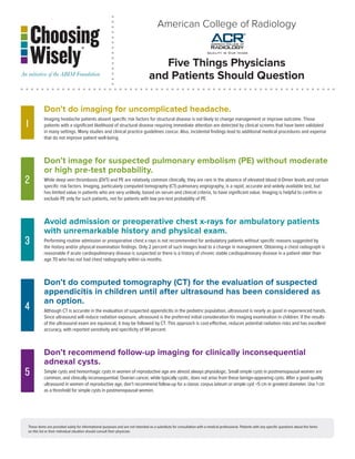
- 1. American College of Radiology Five Things Physicians and Patients Should Question Don’t do imaging for uncomplicated headache. 1 Imaging headache patients absent specific risk factors for structural disease is not likely to change management or improve outcome. Those patients with a significant likelihood of structural disease requiring immediate attention are detected by clinical screens that have been validated in many settings. Many studies and clinical practice guidelines concur. Also, incidental findings lead to additional medical procedures and expense that do not improve patient well-being. Don’t image for suspected pulmonary embolism (PE) without moderate or high pre-test probability. 2 While deep vein thrombosis (DVT) and PE are relatively common clinically, they are rare in the absence of elevated blood d-Dimer levels and certain specific risk factors. Imaging, particularly computed tomography (CT) pulmonary angiography, is a rapid, accurate and widely available test, but has limited value in patients who are very unlikely, based on serum and clinical criteria, to have significant value. Imaging is helpful to confirm or exclude PE only for such patients, not for patients with low pre-test probability of PE. Avoid admission or preoperative chest x-rays for ambulatory patients with unremarkable history and physical exam. 3 Performing routine admission or preoperative chest x-rays is not recommended for ambulatory patients without specific reasons suggested by the history and/or physical examination findings. Only 2 percent of such images lead to a change in management. Obtaining a chest radiograph is reasonable if acute cardiopulmonary disease is suspected or there is a history of chronic stable cardiopulmonary disease in a patient older than age 70 who has not had chest radiography within six months. Don’t do computed tomography (CT) for the evaluation of suspected appendicitis in children until after ultrasound has been considered as an option. 4 Although CT is accurate in the evaluation of suspected appendicitis in the pediatric population, ultrasound is nearly as good in experienced hands. Since ultrasound will reduce radiation exposure, ultrasound is the preferred initial consideration for imaging examination in children. If the results of the ultrasound exam are equivocal, it may be followed by CT. This approach is cost-effective, reduces potential radiation risks and has excellent accuracy, with reported sensitivity and specificity of 94 percent. Don’t recommend follow-up imaging for clinically inconsequential adnexal cysts. 5 Simple cysts and hemorrhagic cysts in women of reproductive age are almost always physiologic. Small simple cysts in postmenopausal women are common, and clinically inconsequential. Ovarian cancer, while typically cystic, does not arise from these benign-appearing cysts. After a good quality ultrasound in women of reproductive age, don’t recommend follow-up for a classic corpus luteum or simple cyst <5 cm in greatest diameter. Use 1 cm as a threshold for simple cysts in postmenopausal women. These items are provided solely for informational purposes and are not intended as a substitute for consultation with a medical professional. Patients with any specific questions about the items on this list or their individual situation should consult their physician.
- 2. How This List Was Created The American College of Radiology (ACR) initially solicited expert opinion from physician leaders with its Board of Chancellors. A working group was then formed to further identify common clinical scenarios in which imaging may be misused and should be reconsidered. Members of the group included the physician chairs or vice chairs of seven ACR commissions such as Quality and Safety, Appropriateness Criteria and Metrics. An initial list of topics was narrowed down based on the highest potential for improvement, representing a broad range of tests and the availability of strong guidelines. Members then researched specific recommendations and evidentiary statements based on their expertise. Recommendations that were too general or were well covered by other existing measures and initiatives were eliminated to identify the final five things list. ACR’s disclosure and conflict of interest policy can be found at www.acr.org. Sources Jordan JE, Wippold FJ II, Cornelius RS, Amin-Hanjani S, Brunberg JA, Davis PC, De La Paz RL, Dormont D, Germano I, Gray L, Mukherji SJ, Seidenwurm DJ, Sloan MA, Turski PA, Zimmerman RD, Zipfel GJ, Expert Panel on Neurologic Imaging. ACR Appropriateness Criteria® headache. [online publication]. Reston (VA): American College of Radiology (ACR); 2009. 8 p. http://www.acr.org/SecondaryMainMenuCategories/ quality_safety/app_criteria/pdf/ExpertPanelonNeurologicImaging/HeadacheDoc6.aspx. Institute for Clinical Systems Improvement (ICSI). Diagnosis and treatment of headache. Bloomington (MN): Institute for Clinical Systems Improvement (ICSI); 2011 Jan. 84 p. 1 Frishberg BM, Rosenberg JH, Matchar DB, et al. Evidence-Based Guidelines in the Primary Care Setting: Neuroimaging in Patients with Nonacute Headache. American Academy of Neurology. 2000. Available online: http://www.aan.com/professionals/practice/pdfs/gl0088.pdf (US Headache Consortium). Stephen D. Silberstein. Practice parameter: Evidence-based guidelines for migraine headache (an evidence-based review): Report of the Quality. Standards Subcommittee of the American Academy of Neurology. 2000;55;754 Neurology. (US Headache Consortium). Edlow JA, Panagos PD, Godwin SA, Thomas TL, Decker WW; American College of Emergency Physicians. Clinical policy: critical issues in the evaluation and management of adult patients presenting to the emergency department with acute headache. Ann Emerg Med. 2008 Oct;52(4):407-36. Torbicki A, Perrier A, Konstantinides S, et al. Guidelines on the diagnosis and management of acute pulmonary embolism: the Task Force for the Diagnosis and Management of Acute Pulmonary Embolism of the European Society of Cardiology (ESC). Eur Heart J. 2008; 29(18):2276-315. 2 Neff MJ. ACEP releases clinical policy on evaluation and management of pulmonary embolism. American Family Physician 2003; 68 (4): 759–60. Stein PD, Woodard PK, Weg JG, Wakefield TW, Tapson VF, Sostman HD, Sos TA, Quinn DA, Leeper KV, Hull RD, Hales CA, Gottschalk A, Goodman LR, Fowler SE, Buckley JD. Diagnostic pathways in acute pulmonary embolism: recommendations of the PIOPED II Investigators. Radiology 2007; 242 (1): 15–21. American College of Radiology. ACR Appropriateness Criteria: routine admission and preoperative chest radiography. http://www.acr.org/SecondaryMainMenuCategories/quality_safety/app_criteria/pdf/ ExpertPanelonThoracicImaging/RoutineAdmissionandPreoperativeChestRadiographyDoc6.aspx. Gomez-Gil E, Trilla A, Corbella B, et al. Lack of clinical relevance of routine chest radiography in acute psychiatric admissions. Gen Hosp Psychiatry 2002; 24(2): 110-113. & colors Archer C, Levy AR, McGregor M. Value of routine preoperative chest x-rays: a meta-analysis. Can J Anaesth 1993; 40(11): 1022-1027. 3 Munro J, Booth A, Nicholl J. Routine preoperative testing: a systematic review of the evidence. Health Technol Assess 1997; 1(12):i-iv; 1-62. Grier DJ, Watson LF, Harnell GG, Wilde P. Are routine chest radiographs prior to angiography of any value? Clin Radiol 1993; 48(2):131-33. and the ABIM Foundation logos FJ, Sen I. Routine chest radiography in the elderly. Age Ageing. 1985; 14(1):11-14. Gupta SD, Gibbins and American College of Radiology. ACR Appropriateness Criteria: routine chest radiographs in ICU patients http://www.acr.org/SecondaryMainMenuCategories/quality_safety/app_criteria/pdf/ he basic building blocks of each ExpertPanelonThoracicImaging/RoutineChestRadiographDoc7.aspx. n’s graphic identity and are the ® Wan MJ, et al. Acute appendicitis in young children: cost-effectiveness of US versus CT in diagnosis-a Markov decision analytic model. Radiology 2009;250:378-86. ual symbols presented to the US or CT for diagnosis of appenditicis in children? A meta-analysis. Radiology 2006;241:83-94. Doria AS, et al. public. Approved 2006 Garcia K, et al. Suspected appendicitis in children: diagnostic importance of normal abdominopelvic CT findings with nonvisualized appendix. Radiology 2009;250:531-537. properly and consistently, a unique 4 Krishnamoorthi R, et al. Effectiveness of a staged US and CT protocol for the diagnosis of pediatric appendicitis: reducing radiation exposure in the age of ALARA. Radiology 2011;259:231-239. ve visual style can be establishedRadiology. ACR Appropriateness Criteria: right lower quadrant pain/suspected appendicitis. http://www.acr.org/SecondaryMainMenuCategories/quality_safety/app_criteria/pdf/ American College of and ExpertPanelonGastrointestinalImaging/RightLowerQuadrantPainDoc12.aspx. sion on the public will be clearKS, Oldham KT. Imaging of acute appendicitis in children: EU versus U.S. or US versus CT? A North American perspective. Pediatr Radiolo. 2009; 39(5):500-5. Frush DP. Frush and . Levine D, Brown DL, Andreotti RF, Management of asymptomatic ovarian and other adnexal cysts imaged at US: Society of Radiologists in Ultrasound Consensus Conference Statement. Radiology 2010 256:943-54. PaNTONe hexadecimal 301 c 005B99 American College of Radiology. ACR Appropriateness Criteria: clinically suspected adnexal masses. http://www.acr.org/SecondaryMainMenuCategories/quality_safety/app_criteria/pdf/ traditional color for ABIM logos. To ExpertPanelonWomensImaging/SuspectedAdnexalMassesDoc11.aspx. 5 R: 0, G: 82 , B: 145 or consistency, ABIM hasGynecol 2002;100(6):1413–1416.Gynecologists. ACOG Committee Opinion: number 280, December 2002. The role of the generalist obstetrician-gynecologist in the early detection of ovarian cancer. chosen the American College of Obstetricians and Obstet C: 100, M: 69, Y: 24, K: 7 olor palettes (pictured on the right) American College of Obstetricians and Gynecologists. ACOG Practice Bulletin. Management of adnexal masses. Obstet Gynecol 2007;110(1):201–214. Timmerman D, Valentin L, Bourne TH, et al. Terms, definitions and measurements to describe the sonographic features of adnexal tumors: a consensus opinion from the International Ovarian Tumor Analysis M and ABIM Foundation Group. Ultrasound Obstet Gynecol 2000;16(5):500–505. (IOTA) logos, based on the Pantone Matching MS), the preferred standard used About the ABIM Foundation: About the American College of Radiology: by graphic artists and printers. can be converted into many Foundation is to advance The mission of the ABIM ApprovedThe mission of the American College of Radiology medical professionalism to improve the health (ACR) is to serve its 34,000 members in advancing the 2006 ch as RGB and Hexadecimal, this by collaborating with care system. We achieve quality, safety, and science of radiology and radiation physicians and physician leaders, medical trainees, oncology. The ACR conducts cutting-edge clinical e preferred color format for systems, payers, policymakers, health care delivery Web and socioeconomic research, establishes quality and consumer organizations and patients to foster a shared safety standards and provides continuing education uter programs, such asof professionalism and how they can understanding Microsoft ® and advocacy for radiologists, radiation oncologists werPoint. adopt the tenets of professionalism in practice. and medical physicists. Since 1923, the ACR has worked to keep medical imaging and radiation To learn more about the ABIM Foundation, visit www.abimfoundation.org. oncology safe, effective and accessible for all. ation of these specific colors will For more information or questions, please visit www.acr.org. ong and consistent identity for both PaNTONe hexadecimal nd the ABIM Foundation. ABIM 3155 c 006d75 For more information or to see other lists of Five Things Physicians and Patients Should Question, visit www.choosingwisely.org. ations should approve all materials R: 0, G: 101, B: 120 BIM or the ABIM Foundation logos. C: 100, M: 44, Y: 43, K: 15