SlideShare ist ein Scribd-Unternehmen logo
1 von 76
Downloaden Sie, um offline zu lesen
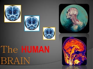
The HUMAN
BRAIN
Protective Structures
Meninges Three layers of
connective tissue that
enclose the brain


Dura Mater
Arachnoid
Pia Mater
Dura Mater
              Outermost layer
              Thickest and
               toughest part of the
               meninges
              There are two
               layers, the outer
               layer is fused to the
               cranial bones.
Dura Mater    In various places,
               the two layers
               separate to allow
               venous channels
               called dural sinuses
               to drain blood from
               the brain.
              Function
               containment of the
               cerebrospinal fluid
               in the brain.
Arachnoid    Middle layer of
              the meninges
             Loosley attached
              to the pia mater by
              weblike fibers.
             Function Allows
              for movement of
              cerebrospinal
              fluid.
Cerebrospinal Fluid
              Fluid that circulates
              in and around the
              brain.
              Protects the brain
              from shock and injury.
              Function-Transports
              nutrients and waste to
              and from cells.
              .
Cerebrospinal Fluid
              Formed in four spaces
               called ventricals.
              The ventricals hold a
               vascular portion called
               the choroid plexus,
               which produces the
               cerebrospinal fluid by
               filtering the blood and
               cellular excretion.
Pia Mater    Innermost layer of
              the meninges
             A delicate connective
              tissue that covers the
              brain and spinal chord
             Function-Holds the
              blood vessels that
              supply nutrients and
              oxygen to the brain
              and spinal cord.
Meninges
Cerebral
Hemispheres
Structure of the Hemispheres
 Two layers
 An outer layer of
  gray matter
  called the
  cerebral
  cortex.
 Supported by an
  inner layer of
  white matter
Structure of the hemispheres
Each hemisphere has four lobes

Frontal
Parietal
Temporal
Occipital
Structure of the hemispheres
Gray Matter – The Cerebral Cortex
 The most highly evolved   portion of the brain.
 Arranged in folds of elevations called
gyri, and grooves
called sulci
Gyri and Sulci
                       The surfaces on
                       which brain cells
                       reside.


More surface = more
complex calculations
Structure of the hemispheres
White Matter
 Connects the gray
  matter areas with
  one another and
  with other parts of
  the brain.
 Dispersed in a tree
  like pattern
 Made of
  myelinated fibers
White and Gray Matter

 White matter


                 Gray Matter
Structure of the hemispheres

Corpus Callosum
 Largest white matter
  structure in the brain
 Facilitates communication
  between the right and left
  hemispheres by electrical
  impulses
Structure of the hemispheres

Basal Ganglia
 Base of forbrain, deep
in each hemisphere.
Functions: helps to re-
gulate body movement
and facial expressions.
Structure of the hemispheres

Internal Capsule
 In between the hemi-
spheres and the brain
stem
Function – Carries im-
pulses between the
cerebral hemispheres
and the brainstem.
Functions of the Cerebral Cortex
The functions of the Cerebral Cortex are
localized according to the four lobes. They
are named for the
overlying cranial
bones.
CEREBRAL CORTEX FUNCTIONS

CEREBRAL CORTEX- Functions
 Conscious deliberation
 Voluntary actions
 Memory
 Association
 Discrimination
 Judgement
Functions of the Cerebral Cortex

The Frontal Lobe lies
anterior to the central
sulcus.
Functions of the Cerebral
Cortex
Some Frontal Lobe Functions
 Contains an area that provides the conscious
  control of skeletal muscles.
 Contains two areas that are important in
    speech
Functions of the Cerebral Cortex

The Parietal Lobe
occupies the superior
part of each hemisphere
and lies posterior to the
central nucleus
Functions of the Cerebral Cortex

Some Parietal Lobe Functions
   Contains a primary sensory area where
    impulses from the skin are interpreted
   Estimates distance and size.
Functions of the Cerebral Cortex

The Temporal Lobe lies
inferior the lateral sulcus
snd folds under the hemi-
sphere on each side.
Functions of the Cerebral Cortex

Some Temporal Lobe Functions
 Responsible for receiving and interpreting

auditory impulses from the ear.
   An olfactory area that concerns the sense
of smell.
Functions of the Cerebral Cortex

The Occipital Lobe
lies posterior to the
parietal lobe and
extends over the
cerebellum.
Functions of the Cerebral Cortex


Some Occipital Lobe Functions
 Visual receiving area and visual ass-
ociation for interpreting impulses from
  the retina of the eye.
Functions of the Cerebral Cortex

The Insula lies deep within each hemisphere
   and
cannot be seen
from the surface.
Functions of the Cerebral Cortex
The Insula Functions
 Visceral reactions and judgments
 Receives, integrates and responds to
  autonomic influx.
Communication areas of the lobes

TEMPORAL LOBE
  Auditory Cortex
 Lies at the posterior
  area of the temporal
  lobe
 Contains the auditory
  receiving and
  association areas.
Communication areas of the lobes

 Auditory Receiving Area
  Detects sound impulses from
   the surrounding environment
 Auditory association area
  Interprets and translates the
   sound impulses.

                       1 1 2
Communication areas of the lobes

FRONTAL LOBE- Motor cortex
 Lies anterior to the most inferior part of the
  frontal lobe
 Contains the Broca area
Communication areas of the lobes
Broca Area – Responsible for spoken and
written communication.
Functions- Controls:
 Muscles in the tongue
 Soft Palate
 The larynyx
 Lies anterior to the area that controls the arm
and hand muscles that produce written speech
Diencephalon
 The area between
the cerebral
  hemispheres
and the brain stem.
 Contains the
  thalamus
and the
  hypothalamus.
The Thalamus
 Sorts and directs sensory
  impulses to areas of the
  cerebral cortex.
 Nearly all sensory
  impulses travel through
  the thalamus.
 Important role in sleep
  and
wakefullness.
Hypothalamus
 Locatedinferior to the
  thalamus
 The Boss of you.
Hypothalamus
 Helps maintain
  homeostasis
 Controls autonomic
  function such as:
  heartbeat, blood flow,
and hormone secretion.
 Controls the pituitary
   gland.
Pituitary Gland
 A „master gland‟ lo-
cated at the bottom
of the hypothalamus.
 Pea sized
 Assists in the regulation
of homeostasis.
Pituitary Gland
Functions Helps to regulate:
 Growth
 Blood pressure
 Child birth
 Sex organ function
 Thyroid gland
 Metabolism
 Temperature regulation
Hypothalamus/ Pituitary
Homeostasis
 Balance
 Maintenance
of body con-
ditions within
set limits.
The Limbic System
 Located between the
   cerebrum and diencephalon
 Includes the Hip-
pocampus
 Links conscious functions
of the cerebral cortex and
autonomic functions of the
brainstem.
The Limbic System
 Located between the
   cerebrum and diencephalon
 Includes the Hip-
pocampus
 Links conscious functions
of the cerebral cortex and
autonomic functions of the
brainstem.
The Limbic System
 Emotional states and
  behavior
 Formation of long
  term memory
The Hippocampus

Long term
memory
Spacial
navigation
The Hippocampus
The Brain Stem
The brainstem
is located in the
anterior region
below the cerebrum
The Brain Stem
Connects the cerebrum and the
diencephalon with the spinal cord.
The brainstem includes:

 The Midbrain
 The Pons
 The Medulla Oblongata
The Midbrain
  The midbrain is located below the center of the
cerebrum.
 The midbrain forms the superior part of the brain
stem.
The Midbrain
The midbrain consists of centers that are
  concerned with aspects of vision and
  hearing.
The Pons
 Located anterior to the cerebellum.
 Lies between the midbrain and the medulla
 Connects the two halves of the cerebellum
with the brainstem.
Medulla Oblongata
 Located between the pons and
  spinal cord.
 Contains gray matter which
  has centers that play an
  important role in many
  involuntary actions such as
  respiration.
 The centers are called vital
  centers.
The Medulla Vital Centers
 The respiratory center
 The cardiac center
 The vasomotor center
The Cerebellum “Little Brain”
 Divided into two hemispheres,
  and one middle part(vermis).
 Outer layer gray matter, inner
  layer white matter.
 Located above the brainstem,
  and beneath the occipital
  lobes.
The Cerebellum
Functions:
 Coordination in voluntary
  movement.
 Helps maintain balance
  and equilibrium.
 Helps maintain muscle
  tone.
Brain Studies

Imaging used to study the brain
without physically entering it
Brain Studies
CT Scan Provides photographs of the bone,
soft tissue, and cavities of the brain. Used to de-
tect:
 Scar tissue accumulations
 Anatomic lesions such as tumors
Brain Studies
MRI Magnetic Resonance Imaging
 Shows more views than CT scans
 Often reveals issues that the CT
scan can miss such as tumors, scar
tissue, and hemorrhaging.
Brain Studies EEG
The Electroencephalograph records
Electrical currents produced by the brain‟s
Nerve cells.
Cranial Nerves
Cranial Nerves
 12 pairs of cranial nerves
 Numbered 1- 12 based on
connection with the brain.
 Originate in the brain stem
 Divided into 4 categories
based on the impulses they
send.
Cranial Nerves Types
Special sensory impulses
Located in special sense
organs in the head, responsible
for:
 Smell
 Taste
 Vision
 Hearing
Cranial Nerves Types
General sensory impulses
Originate from receptors through the body.
 Pain
 Touch
 Temp
 Pressure
 Vibration
 Deep muscle sense
Cranial Nerves Types
Somatic Motor Impulses
 Voluntary control of skeletal muscles
Cranial Nerves Types
VISCERAL MOTOR IMPULSES
 Part of autonomic nervous system
 Involuntary control of glands
and involuntary muscle
Aging and the Nervous System
Aging of the brain
The brain loses 5-10 % of it’s
volume between
the ages of 20-90.
The grooves widen and the
surface shrinks.
Agingof the brain
 Aging in
 the Brain


Senile plaque forms
Synapses and neurons
decrease, esp in the cere-
bral cortex.
Aging in the brain
 Movement is slowed
 Information processing slows
 Chance of stroke, and alzheimers increases
Dementia
Loss of cognitive ability in a previously-unimpaired
  person, beyond what might be expected from
  normal aging.

 Massage - Yes
Alzheimers
 Alzheimer’s
 disease leads to
 nerve cell death
 and tissue loss
 throughout the
 brain. Over time,
 the brain shrinks
 dramatically,
 affecting nearly all   Massage –Yes, best to
 its functions.         start in early stages
Multiple Sclerosis
Eats away at the protective
sheath that covers your
nerves. This interferes with
the communication between
your brain and the rest of
your body.

Massage- During
remissions

Weitere ähnliche Inhalte

Was ist angesagt?

Brain and cranial nerves
Brain and cranial nervesBrain and cranial nerves
Brain and cranial nervesPave Medicine
 
Anatomy of meninges, ventricles, cerebrospinal fluid
Anatomy of meninges, ventricles, cerebrospinal fluidAnatomy of meninges, ventricles, cerebrospinal fluid
Anatomy of meninges, ventricles, cerebrospinal fluidMBBS IMS MSU
 
The cerebral hemispheres - Gross Anatomy & Connections
The cerebral hemispheres - Gross Anatomy & ConnectionsThe cerebral hemispheres - Gross Anatomy & Connections
The cerebral hemispheres - Gross Anatomy & ConnectionsDr. Ashik Ahsan
 
Cerebellum Anatomy and Physiology
Cerebellum Anatomy and PhysiologyCerebellum Anatomy and Physiology
Cerebellum Anatomy and PhysiologyPS Deb
 
Ascending tracts of spinal cord
Ascending tracts of spinal cordAscending tracts of spinal cord
Ascending tracts of spinal cordDr. sana yaseen
 
Anatomy of the spinal cord
Anatomy of the spinal cordAnatomy of the spinal cord
Anatomy of the spinal cordRanjeet Singha
 

Was ist angesagt? (20)

Brain and cranial nerves
Brain and cranial nervesBrain and cranial nerves
Brain and cranial nerves
 
Anatomy of meninges, ventricles, cerebrospinal fluid
Anatomy of meninges, ventricles, cerebrospinal fluidAnatomy of meninges, ventricles, cerebrospinal fluid
Anatomy of meninges, ventricles, cerebrospinal fluid
 
NEUROANATOMY - I
NEUROANATOMY - INEUROANATOMY - I
NEUROANATOMY - I
 
Cerebellum ppt
Cerebellum pptCerebellum ppt
Cerebellum ppt
 
Nervous system
Nervous systemNervous system
Nervous system
 
Ventricles of the brain
Ventricles of the brainVentricles of the brain
Ventricles of the brain
 
Csf & ventricles
Csf & ventriclesCsf & ventricles
Csf & ventricles
 
Cerebral circulation
Cerebral circulationCerebral circulation
Cerebral circulation
 
The cerebral hemispheres - Gross Anatomy & Connections
The cerebral hemispheres - Gross Anatomy & ConnectionsThe cerebral hemispheres - Gross Anatomy & Connections
The cerebral hemispheres - Gross Anatomy & Connections
 
Cerebellum Anatomy and Physiology
Cerebellum Anatomy and PhysiologyCerebellum Anatomy and Physiology
Cerebellum Anatomy and Physiology
 
HUMAN BRAIN
HUMAN BRAINHUMAN BRAIN
HUMAN BRAIN
 
Nervous system
Nervous systemNervous system
Nervous system
 
Nervous system
Nervous systemNervous system
Nervous system
 
Anatomy of brain
Anatomy of brainAnatomy of brain
Anatomy of brain
 
Anatomy of the Cerebrum
Anatomy of the CerebrumAnatomy of the Cerebrum
Anatomy of the Cerebrum
 
Human brain
Human brainHuman brain
Human brain
 
Ascending tracts of spinal cord
Ascending tracts of spinal cordAscending tracts of spinal cord
Ascending tracts of spinal cord
 
Nervous system
Nervous systemNervous system
Nervous system
 
Anatomy of the spinal cord
Anatomy of the spinal cordAnatomy of the spinal cord
Anatomy of the spinal cord
 
Cerebellum
CerebellumCerebellum
Cerebellum
 

Ähnlich wie Pdf human brain

Kkkkkkkkkkkkkkkkkkkkkkkkkkkkkkkkkk
KkkkkkkkkkkkkkkkkkkkkkkkkkkkkkkkkkKkkkkkkkkkkkkkkkkkkkkkkkkkkkkkkkkk
Kkkkkkkkkkkkkkkkkkkkkkkkkkkkkkkkkkfridelbabe
 
Control and coordination
Control and coordinationControl and coordination
Control and coordinationChetan Kute
 
Central Nervous system CNS
Central Nervous system CNSCentral Nervous system CNS
Central Nervous system CNSEduFlash
 
CNS-BRAIN.pptx
CNS-BRAIN.pptxCNS-BRAIN.pptx
CNS-BRAIN.pptxNPRNRI
 
Unit iv -NERVOUS SYSTEM- SANJAY SIR
Unit  iv -NERVOUS SYSTEM- SANJAY SIRUnit  iv -NERVOUS SYSTEM- SANJAY SIR
Unit iv -NERVOUS SYSTEM- SANJAY SIRSANJAY SIR
 
Anatomy of the brain
Anatomy of the brainAnatomy of the brain
Anatomy of the brainSaika Lashari
 
Cns Compressed 2 Nd
Cns Compressed 2 NdCns Compressed 2 Nd
Cns Compressed 2 Ndmycomic
 
Fight or flee artifact
Fight or flee artifactFight or flee artifact
Fight or flee artifactzs4033bn
 
Nervous System-class 6.pdf
Nervous System-class 6.pdfNervous System-class 6.pdf
Nervous System-class 6.pdfRachaelAdeleke
 
Nervous system physiology.pdf
Nervous system physiology.pdfNervous system physiology.pdf
Nervous system physiology.pdfIshita60889
 
NERVOUS_SYSTEM.pptx
NERVOUS_SYSTEM.pptxNERVOUS_SYSTEM.pptx
NERVOUS_SYSTEM.pptxRishavDhar3
 
10 Brain.ppt
10 Brain.ppt10 Brain.ppt
10 Brain.pptShama
 
13 Brain & Five Seneses part-1.ppt
13 Brain & Five Seneses part-1.ppt13 Brain & Five Seneses part-1.ppt
13 Brain & Five Seneses part-1.pptShama
 
Presentation1.pptx, radiological anatomy of the brain.
Presentation1.pptx, radiological anatomy of the brain.Presentation1.pptx, radiological anatomy of the brain.
Presentation1.pptx, radiological anatomy of the brain.Abdellah Nazeer
 
Cns Presentatio
Cns PresentatioCns Presentatio
Cns Presentatiomycomic
 

Ähnlich wie Pdf human brain (20)

Kkkkkkkkkkkkkkkkkkkkkkkkkkkkkkkkkk
KkkkkkkkkkkkkkkkkkkkkkkkkkkkkkkkkkKkkkkkkkkkkkkkkkkkkkkkkkkkkkkkkkkk
Kkkkkkkkkkkkkkkkkkkkkkkkkkkkkkkkkk
 
Nervous system ppt
Nervous system pptNervous system ppt
Nervous system ppt
 
Control and coordination
Control and coordinationControl and coordination
Control and coordination
 
Central Nervous system CNS
Central Nervous system CNSCentral Nervous system CNS
Central Nervous system CNS
 
CNS-BRAIN.pptx
CNS-BRAIN.pptxCNS-BRAIN.pptx
CNS-BRAIN.pptx
 
Unit iv -NERVOUS SYSTEM- SANJAY SIR
Unit  iv -NERVOUS SYSTEM- SANJAY SIRUnit  iv -NERVOUS SYSTEM- SANJAY SIR
Unit iv -NERVOUS SYSTEM- SANJAY SIR
 
Anatomy of the brain
Anatomy of the brainAnatomy of the brain
Anatomy of the brain
 
Cns Compressed 2 Nd
Cns Compressed 2 NdCns Compressed 2 Nd
Cns Compressed 2 Nd
 
Fight or flee artifact
Fight or flee artifactFight or flee artifact
Fight or flee artifact
 
The human brain
The human brainThe human brain
The human brain
 
Neural control
Neural  control Neural  control
Neural control
 
Nervous system ppt.pptx
Nervous system ppt.pptxNervous system ppt.pptx
Nervous system ppt.pptx
 
CNS
CNSCNS
CNS
 
Nervous System-class 6.pdf
Nervous System-class 6.pdfNervous System-class 6.pdf
Nervous System-class 6.pdf
 
Nervous system physiology.pdf
Nervous system physiology.pdfNervous system physiology.pdf
Nervous system physiology.pdf
 
NERVOUS_SYSTEM.pptx
NERVOUS_SYSTEM.pptxNERVOUS_SYSTEM.pptx
NERVOUS_SYSTEM.pptx
 
10 Brain.ppt
10 Brain.ppt10 Brain.ppt
10 Brain.ppt
 
13 Brain & Five Seneses part-1.ppt
13 Brain & Five Seneses part-1.ppt13 Brain & Five Seneses part-1.ppt
13 Brain & Five Seneses part-1.ppt
 
Presentation1.pptx, radiological anatomy of the brain.
Presentation1.pptx, radiological anatomy of the brain.Presentation1.pptx, radiological anatomy of the brain.
Presentation1.pptx, radiological anatomy of the brain.
 
Cns Presentatio
Cns PresentatioCns Presentatio
Cns Presentatio
 

Pdf human brain

  • 3. Meninges Three layers of connective tissue that enclose the brain Dura Mater Arachnoid Pia Mater
  • 4. Dura Mater  Outermost layer  Thickest and toughest part of the meninges  There are two layers, the outer layer is fused to the cranial bones.
  • 5. Dura Mater  In various places, the two layers separate to allow venous channels called dural sinuses to drain blood from the brain.  Function containment of the cerebrospinal fluid in the brain.
  • 6. Arachnoid  Middle layer of the meninges  Loosley attached to the pia mater by weblike fibers.  Function Allows for movement of cerebrospinal fluid.
  • 7. Cerebrospinal Fluid Fluid that circulates in and around the brain. Protects the brain from shock and injury. Function-Transports nutrients and waste to and from cells. .
  • 8. Cerebrospinal Fluid  Formed in four spaces called ventricals.  The ventricals hold a vascular portion called the choroid plexus, which produces the cerebrospinal fluid by filtering the blood and cellular excretion.
  • 9. Pia Mater  Innermost layer of the meninges  A delicate connective tissue that covers the brain and spinal chord  Function-Holds the blood vessels that supply nutrients and oxygen to the brain and spinal cord.
  • 12. Structure of the Hemispheres  Two layers  An outer layer of gray matter called the cerebral cortex.  Supported by an inner layer of white matter
  • 13. Structure of the hemispheres Each hemisphere has four lobes Frontal Parietal Temporal Occipital
  • 14. Structure of the hemispheres Gray Matter – The Cerebral Cortex  The most highly evolved portion of the brain.  Arranged in folds of elevations called gyri, and grooves called sulci
  • 15. Gyri and Sulci The surfaces on which brain cells reside. More surface = more complex calculations
  • 16. Structure of the hemispheres White Matter  Connects the gray matter areas with one another and with other parts of the brain.  Dispersed in a tree like pattern  Made of myelinated fibers
  • 17. White and Gray Matter White matter Gray Matter
  • 18. Structure of the hemispheres Corpus Callosum  Largest white matter structure in the brain  Facilitates communication between the right and left hemispheres by electrical impulses
  • 19. Structure of the hemispheres Basal Ganglia  Base of forbrain, deep in each hemisphere. Functions: helps to re- gulate body movement and facial expressions.
  • 20. Structure of the hemispheres Internal Capsule  In between the hemi- spheres and the brain stem Function – Carries im- pulses between the cerebral hemispheres and the brainstem.
  • 21. Functions of the Cerebral Cortex The functions of the Cerebral Cortex are localized according to the four lobes. They are named for the overlying cranial bones.
  • 22. CEREBRAL CORTEX FUNCTIONS CEREBRAL CORTEX- Functions  Conscious deliberation  Voluntary actions  Memory  Association  Discrimination  Judgement
  • 23. Functions of the Cerebral Cortex The Frontal Lobe lies anterior to the central sulcus.
  • 24. Functions of the Cerebral Cortex Some Frontal Lobe Functions  Contains an area that provides the conscious control of skeletal muscles.  Contains two areas that are important in speech
  • 25.
  • 26. Functions of the Cerebral Cortex The Parietal Lobe occupies the superior part of each hemisphere and lies posterior to the central nucleus
  • 27. Functions of the Cerebral Cortex Some Parietal Lobe Functions  Contains a primary sensory area where impulses from the skin are interpreted  Estimates distance and size.
  • 28. Functions of the Cerebral Cortex The Temporal Lobe lies inferior the lateral sulcus snd folds under the hemi- sphere on each side.
  • 29. Functions of the Cerebral Cortex Some Temporal Lobe Functions  Responsible for receiving and interpreting auditory impulses from the ear.  An olfactory area that concerns the sense of smell.
  • 30. Functions of the Cerebral Cortex The Occipital Lobe lies posterior to the parietal lobe and extends over the cerebellum.
  • 31. Functions of the Cerebral Cortex Some Occipital Lobe Functions  Visual receiving area and visual ass- ociation for interpreting impulses from the retina of the eye.
  • 32. Functions of the Cerebral Cortex The Insula lies deep within each hemisphere and cannot be seen from the surface.
  • 33. Functions of the Cerebral Cortex The Insula Functions  Visceral reactions and judgments  Receives, integrates and responds to autonomic influx.
  • 34. Communication areas of the lobes TEMPORAL LOBE Auditory Cortex  Lies at the posterior area of the temporal lobe  Contains the auditory receiving and association areas.
  • 35. Communication areas of the lobes Auditory Receiving Area  Detects sound impulses from the surrounding environment Auditory association area  Interprets and translates the sound impulses. 1 1 2
  • 36. Communication areas of the lobes FRONTAL LOBE- Motor cortex  Lies anterior to the most inferior part of the frontal lobe  Contains the Broca area
  • 37. Communication areas of the lobes Broca Area – Responsible for spoken and written communication. Functions- Controls:  Muscles in the tongue  Soft Palate  The larynyx  Lies anterior to the area that controls the arm and hand muscles that produce written speech
  • 38. Diencephalon  The area between the cerebral hemispheres and the brain stem.  Contains the thalamus and the hypothalamus.
  • 39. The Thalamus  Sorts and directs sensory impulses to areas of the cerebral cortex.  Nearly all sensory impulses travel through the thalamus.  Important role in sleep and wakefullness.
  • 40. Hypothalamus  Locatedinferior to the thalamus  The Boss of you.
  • 41. Hypothalamus  Helps maintain homeostasis  Controls autonomic function such as: heartbeat, blood flow, and hormone secretion.  Controls the pituitary gland.
  • 42. Pituitary Gland  A „master gland‟ lo- cated at the bottom of the hypothalamus.  Pea sized  Assists in the regulation of homeostasis.
  • 43. Pituitary Gland Functions Helps to regulate:  Growth  Blood pressure  Child birth  Sex organ function  Thyroid gland  Metabolism  Temperature regulation
  • 44. Hypothalamus/ Pituitary Homeostasis  Balance  Maintenance of body con- ditions within set limits.
  • 45. The Limbic System  Located between the cerebrum and diencephalon  Includes the Hip- pocampus  Links conscious functions of the cerebral cortex and autonomic functions of the brainstem.
  • 46. The Limbic System  Located between the cerebrum and diencephalon  Includes the Hip- pocampus  Links conscious functions of the cerebral cortex and autonomic functions of the brainstem.
  • 47. The Limbic System  Emotional states and behavior  Formation of long term memory
  • 50. The Brain Stem The brainstem is located in the anterior region below the cerebrum
  • 51. The Brain Stem Connects the cerebrum and the diencephalon with the spinal cord. The brainstem includes:  The Midbrain  The Pons  The Medulla Oblongata
  • 52. The Midbrain  The midbrain is located below the center of the cerebrum.  The midbrain forms the superior part of the brain stem.
  • 53. The Midbrain The midbrain consists of centers that are concerned with aspects of vision and hearing.
  • 54. The Pons  Located anterior to the cerebellum.  Lies between the midbrain and the medulla  Connects the two halves of the cerebellum with the brainstem.
  • 55. Medulla Oblongata  Located between the pons and spinal cord.  Contains gray matter which has centers that play an important role in many involuntary actions such as respiration.  The centers are called vital centers.
  • 56. The Medulla Vital Centers  The respiratory center  The cardiac center  The vasomotor center
  • 57. The Cerebellum “Little Brain”  Divided into two hemispheres, and one middle part(vermis).  Outer layer gray matter, inner layer white matter.  Located above the brainstem, and beneath the occipital lobes.
  • 58. The Cerebellum Functions:  Coordination in voluntary movement.  Helps maintain balance and equilibrium.  Helps maintain muscle tone.
  • 59. Brain Studies Imaging used to study the brain without physically entering it
  • 60. Brain Studies CT Scan Provides photographs of the bone, soft tissue, and cavities of the brain. Used to de- tect:  Scar tissue accumulations  Anatomic lesions such as tumors
  • 61. Brain Studies MRI Magnetic Resonance Imaging  Shows more views than CT scans  Often reveals issues that the CT scan can miss such as tumors, scar tissue, and hemorrhaging.
  • 62.
  • 63. Brain Studies EEG The Electroencephalograph records Electrical currents produced by the brain‟s Nerve cells.
  • 65. Cranial Nerves  12 pairs of cranial nerves  Numbered 1- 12 based on connection with the brain.  Originate in the brain stem  Divided into 4 categories based on the impulses they send.
  • 66. Cranial Nerves Types Special sensory impulses Located in special sense organs in the head, responsible for:  Smell  Taste  Vision  Hearing
  • 67. Cranial Nerves Types General sensory impulses Originate from receptors through the body.  Pain  Touch  Temp  Pressure  Vibration  Deep muscle sense
  • 68. Cranial Nerves Types Somatic Motor Impulses  Voluntary control of skeletal muscles
  • 69. Cranial Nerves Types VISCERAL MOTOR IMPULSES  Part of autonomic nervous system  Involuntary control of glands and involuntary muscle
  • 70. Aging and the Nervous System
  • 71. Aging of the brain The brain loses 5-10 % of it’s volume between the ages of 20-90. The grooves widen and the surface shrinks.
  • 72. Agingof the brain Aging in the Brain Senile plaque forms Synapses and neurons decrease, esp in the cere- bral cortex.
  • 73. Aging in the brain  Movement is slowed  Information processing slows  Chance of stroke, and alzheimers increases
  • 74. Dementia Loss of cognitive ability in a previously-unimpaired person, beyond what might be expected from normal aging. Massage - Yes
  • 75. Alzheimers Alzheimer’s disease leads to nerve cell death and tissue loss throughout the brain. Over time, the brain shrinks dramatically, affecting nearly all Massage –Yes, best to its functions. start in early stages
  • 76. Multiple Sclerosis Eats away at the protective sheath that covers your nerves. This interferes with the communication between your brain and the rest of your body. Massage- During remissions