2. VOL. 44, 2006 CITRATED SHEEP AND HUMAN BLOOD AGAR 3347
blood donor packs (which come together with a venipuncture TABLE 1. Colony counts of strains tested on four
needle and relevant tubing) containing citrate phosphate dex- different blood agar plates
trose. We therefore aimed to compare blood agar prepared by Colony counts on blood agar plates (range
using citrated human blood, citrated sheep blood, and defi- Strain and dilutiona for triplicate plates)
brinated, noncitrated sheep blood with commercially available HBA CSBA DSBA HuBA
defibrinated, noncitrated horse blood agar. We tested each of
S. pneumoniae ATCC 6305
these agars for their performance characteristics for the growth
1.4 103 100 100 100 100
and antibiotic susceptibility testing of reference and clinical 1.4 104 1,000 1,000 1,000 1,000
strains of S. pneumoniae, S. pyogenes, and S. aureus. 1.5 102 in CSF 100 100 100 100
1.1 104 in CSF 1,000 1,000 1,000 1,000
MATERIALS AND METHODS S. pneumoniae ATCC 49619
This study was duplicated at two sites, the Colonial War Memorial Hospital
2.1 103 100 100 100 100
(CWMH) laboratory, Suva, Fiji, and the Royal Children’s Hospital (RCH), 2.1 104 1,000 1,000 1,000 1,000
Melbourne, Australia. Photographs were taken from the RCH results. 1.6 102 in CSF 100 100 100 100
Laboratory methods. Each strain was cultured in serial dilutions prepared in
1.6 104 in CSF 1,000 1,000 1,000 1,000
brain heart infusion on each of four agars: defibrinated horse blood agar (HBA;
S. aureus ATCC 25923
Oxoid Ltd., Hampshire, United Kingdom), defibrinated sheep blood agar
2 102 18–21 22–29 22–32 13–32
(DSBA), citrated sheep blood agar (CSBA), and citrated human blood agar
2 104 1,000 1,000 1,000 1,000
(HuBA) plates. Each dilution of each organism on each blood agar was tested in
1.1 102 in CSF 100 100 100 100
triplicate at both laboratories. In an attempt to mimic the isolation of bacteria
1.1 104 in CSF 1,000 1,000 1,000 1,000
from a clinical sample, we repeated the same experiments using serial dilutions
of the same organisms in a sample of sterile cerebrospinal fluid (CSF) obtained
S. aureus ATCC 29213
from a patient who had been admitted to RCH for a revision of his ventriculo-
2.5 102 18–38 9–21 24–28 9–14
peritoneal shunt. At CWMH, the CSF, obtained from a patient with suspected
2.5 104 1,000 1,000 1,000 1,000
meningitis, had a normal appearance by microscopy and no growth on culture.
1.6 102 in CSF 100 100 100 100
Finally, we compared DSBA, CSBA, and HuBA for use in antibiotic suscepti-
1.6 104 in CSF 1,000 1,000 1,000 1,000
bility testing for S. pneumoniae and S. pyogenes.
Blood collection and medium preparation. At CWMH, citrated and defi- S. pyogenes ATCC 19615
brinated sheep blood were collected by an aseptic technique from healthy sheep 7 101 6–8 6–22 8–24 1–14
who were not receiving antibiotics at the time of blood collection. The defi- 7 103 100 100 100 100
brinated blood was collected in a sterile glass bottle containing glass rods that 1 102 in CSF 100 100 100 100
were manually rotated. Four hundred fifty milliliters of sheep blood was collected 1 104 in CSF 100 100 100 100
in a standard, sterile blood donor bag (Baxter Healthcare) containing 63 ml of
citrate phosphate dextrose as an anticoagulant. Following collection, the blood S. pyogenes strain JC20
was immediately chilled and transported to the CWMH laboratory, where 10 ml 1.2 102 6–16 2–15 2–13 12–15
of blood was transferred aseptically into BacTalert (Biomerieux, Inc, Durham, 1.2 104 1,000 1,000 1,000 1,000
NC) bottles and incubated for 7 days to ensure sterility. Expired human blood 4.5 101 in CSF 14–20 7–10 10–12 2–6
that had been collected from numerous donors in an aseptic manner and that 4.5 103 in CSF 100 100 100 70
had been stored at between 2 and 8°C was pooled and obtained from both
a
hospitals’ blood banks. Fifty-six millimeters of citrated sheep blood was added to Results are presented for the upper and the lower dilutions only. In many
1 liter of agar. The blood agar plates were prepared by standard methods (Oxoid cases (e.g., for all S. aureus and S. pyogenes strains) intermediate dilutions were
Australia Pty. Ltd). Columbia (Oxoid Ltd.) agar base was used for initial growth, also tested, with identical results for each agar. A clinical isolate of S. pneu-
moniae and a clinical isolate of S. aureus were also tested (results not shown).
and Mueller-Hinton (Oxoid Ltd.) agar was used for sensitivity testing. The agar
plates were stored at 2 to 8°C until they were required.
At RCH, an expired human blood pack containing 63 ml of citrate phosphate
dextrose was obtained from one donor. The citrated sheep blood and the defi-
brinated sheep blood were obtained from a commercial supplier (Institute of (6, 7), each sample was inoculated onto three different blood Mueller-Hinton
Medical and Veterinary Science, South Australia). The medium was prepared (Oxoid Ltd.) plates. Disks containing oxacillin at 1 g, erythromycin at 15 g,
according to the methods of CWMH (Oxoid Australia Pty. Ltd.). The defi- chloramphenicol at 30 g, and co-trimoxazole at 1.25 g (Difco, Sparks, MD) were
brinated HBA was made commercially (Oxoid Ltd.). applied along with an optochin disk for the pneumococcal isolates. Disks con-
Strains and dilutions. Three S. pneumoniae strains were used: strains ATCC taining penicillin at 1 g, erythromycin at 15 g, chloramphenicol at 30 g, and
49619 (serotype 19F) and ATCC 6305 (serotype 5) and a clinical isolate (sero- vancomycin at 30 g (Oxoid Ltd.) were applied for the S. pyogenes isolates. All
type 1, isolated from a blood culture). Three S. aureus strains were used: strains plates were incubated overnight in 5% CO2 at 35°C. Zone diameters were read
ATCC 25923 and ATCC 29213 and a clinical isolate (isolated from a blood following 24 h if incubation. Two scientists read the plates independently.
culture). Two S. pyogenes strains were used: strain ATCC 19615 (emm type 80)
and a clinical isolate (emm type 1, isolated from a blood culture). Each strain was
inoculated in brain heart infusion (Oxoid Ltd.) broth to make a suspension equal RESULTS
to a 0.5 McFarland standard ( 1.2 108 CFU/ml). These were serially diluted
before 100 l was plated in triplicate on each agar. Similarly, each strain was The results of the isolation of all organisms were combined
serially diluted in CSF before it was plated in triplicate on each agar. The and the results from both CWMH and RCH were similar.
inoculated plates were incubated at 35°C in 5% CO2 for 18 to 20 h. The results
S. pneumoniae. The numbers of colonies of the S. pneu-
were recorded in terms of viable counts, colony morphology (by description and
photographs), and colony size (recorded in millimeters at the widest margin). moniae reference strains were similar on all blood agars at all
Antibiotic susceptibility testing. Susceptibility testing was performed at dilutions, including the strains in CSF (Table 1). In addition,
CWMH only. Susceptibility testing for S. aureus was not performed, as blood is the growth of a clinical isolate of S. pneumoniae was similar on
not required as a medium supplement for this organism. To test susceptibility, 0.5 all blood agars at all dilutions and in CSF (data not shown).
McFarland standard concentrations were made in triplicate from S. pneumoniae
strains ATCC 49619 and ATCC 6305 and a clinical isolate from a normally sterile
The morphological appearances were similar for colonies on
site and from S. pyogenes strain ATCC 19615 and a clinical isolate from a HBA, CSBA, and DSBA. However, colonies were much
normally sterile site. According to the methods of the CLSI (formerly the NCCLS) smaller and alpha-hemolysis was not obvious for any strain on
3. 3348 RUSSELL ET AL. J. CLIN. MICROBIOL.
TABLE 2. Appearances of colonies and zones of hemolysis for tested strains on four different blood agar plates
Appearance of colonies and hemolysis on blood agar plates
Strain and characteristic a
HBA CSBA DSBA HuBA
S. pneumoniae ATCC 6305
Colony appearance Shiny, grey Mucoid, grey Dull, grey Dull, grey
Colony size (mm) 1 1 1 Pinpoint
Alpha-hemolysis Small, 1 mm Obvious, 1–5 mm Obvious, 1 mm Absent
S. pneumoniae ATCC 49619
Colony appearance Shiny, mucoid grey Dry, grey Dry, grey Shiny, grey
Colony size (mm) 1 1.5 1 Pinpoint
Alpha-hemolysis Obvious, 1 mm Obvious, 1–5 mm Obvious, 1 mm Absent
S. aureus ATCC 25923
Colony appearance Opaque white glossy Opaque white glossy Opaque white glossy Opaque white glossy
Colony size (mm) 1.5 1.5 1.5 1.5
Beta hemolysis Faint, 1 mm Obvious, 1 mm Obvious, 1 mm Absent
S. aureus ATCC 29213
Colony appearance Opaque yellow glossy Opaque yellow glossy Opaque yellow glossy Opaque yellow glossy
Colony size (mm) 1.5 1.5 1.5 1
Beta-hemolysis Faint, 1 mm Obvious, 1.8 mm Obvious, 1.8 mm Faint, 1 mm
S. pyogenes ATCC 19615
Colony appearance Glossy white Dry grey-white Dry grey Glossy white
Colony size (mm) 1 1–2 1 Pinpoint
Beta-hemolysis Obvious, 5 mm Faint, 1 mm Obvious, 1 mm Absent
S. pyogenes strain JC20
Colony appearance Glossy white Glossy white Glossy white Glossy white
Colony size (mm) 1.5–1.8 1 1 Pinpoint
Beta-hemolysis Obvious, 1–5 mm Obvious, 1 mm Obvious, 1 mm Absent
a
A clinical isolate of S. pneumoniae and a clinical isolate of S. aureus were also tested (results not shown).
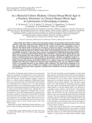
FIG. 1. Growth of S. pneumoniae ATCC 6305 on the four different blood agars at a dilution of 1 101 CFU/ml.
4. VOL. 44, 2006 CITRATED SHEEP AND HUMAN BLOOD AGAR 3349
FIG. 2. Growth of S. aureus ATCC 25923 on the four different blood agars at a dilution of 1 102 CFU/ml.
HuBA (Table 2; Fig. 1). Additional photographs are available plates. The hemolysis on these plates was subtle, and it was
on the http://www.rch.org.au/cich/pubs/ website. difficult to discern where growth began.
S. aureus. The numbers of colonies of the S. aureus reference The antibiotic susceptibility results for S. pyogenes showed
strains were similar on all blood agars at all dilutions, including that the citrated sheep Mueller-Hinton blood agar (CSB
the strains in CSF (Table 1). In addition, the growth of a MHA) and the Hu MHA plates displayed larger zone sizes
clinical isolate of S. aureus was similar on all blood agars at all than the defibrinated Mueller-Hinton blood agar (DSB MHA)
dilutions and in CSF (data not shown). The appearances and plate (Table 4). It was much easier to read the defibrinated and
the sizes of the colonies were similar on all agars, but hemolysis citrated sheep blood agars due to the hemolysis present. The
was not obvious for any strain on HuBA and was only faint on Hu MHA plates were difficult to read due to the lack of
HBA (Table 2; Fig. 2). Additional photographs are available beta-hemolysis.
on the http://www.rch.org.au/cich/pubs/ website.
S. pyogenes. The numbers of colonies of the S. pyogenes DISCUSSION
reference and clinical strains were similar on all blood agars at
all dilutions, including the strains in CSF (Table 1). The ap- The results presented here confirm that although S. pneu-
pearances and the sizes of the colonies were similar on HBA, moniae, S. aureus, and S. pyogenes grow on HuBA, the colony
CSBA, and DSBA. Hemolysis was most obvious on HBA and sizes tend to be very small, the morphology often varies from
DSBA, although it was still present on CSBA. On HuBA, S. that present on animal blood agar, and hemolysis is minimal.
pyogenes colonies were pinpoint in size and hemolysis was As colony size, colony morphology, and hemolysis are all crit-
absent (Table 2, Fig. 3). Additional photographs are available ical to the identification of these organisms, there is a much
on the http://www.rch.org.au/cich/pubs/ website. greater chance these organisms from human specimens cul-
Antibiotic susceptibility testing results. The antibiotic sus- tured on HuBA will be overlooked or misidentified, especially
ceptibility testing results for S. pneumoniae were similar for when other organisms may be present (e.g., for samples from
both sheep Mueller-Hinton blood agars, but the human Muel- the upper respiratory tract or skin swabs). HuBA also per-
ler-Hinton blood agar (Hu MHA) demonstrated larger zone formed poorly compared to the performance of sheep blood
sizes for all antibiotics (Table 3). The susceptibility reference agar for antibiotic susceptibility testing. These findings have
strain, ATCC 49619, failed quality control on the Hu MHA profound implications for developing countries, where expired
5. 3350 RUSSELL ET AL. J. CLIN. MICROBIOL.
FIG. 3. Growth of Streptococcus pyogenes ATCC 19615 on the four different blood agars at a dilution of 1 102 CFU/ml.
human blood is commonly used as a medium supplement. ing of S. pneumoniae, S. aureus, and S. pyogenes. The only
Aside from the implications for clinical care, it is likely that published comparison of HBA, DSBA, and anticoagulated hu-
measurements of the disease burden due to these organisms man blood agar that we could find was a study that compared
are underestimates in countries that routinely use HuBA. the isolation of Bordetella pertussis on different blood agars (4).
To our knowledge, this is the first time that HuBA has been That study demonstrated that HuBA was inferior to defi-
shown to be suboptimal for the growth and susceptibility test- brinated horse and sheep blood agar. It is not clear why HuBA
TABLE 3. Antibiotic disk susceptibility results for three S. pneumoniae strains
Zone diam (mm)a
S. pneumoniae strain
and medium Trimethoprim-
Oxacillin Erythromycin Chloramphenicol Optochin
sulfamethoxazole
ATCC 6305
CSB MHA 32–35 31–33 32–33 26–32 13–20
DSB MHA 32–33 31–33 30–34 29–32 15–17
Hu MHA 32–39 30–35 30–39 28–40 14–17
ATCC 49619 a
CSB MHA 10–11 28–29 27–29 26–28 10–13
DSB MHA 10–11 27–29 26–27 25–27 10–13
Hu MHA 13–14 30–32 33–36 32–36 10–13
Clinical isolate 42796
CSB MHA 29–33 29–30 29–31 25–28 12–15
DSB MHA 28–31 29–32 29–32 28–31 12–14
Hu MHA 30–35 30–42 32–38 27–38 14–17
a
Values are ranges. Acceptable results for CLSI susceptibility quality control: oxacillin, 12 mm; erythromycin, 25 to 30 mm; chloramphenicol, 23 to 27 mm;
trimethoprim-sulfamethoxazole, 20 to 28 mm.
6. VOL. 44, 2006 CITRATED SHEEP AND HUMAN BLOOD AGAR 3351
TABLE 4. Antibiotic disk sensitivity results for the organisms; therefore, care needs to be taken at the point of
three S. pyogenes strains collection to ensure that the correct ratio of blood is collected.
S. pyogenes Zone diam (mm) Hu MHA plates, which are used routinely in developing
strain and countries, cannot be recommended for use for the isolation or
medium Penicillin Erythromycin Chloramphenicol Vancomycin
susceptibility testing of any of these organisms, which supports
ATCC 19615 existing recommendations (1, 2, 3, 5). A strategy is required to
CSB MHA 38–42 35–36 28 22–23 phase out its use. Further work is required to assess whether
DSB MHA 32–33 25–27 26–27 22–23 CSB MHA is suitable for antibiotic susceptibility testing for S.
Hu MHA 35–37 24–25 27–28 23–24
pneumoniae. Preliminary data suggest that it is likely to per-
JC 20 form better than Hu MHA. Our results did not confirm that
CSB MHA 33–34 27–29 22–23 20–21 CSB MHA plates could be used for susceptibility testing for S.
DSB MHA 30–31 24–25 22–23 18–19 pyogenes. Further work is required to assess whether CSBA
Hu MHA 35–36 24–25 22–24 20–22
and CSB MHA are suitable for the isolation and susceptibility
Clinical isolate testing, respectively, of a larger range of organisms. This will
CSB MHA 38–39 28–29 27–28 21–22 determine whether they could be recommended as practical
DSB MHA 38–40 28 28–29 19–20 alternatives for universal use.
Hu MHA 31–34 22–25 27–28 18–21
REFERENCES
1. Anand, C. R., H. Gordon, H. Shaw, K. Fonseca, and M. Olsen. 2000. Pig and
is inferior to agar with other animal blood. It has been sug- goat blood as substitutes for sheep blood in blood-supplemented agar media.
J. Clin. Microbiol. 38:591–594.
gested that human blood may contain antibiotics, antibodies, 2. Centers for Disease Control and Prevention. 1998. Laboratory methods for
or other anti-infective agents (5). The lack of hemolysis on the diagnosis of meningitis caused by Neisseria meningitidis, Streptococcus
human blood agar may be due to the age of the red cells in the pneumoniae, and Haemophilus influenzae. Centers for Disease Control and
Prevention, Atlanta, Ga.
expired human blood or some other factor. 3. Gratten, M., D. Battistutta, P. Torzillo, J. Dixon, and K. Manning. 1994.
By contrast, we were able for the first time to describe a Comparison of goat and horse blood as culture medium supplements for
isolation and identification of Haemophilus influenzae and Streptococcus pneu-
practical alternative to human blood—citrated sheep blood— moniae for upper respiratory tract secretions. J. Clin. Microbiol. 32:2871–
that most countries should be able to adapt without requiring 2872.
a major investment in infrastructure. Citrate is said to have 4. Hoppe, J. E., and M. Schlagenhauf. 1989. Comparison of three kinds of blood
and two incubation atmospheres for cultivation of Bordetella pertussis on
antibacterial characteristics (8, 9), but we found that CSBA charcoal agar. J. Clin. Microbiol. 27:2115–2117.
performed similarly in most aspects to DSBA. Other studies 5. Johnson, D. R., E. L. Kaplan, J. Sramek, R. Bicova, J. Havlicek, H.
have demonstrated that defibrinated pig blood and goat blood Havilickova, J. Moliova, and P. Kriz. 1996. Laboratory diagnosis of group A
streptococcal infections. World Health Organization, Geneva, Switzerland.
are suitable alternatives for medium supplements for S. pneu- 6. National Committee for Clinical Laboratory Standards. 2003. Performance
moniae (1, 3), raising the possibility that citrated blood from standards for antimicrobial disk susceptibility tests; approved standard, 8th
animals other than sheep may also be acceptable. In our ex- ed. M2-A8, vol. 23, no. 1. National Committee for Clinical Laboratory Stan-
dards, Wayne, Pa.
perience, it is the collection of defibrinated blood rather than 7. National Committee for Clinical Laboratory Standards. 2003. Performance
the availability of the animal that has posed the most difficult standards for antimicrobial susceptibility testing. NCCLS document M100-
S13 (M2). National Committee for Clinical Laboratory Standards, Wayne, Pa.
hurdle in providing a sustainable supply of blood. 8. Phillips, C. A. 1999. The effects of citric acid, lactic acid, sodium citrate and
We are confident that CSBA may be used for the isolation of sodium lactate, alone and in combination with nisin, on the growth of Arco-
all three organisms tested in this study. Our study evaluated a bacter butzleri. Lett. Appl. Microbiol. 29:424–428.
9. Young, K. M., and P. M. Foegeding. 1993. Acetic, lactic, and citric acids and
citrate to blood ratio of 1:10. It is not known whether a ratio pH inhibition of Listeria monocytogenes Scott A and the effect on intracel-
lower than this may affect the growth and sensitivity pattern of lular pH. J. Appl. Bacteriol. 74:515–520.