11 feb lect. reflexes, Ashok Solanki, asso. prof physio,
•Als PPTX, PDF herunterladen•
15 gefällt mir•3,078 views

classification, ref arc, clinical significance of reflexes.

Empfohlen
Weitere ähnliche Inhalte
Was ist angesagt?
Was ist angesagt? (20)
Andere mochten auch
Andere mochten auch (14)
Ähnlich wie 11 feb lect. reflexes, Ashok Solanki, asso. prof physio,
Ähnlich wie 11 feb lect. reflexes, Ashok Solanki, asso. prof physio, (20)
Mehr von Smt. N.H.L. Municipal Medical college
Mehr von Smt. N.H.L. Municipal Medical college (13)
Kürzlich hochgeladen
Kürzlich hochgeladen (20)
11 feb lect. reflexes, Ashok Solanki, asso. prof physio,
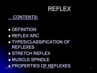
- 1. REFLEX CONTENTS: DEFINITION REFLEX ARC TYPES/CLASSIFICATION OF REFLEXES STRETCH REFLEX MUSCLE SPINDLE PROPERTIES OF REFLEXES 24-Feb-14 Dr. Ashok Solanki 1
- 4. Nerve pathways Ascending Tracts Tract Signal function Dorsal columns Vibration, tactile sensation, conscious proprioception Spinocerebeller Spinothalamic (lateral and Proprioception anterior) Spinoreticular Spinomesencephalic Spino-cervico-thalamic Spinohypothalamic Pain, temperature, itch (lateral), crude touch (anterior) Pain Pain Pain (touch?) Pain
- 5. Structure of spinal cord 24-Feb-14 Dr. Ashok Solanki 5
- 6. Spinal cord Fetal 3rd month: ends at coccyx Birth: ends at L3 Adult position at approx L1-2 during childhood End: conus medullaris This tapers into filum terminale of connective tissue, tethered to coccyx Spinal cord segments are superior to where their corresponding spinal nerves emerge through intervetebral foramina (see also fig 17.5, p 288) Denticulate ligaments: lateral shelves of pia mater anchoring to dura (meninges: more later) http://www.apparelyzed.com/spinalcord.html
- 7. Spinal nerves continued Divided based on vertebral locations 8 cervical 12 thoracic 5 lumbar 5 sacral 1 coccygeal Cauda equina (“horse’s tail”): collection of nerve roots at inferior end of vertebral canal
- 8. Classified as According to centre IN THE SPINAL CORDseg, inter, supra. According to functionflexor, extensor, postural R. Clinicallysupreficial, deep, visceral No. of synapse involved. Mono and polysynaptic According to origin– spinal cord, brain stem, cortical etc. Conditional and unconditional – since birth Rapid, stereotyped, invountary response to a sensory stimuli Dr. Ashok . consciouslly or unconsciousllySolanki 24-Feb-14 8
- 9. CLASSIFICATION CONDITIONED (ACQUIRED)/ UNCONDITIONED(SINCE BIRTH) CEREBELLER, CORTICAL, MIDBRAIN, SPINAL SOMATIC:FLEXOR , EXTENSOR VISCERAL: AUTONOMIC MONOSYNAPTIC , POLYSYNAPTIC SUPERFICIAL, DEEP, VISCERAL, PATHOLOGICAL SEGMENTAL, INTERSEGMENTAL, SUPRASEGMENTAL 24-Feb-14 Dr. Ashok Solanki 9
- 12. Functions or reflex action Maintain the homeostasis- b.p regulation, heart rate, digestive , autonomic reflexes Automatic actions Balance and posture Reflex maintining the movements -eyes 24-Feb-14 Dr. Ashok Solanki 12
- 13. REFLEX ARC ANATOMICAL NERVOUS PATHWAY OF REFLEX IS CALLED REFLEX ARC. RECEPTOR SENSORY / AFFERENT NERVE CENTER EFFERENT / MOTOR NERVE EFFECTOR ORGAN * BELL-MAGENDIE LAW: DORSAL ROOTS ARE SENSORY & VENTRAL ROOTS ARE MOTOR. 24-Feb-14 Dr. Ashok Solanki 13
- 14. SUPERFICIAL REFLEXES CORNEAL AND CONJUNCTIVAL REFLEX PHARYNGEAL REFLEX PALATAL REFLEX ABDOMINAL RELEX PLANTAR REFLEX: Scratch over the outer edge of sole cause plantar flexion and adduction of all toes and dorsiflexion and inversion of foot.( L5,S1) ANAL REFLEX 24-Feb-14 Dr. Ashok Solanki 14
- 15. DEEP REFLEXES JAW JERK: 5TH CRANIAL NV NUCLEI BICEPS JERK: C5,6 TRICEPS JERK: C6,7 SUPINATOR JERK: C5,6 KNEE JERK: L2,3,4 ANKLE JERK: S1,2 24-Feb-14 Dr. Ashok Solanki 15
- 16. Reflex Arc Specific nerve impulse pathway 5 components of reflex arc receptor sensory neuron integrating center motor neuron effector 24-Feb-14 Dr. Ashok Solanki 16
- 17. PROPERTIES ONE WAY CONDUCTION SUMMATION: SPATIAL, TEMPORAL OCCLUSION SUBLIMINAL FRINGE RECRUITMENT AFTERDISCHARGE REBOUND PHENOMENON FATIGUE RECIPROCAL INNERVATION AND RECIPROCAL INHIBITION 24-Feb-14 Dr. Ashok Solanki 17
- 18. Flexor (withdrawal) Reflex 24-Feb-14 Dr. Ashok Solanki Step on tack (pain fibers send signal to spinal cord Interneurons branch to different spinal cord segments Motor fibers in several segments are activated More than one muscle group activated to lift foot off of tack 18
- 19. Crossed Extensor Reflex 24-Feb-14 Dr. Ashok Solanki Lifting left foot requires extension of right leg to maintain one’s balance Pain signals cross to opposite spinal cord Contralateral extensor muscles are stimulated by interneurons to hold up the body weight Reciprocal innervation - when extensors contract flexors relax, etc 19
- 20. Clinical Considerations Checking a patient’s reflexes may help to detect disorders/injury Plantar flexion reflex -- stroke the lateral margin of the sole normal response is curling under the toes abnormal response or response of children under 18 months is called Babinski sign (upward fanning of toes due to incomplete myelination in child) 24-Feb-14 Dr. Ashok Solanki 20
- 21. Inverse stretch reflex Golgi tendon organ- 2 to 15 in each muscle. Responds to tension and not the length The Golgi tendon reflex is a protective reflex rise in tension is sensed by the Golgi tendon a which stimulates the Ib stimulates the I-b afferents stimulate the inhibitory interneurons inhibit the α-motoneuron discharge to 24-Feb-14 Dr. Ashok Solanki 21
- 22. INVERSE STRETCH REFLEX/ AUTOGENIC INHIBITION WHEN A MUSCLE IS STRETCHED, IT CONTRACTS BUT IF THE STRETCH IS MAINTAINED (CONTINUED), THE MUSCLE RELAXES. 24-Feb-14 Dr. Ashok Solanki 22
- 23. UMN lesions LMN lesions •weakness, paralysis •weakness, paralysis •spasticity •flaccidity, hypotonia • tendon reflexes •Hypo- /no tendon •+ Babinski sign reflex •little,if any,muscle • - Babinski sign atrophy •muscle atrophy •no fasiculation •fasiculation of involved muscle 24-Feb-14 Dr. Ashok Solanki 23
- 24. VISCERAL REFLEXES PUPILLARY REFLEXES: DIRECT LIGHT REFLEX INDIRECT OR CONSENSUAL LIGHT REFLEX ACCOMODATION REFLEX: CONSTRICTION OF PUPIL, CONVERGENCE OF EYE BALLS, INCREASE IN ANTERIOR CURVATURE OF LENS CILIOSPINAL REFLEX: STIMULATION OF SKIN IN NECK –DILATATION OF PUPILS OCULOCARDIAC REFLEX: PRESSURE OVER EYEBALLS - BRADYCARDIA 24-Feb-14 Dr. Ashok Solanki 24
- 25. PATHOLOGICAL REFLEXES BABINSKI’S SIGN + Dorsiflexion of great toe and fanning of other toes. CLONUS PENDULAR MOVEMENTS 24-Feb-14 Dr. Ashok Solanki 25
- 29. 1. receptors sense Flexor reflex (Withdrawal, "hot stove") pain 2. sensory impulse to spinal cord 3. synapse to association neuron, synapse to motor neurons polysynaptic 4. motor neurons to flexor muscles to 5. withdraw offended body part from stimulus 24-Feb-14 Dr. Ashok Solanki 29
- 31. Spinal reflexes Static stretch reflex- maintain the tone Maintain constant degree of muscle contraction (Tone) Continuous static receptor signal → transmitted via both primary and secondary neurons → S.C → continuous command by static gamma motor neurons → Tone. 24-Feb-14 Dr. Ashok Solanki Normal tone is due to continuous dischrge 31
- 32. Stretch reflex 2 types -Response that is transmitted: Dynemic: -when there is change in the length of the spindle receptor (stretching of the sensory receptor area of the muscle spindle by stretching of the muscle spindle or the whole muscle). Detect Change in length. -transmitted by the primary fiber Aα type Static continuous information about the length of the muscle (not the change in length). transmitted by both the primary Aα and secondary (Aβ and Aγ) 24-Feb-14 Dr. Ashok Solanki 32
- 33. APPLIED: Decreased (hypoactive) stretch reflex: Destruction of sensory or motor nerve to the muscle Stimulation of inhibitory areas in brain Inhibition of facilitatory areas in the brain Hypothyroidism 24-Feb-14 Dr. Ashok Solanki 33
- 34. Importance or use of stretch reflex: 1. Tone maintenance 2. Maintenance of posture 3. Control of voluntary movements 24-Feb-14 Dr. Ashok Solanki 34
- 35. What are the components of reflex action? Components of reflex forms reflex arc involving 1. receptor- sensory organ 2. afferent neuron3. centre 4. efferent neuron 5. effector organ 24-Feb-14 Dr. Ashok Solanki 35
- 36. Reflex arc 24-Feb-14 Diagram showing complete reflex arc Dr. Ashok Solanki 36
- 39. 2. 5 Essential Components of the Reflex Arc Skin Stimulus at distal end of neuron Spinal cord (in cross section) Sensory neuron Receptor Motor neuron (a) Integration center Interneuron Effector Dr. Ashok Solanki 24-Feb-14 Dr. Ashok Solanki 39