SlideShare ist ein Scribd-Unternehmen logo
1 von 122
SURGICAL SUTURES AND
MESH
            PRESENTED BY DR ANKUR GOEL
            GUIDE DR T.R.CHIKKANNACHARI
     CO-GUIDE DR APARAJITA MOOKHERJEE
Abdominal Incisions
History of Sutures

 Animal sinew such as tendons and
  ligaments were the first recorded sutures.
 Egyptian literature mentions the use of
  linen strips coated with a mixture of honey
  and flour
 Sushruta described sutures and needles
  for the first time
History of Sutures

 Early wound closure devices were made
  of natural materials such as flax, silk, linen
  strips, and cotton. These natural
  materials were lubricated in oil and wine
  prior to application so as to reduce tissue
  drag and create a cleaner wound closure
  procedure
 Another early form of a wound closure
  technique involved the use of the
  mandibles of soldier ants
History of Sutures

 Galen, a Roman surgeon, first commented
  on using catgut as a suture material
 Lister first used sterilized suture material by
  using carbolic acid and then presenting the
  suture material in a glass tube
 Halsted is still remembered for his rules of
  surgery advocating gentle handling of
  tissues, meticulous haemostasis, and
  interrupted silk sutures
History of Sutures

 By 1900 the catgut industry was firmly
  established in Germany due to the use of
  sheep intestines in their sausage industry
 George Merson developed atraumatic
  sutures
 1940s- uniform preparation and sterilization
  of sutures
 1950s- sutures attached to pre attached
  needles
 1960s- synthetic absorbable sutures
  introduced
Definition of Suture

 The word suture describes any material
  used to approximate tissue edges
  together or ligate blood vessels.
 A suture attached to a needle is called as
  a ‘Stick tie’
 A single strand of suture handed to the
  surgeon is called a ‘Free tie’
 A free tie placed on the tip of a forceps or
  an instrument is called a ‘Tie on a passer’
Atraumatic sutures

 When a suture is attached to an eyeless
  needle, it is called an atraumatic suture.
 Developed first by George Merson of
  Edinburgh. Hence these sutures are also
  called as ‘Mersutures’ in his memory.
Suture size

 Defined by US pharmacopoiea
 The size reflects suture diameter
 As the suture number increases, suture
  diameter decreases
 Suture size is selected on the basis of
  tissue being sutured
 The smallest size required should always
  be chosen to reduce injury to the tissue.
Knot strength

 Measured as force in pounds a suture will
  withstand before it breaks when knotted
 The tensile strength of the suture material
  should not exceed the tensile strength of
  the tissue.
Characteristic of Ideal suture material

 Sterile and cost efficient
 Non electrolytic, not allergenic, non carcinogenic
 Inert
 Easy to handle
 Capable of securely holding the tissue layers
  throughout the healing process
 Minimally reactive to tissue
 Absorbed completely with minimal tissue reaction once
  the wound is healed
 Should have uniform diameter
 Should have adequate tensile strength until the
  purpose is over
Unfortunately an ideal suture material does not exist.
Suture strands

Monofilament                  Multifilament
 Goes through the tissue      Several strands braided
  with less drag or             together
  resistance                   Greater tensile strength,
 Less chance of suture
                                pliability, flexibility
  line infection- hence        Are coated to reduce the
  suture of choice for          drag
  vascular surgeries           May act as a cove for
                                organisms, hence never
 Greater chance of getting     used in infected wounds
  crushed
Suture strands

Monofilament                  Multifilament
 Due to its smoothness,       Knots are more secure
  knots more likely to slip    Fraying on cutting edges
 No fraying of the ends on
  cutting
 Less tissue reaction

                               Eg Vicryl, Silk
 Eg. Prolene, PDS II
Cross section of sutures
Sutures

Absorbable sutures              Non absorbable sutures
 Are digested by enzymes        Are used to suture at
  and phagocytosed eg.
  Catgut                          sites where tensile
 Or get hydrolysed and           strength needs to be
  removed by phagocytosis         maintained for at least a
  eg. Vicryl                      year
 Are used to hold the wound
  edges in approximation         Made of non
  temporarily until the wound     biodegradable materials
  is healed sufficiently to
  withstand normal stress         that are ultimately
                                  encapsulated or walled
                                  off by body’s fibroblasts
Sutures

Absorbable sutures             Non absorbable sutures
 Are used to suture            Are used at exposed
  internal structures in the     sites in the body from
  body where suture              where they can be
  removal is not                 removed whenever
  possible/feasible              needed eg. Skin

 Eg. Catgut, Vicryl            Eg. Nylon, Silk,
                                 polypropylene
Classification of suture materials
 Monofilament                    Polyfilament
   Natural Absorbable              Absorbable
     Catgut                          Natural
   Synthetic Absorbable              Synthetic
     Monocryl                          Polyglactin (Vicryl)
     Caprosyn                          Dexon II
                                        Bondek
     Biosyn
     Maxon                         Non absorbable
     PDS II                          Natural
                                        Cotton
   Non absorbable                      Silk
     Synthetic                       Synthetic
       Polypropylene(prolene)          Nurolon
       Nylon (ethilon)                 Mersilene
       Stainless steel                 Tevdek
Catgut
Catgut

 Natural absorbable surgical suture
 It is a brand name- manufactured by
  Ethicon division f Johnson and Johnson;
  other similar sutures include Trugut,
  Progut, etc.
 It is 99% collagen
 Absorbed by enzymatic digestion by
  proteolytic enzymes
 Easy to handle and knots well
Catgut

 Derived from submucosa of sheep’s
 intestine or serosa of cattle’s intestine.
 The layers of intestine are scraped off to
 leave only submucosa. It is then treated
 by a fat solvent to wash of any fat. It is
 then dried and cut into various size. This
 is plain catgut.
Catgut

 If this is further treated with 20% chromic
  acid, it is called chromic catgut. Chromic
  acid increases the tissue life of the suture
  and thereby the tensile strength of the
  suture is maintained for a longer time.
 Sterilized with gamma radiation and
  packed with isopropyl alcohol.
Plain Catgut

 Loses 50% tensile strength in 3 days and 100%
  strength by 15 days
 Gets completely absorbed in tissue by 60 days
Uses:
1. To tie small subcutaneous vessels
2. To approximate subcutaneous tissue during
   closure of incision
3. In circumcision to suture the cut margins of the
   prepuce
4. To repair the wound of lip or the oral cavity.
Chromic Catgut

 Loses 50% tensile strength in 7 days and 100%
  strength by 28 days
 Gets completely absorbed in tissue in 90-100
  days
 Uses
1. To suture muscles, bowel anastomosis,
   peritoneum
2. During appendicectomy, to tie the
   mesoappendix and the base of appendix
3. The inner layer during two layer anastomosis of
   the small gut or anastomosis during
   gastrojejunostomy
Chromic Catgut

4. During cholecystectomy, to stop bleeding
   from gall bladder bed.

For most of the above said uses however,
   polygalactin sutures are replacing catgut.
Polyglecaprone 25 (Monocryl)

 Monofilament
 Copolymer of 75%
  glycolide and 25%
  caprolactone
 Undyed or dyed violet
 Double the strength of
  chromic catgut
 Good handling
  properties and smooth
  surface- easy passage
  through tissue
Monocryl

 Tensile strength             Uses
  maintained for 21 days      1. Can be used instead of
  and absorbed in 90-120           catgut
  days by hydrolysis
                              2.   Intestinal anastomosis
 Is sterilized by ethylene
  oxide                       3.   For closure of
                                   peritoneum
                              4.   Subcutaneous tissue
                                   apposition
                              5.   Urological procedures-
                                   pyeloplasty, ureter
                                   repair
Polydioxanone Suture (PDS-II)
 Polymer of paradioxanone
 Is dyed violet
 Is soft pliable and smooth
  and allows easy passage
  through tissues.
 Tensile strength maintained
  for about 8 weeks and
  complete absorption in
  180-210 days
 Well suited for paediatric,
  CVS, orthopaedic,
  ophthalmic, plastic and
  gastro-intestinal surgeries
Polypropylene( Prolene)

 Synthetic
  monofilament non
  absorbable suture
 Is inert with
  extremely low tissue
  reactivity
 Low coefficient of
  friction, so it passes
  through the tissue
  plane easily.
Polypropylene( Prolene)
 The suture can extend upto
  30% before breaking and
  hence is useful in situations
  where post operatively
  some elasticity is required
  on the part of the suture to
  accommodate post
  operative swelling and
  thereby helps prevent
  tissue strangulation.
 High tensile strength for an
  indefinite period of time
Polypropylene( Prolene)

 Uses
   In repair of posterior inguinal wall in hernia
      operations
     Closure of skin incisions
     Repair tendon injuries
     Single layer closure of abdomen in case of
      intra abdominal infections/ obese patients
     To secure prolene meshes
Polyamide (Ethilon)

 Synthetic, non             High tensile strength
  absorbable,                Loses 25 % tensile
  monofilamentous             strength after 1 year
 Very low coefficient of    Knot security is poor
  friction; hence
  passes through the
  tissues easily
 Minimal tissue
  reaction
Ethilon

 Uses
   Closure of skin
    incision
   For hernioraphhy
   Vascular surgery
Stainless Steel Suture

 Is composed of 316L      Non needled or
  stainless steel           affixed to needles
  conforming to             with permanent
  American Society for      attachment
  Testing and Materials     techniques or by
 Available in precut       ROTOGRIP
  lengths                  Get encapsulated by
 Very high tensile         fibroblast inside the
  strength                  body
Stainless Steel Suture

 Indicated for             Contraindicated in
  abdominal wound            people with known
 closure, intestinal         metal allergies
 anastomosis, hernia        May interfere with
 repair, sternal closure     certain
 and some orthopedic         radiodiagnostics
 procedures like
 tendon repair
Stainless Steel Suture

 Requires expert
  technique for suturing
 Sterilized by
  autoclaving
Polyglycolic acid suture (dexon)
 Delayed absorbable
  polyfilament
 Green in colour
 Maintains tensile
  strength for about 30
  days and gets absorbed
  in 80-90 days
 Dexon is a brand name
  for the suture
  synthesized by US
  surgical. Other brand
  names are Petcryl and
  Maxon.
Polyglactin 910 Sutures (Vicryl)
Polygalactin Sutures (Vicryl)

 Is a copolymer of glycolide (90%) and lactide
  (10%)
 Is digested by hydrolysis and not enzymatic
  reaction, hence there is less tissue reaction
 Tensile strength lasts for 28-30 days and gets
  completely absorbed by 80-90 days.
 Vicyl is a brand name for the polyglactin
  suture made by Ethicon. Other analogues
  are Truglyde, Centricryl and Safil.
Dexon and Vicryl

Uses
1. Indicated in all the situations where
   catgut is used
2. Better suited than catgut to suture
   infected wounds

Disadvantages
1. Fraying of ends
2. Roughness
Vicryl Rapide Suture

 Polyglactin 910
  irradiated with
  gamma rays
 This leads to a suture
  material with a lower
  molecular weight and
  hence faster
  absorption
Vicryl Rapide Suture
 Maintains tensile
  strength for 10-12 days
  and absorbed by 42
  days
 Is undyed
 Use
1. Subcuticular suturing
2. Mucosal sutures
3. In circumcision
4. Episiotomy repair
Silk
 Natural non absorbable
  braided suture
 Dyed black
 Derived from cocoon of
  silk worm larva. Is
  covered with a an
  albuminous layer which
  is removed during
  processing by a
  process called
  degummation.
Silk
 Handling property is very     Causes tissue reaction
    good and knots securely      leading to PMN
   Maintains tensile            infiltration and fibrous
    strength for a long time     capsule formation
    (2 years)                    around the silk
   Is supplied as a reel or    Although classified as
    over atraumatic needles      non absorbable, it loses
    (as Mersilk)                 its tensile strength in 1
                                 year and cannot be
   Sterilized by gamma          detected after 2 years,
    radiation/ autoclaving       hence may be re-
   Loses tensile strength       classified as delayed
    when wet                     absorbable suture
Silk

 Uses
   In cholecystectomy to ligate cystic duct and cystic
      artery
     Ligating mesenteric vessels during bowel
      resection
     To ligate pedicles in nephrectomy and
      splenectomy
     Ligation of Vagus nerve trunks during trunkal
      vagotomy
     May be used for skin closure
     To repair posterior wall of inguinal canal during
      hernioraphy
     To secure drains/ tubes
Cotton

 Weakest non absorbable suture
 Gains tensile strength when wet
 Indicated for retracting vessels in CVS or
  pediatric surgeries
 For tying off umbilicus in newborns
SOFSILK

 Multifilament, non absorbable, natural
 Composed of natural, proteinaceous silk
    fibres called fibroin obtained from silk worm
    (Bombyx mori)
   Black colour
   Lubricated with wax or silicon
   Used in CVS, ophthalmic or neuro-surgery
   CI- Kidney, Urinary bladder surgeries as it is
    a nucleus for stone formation
Misc multifilament non absorbable
sutures
 Ethibond excel suture- ethylene
    terephthalate
   Mersilene- ethylene terephthtalate
   Surgilon- Nylon 6 coated with silicon
   Nurolon- Nylon 6
   Ti-Cron- Braided polyester suture

 Used in CVS, ophthalmic and neuro-
    surgeries
Tevdek/ Polydek

 Made of PTFE
 Makes the suture
  inert and reduces
  tissue reactivity
 Originally designed
  for heart valves
  implantation
 Dyed green and
  white
Tevdek/ Polydek
Surgidac

 Braided, polyester, non absorbable made
  of ethelyne terephthalate
 Used for general tissue approximation,
  cardiovascular, ophthtalmic and neuro
  surgeries
 Undyed or dyed green
SUTURE NEEDLES
Things to consider…

 … while chosing a needle type for suturing


 Type of needle- Type of tissue being
  closed
 Curvature of the needle- based on
  working space in operative field
Needle material

 Initially stainless steel needles were being
  used
 Now we use Surgalloy- a high nickel
  stainless steel
   Gives it better resistance to bending and
    breakage
Anatomy of the needle

 Chord length- straight line distance from
  point of curve to swage
 Needle length- the entire length of the
  needle
 Radius- distance between the centre of
  the circle to the body of the needle if the
  curved needle were to make a full circle
 Diameter- thickness of the needle
Anatomy of the needle

 Swage: This is the suture attachment end
  creates a single, continuous unit of suture
  and needle. This may be designed to
  allow easy release of the needle and
  suture material (pop-off)
 The surgical needle may be coated
  with silicone to allow easier tissue
  passage.
Structure of suturing needle
Types of suture needles

       Round bodied
         minimises the tissue trauma because the needle
          pierces the tissue without cutting it
         used for suturing peritoneum, abdominal
          viscera, myocardium, dura
       Cutting needle
         Have two opposing cutting edges
         Designed to cut through tough difficult to
          penetrate structures
         Used in plastic surgery, especially of the face
Types of suture needles
 Reverse cutting needle
   Designed with cutting
    edge on the outer
    convex side
   For skin, oral mucosa,
    tendon sheaths
 Blunt needles
   To dissect to friable
    tissue rather than cutting
    through it
   For suturing liver and
    kidney
Types of suture needles

 DermaX needle
   By Syneture
   Contains four cutting
    edges with a double
    point tip
   Moves smoothly with
    precision
   Better control through
    subcuticular/ cuticular
    tissues like forehead,
    eyelid, nose, chin
    breast
Shapes of sututre needles
Packaging of sutures

 Two layer cover to allow sterile field
 Outer cover is made of laminate packing
  and is heat sealed
 Inner cover is different for absorbable and
  non absorbable sutures
 Because absorbable sutures need to be
  protected from atmospheric moisture, they
  are heat sealed in aluminium foils.
SUTURING TECHNIQUES
Interrupted suturing
Simple suture
Interrupted suturing
Vertical Mattress suture
Interrupted suturing
Horizontal Mattress Suture
Continuous suturing
Subcuticular suturing

                    Particularly useful in
                     patients where keloid
                        formation may be
                        suspected.
Running closure (Baseball stitch)

                     Where wound edges
                      approximate easily
                      and where a straight
                      incision is being
                      approximated
                     Not done where
                      collection is
                      suspected
Running lock closure

                    Similar to Running
                     closure except that
                       the stitch is locked
                       before giving the next
                       stitch
Purse string suture

                       Placed in a circular
                        motion around a lumen
                        and then tightened to
                        invert the opening
                       Given around the
                        stump of appendix
                        once appendicectomy
                        has been done
                       In bowel end to end
                        anastomosis
Traction sutures

                    Used to retract
                     tissues that is not
                     easily held back with
                     a conventional
                     retractor
                    Eg. In myocardium,
                     sclera of the eye,
                     tongue
Retention suture/ Stay suture

 Placed at a distance     Used as preventive
  from primary suture       to avoid primary
  to relieve tension on     wound disruption
  the primary line         Removed after the
 Placed on skin,           tension in the
  subcutaneous tissue       affected area is
  or fascia                 decreased
 Used for those
  patients where slow
  healing is expected
Secondary line suture

 Used to support the primary line suture,
  eliminate dead space or prevent fluid
  accumulation in an abdominal wound
Figure of eight stick tie

 Indicated for larger vessels or tissue
  pedicels
 To prevent knot slippage
 To stop haemorrhage
Timing of Suture Removal

Wound location   Timing of removal (days)

Face             Three to five
Scalp            Seven to 10
Arms             Seven to 10
Trunk            10 to 14
Legs             10 to 14
Hands or feet    10 to 14
Palms or soles   14 to 21
TYPES OF KNOTS
Reef Knot
Surgeon’s knot
Granny knot
Principles of knot tying

 Ensure that the knot is firm enough to
    eliminate slippage
   Make sure that the knot is as small as
    possible and the free ends are as short as
    feasible
   Avoid damaging suture material while
    handling
   Avoid excessive tension
   Do not tie the knots too tightly- prevent
    strangulation of tissue
NEWER TECHNIQUES
Surgical Staples
Surgical Staples

 Basic principles
       Preservation of adequate tissue vascularization
       Creation of adequate lumen
       Prevention of leaks and fistulas
       Evading of tissue tension
       Haemostasis
Surgical Staples

 Advantages
      Less tissue reaction
      Accelerated wound healing
      Efficiency
      Less anesthesia and intra operative time
Surgical Staples

 Types of staplers
 Linear stapler- used for closure of a linear
   wound
 Circular or intraluminal staplers- used in GI
   surgeries for end-to-end, end-to-side, side-
   to-end or side-to-side anastomosis. They
   offer a circular, double staggered row of
    staples.
   Ligating and dividing stapler- issues a
    double row of two staples and ligates the
    tissue. The tissue can then be divided
    between the staple lines.
Surgical Staples

 Purse string suture clamps- for
  placement of purse string sutures.
 Skin staplers- to approximate skin edges.
 Endoscopic staplers
Skin stapler
Purse string stapler
Fibrin Glue

 Synthesized from bovine blood
• Uses
  • Used to control bleeding and approximating
      tissues that are difficult to approximate by
      suturing, eg. Liver, spleen and lung.
  •   Microsurgical anastomosis of blood vessels
  •   Used in Cardiopulmonary bypass surgery
  •   Repair ocular implants
  •   Close superficial lacerations and fistula
  •   Repair dural tears
Surgical Glue

                 Made of
                  cyanoacrylate
                 For adhesion of
                  superficial lacerations
III COSEAL Surgical sealant

 Completely surgical vascular sealant
 Used in vascular surgeries as an
    adjunctive haemostat
   Seals immediately
   It is not a substitute for sutures, staples
    but an adjunct
   No contraindication
   Should not be injected into vessels
IV Vessel Sealing technology
(LigaSure)
Vessel Sealing technology (LigaSure)

 For ligating vessels and tissue bundles
 Works by fusing the collagen and elastin
  fibres in vessels to seal the blood flow
 It uses ‘Instant Response Technology’- It is a
  feedback controlled response system that
  diagnoses the tissue type in the jaws of the
  instrument and delivers the appropriate
  amount of energy to effectively seal the
  vessel or tissue bundle.
 Thermal spread upto 1mm
 No sticking or charring of tissue.
Ligasure
LigaSure
Adhesive skin closure tapes/strips
 Made of nylon or polypropelene
 Used to reinforce subcuticular skin closure
    or to approximate wound edges of small
    incisions or lacerations
   Minimal tissue reactivity
   Low rate of infection
   No ischemia or necrosis
   Is gentler to the skin than needle
   May have an additional microbiocidal
Adhesive skin closure tapes/strips

 Disadvantage-
  cannot be used in
  presence of moisture/
  infection/ oily surface/
  hairy surface/ skin
  under tension
 Eg Dermabond/
  Indermil
SURGICAL MESH
Surgical Mesh

 Surgical mesh is a woven fabric used for
  chest wall reconstruction, strengthening
  tissues, provide support for internal
  organs, and to treat surgical or traumatic
  wounds.
 The most common types of surgical
  mesh are hernia mesh, stress
  urinary incontinence slings and mesh for
  treating prolapse.
Surgical Mesh

 Titanium mesh has been used in some
  back surgeries
 As with any surgical implant, some
  complications can occur, including
  infection, inflammation, tissue damage,
  and septic shock.
Surgical Mesh

 Hernioplasty with living Fascia
     MacArthur’s operation
      A strip of fascia 1.25 cm wide was cut from
        external oblique aponeurosis
      The strip was left attached to at the pubic end
        and cut from where the aponeurosis
        becomes muscular
      The free end was attached to a needle with a
        wide eye ( Gallie’s needle)
      The living mesh was darned into the posterior
        wall of the inguinal canal
Surgical Mesh

       Gallie’s Graft
              If external oblique aponeurosis is
    thin/friable, tensor fascia lata cut from the
    outer side of the thigh and grafted over the
    posterior wall of the inguinal canal

        Tantalum Gauze- made of wire darn
    (filigree)
Surgical Mesh

  Hernioplasty with a skin graft (Mair’s
   procedure)- A full or a partial thickness graft
   was harvested and sutured to the posterior
   wall of inguinal canal.

 Side effects: formation of cysts/ dermoid cyst.
Synthetic meshes- Non absorbable

 Polypropylene mesh
     Monofilament
     Inert; can be used in presence of infection
     Good elasticity and high tensile strength
     Non absorbable
     Used for
       Hernia repair
       Abdominal wall construction
   Sterilized by gamma radiations
   Fixed with prolene sutures
Synthetic meshes- Non absorbable

 Polyglactin 910(vicryl) mesh
   absorbable mesh that offers temporary
    support during wound healing
 PTFE or Gore-Tex
   Soft, flexible non absorbable
   Impervious to fluids
   Should not be used in presence of infection
Synthetic meshes- Non absorbable

 Expanded PTFE (ePTFE)
   Greatest expansion of all the meshes
   Useful in abdominal and vascular surgeries
 Stainless steel mesh
   Rigid and difficult to work with
   Causes discomfort to patient
Synthetic meshes- non absorbable

 Polyester mesh (Mersilene)
   Least inert synthetic mesh
   Never be used in presence of infection
   Fibres may harbor bacteria
 Polyethylene mesh
   multifilament,; made of polyester fibre
   Used for abdominal wall repair and hernia
    repair
Synthetic meshes- absorbable

 Polyglycolic acid; Polyglactin 910
   Used for wound closure and organ support
Mesh for inguinal hernia
Stents

 A stent is a small mesh      Stents usually are made
  tube that's used to treat     of metal mesh, but
  narrowed or weakened          sometimes they're made
  arteries in the body.         of fabric. Fabric stents,
 Stents help prevent the       also called stent grafts,
  arteries from becoming        are used in larger
  narrowed or blocked           arteries.
  again in the months or       Some stents are coated
  years after angioplasty       with medicines that are
                                slowly and continuously
                                released into the artery.
                                These stents are called
                                drug-eluting stents.
Stents according to location

                     The most widely
                      known stent use is in
                      the coronary
                      arteries with a bare-
                      metal stent, a drug-
                      eluting stent or
                      occasionally a
                      covered
                      stent.Coronary
                      stents are placed
                      during angioplasty.
Stents according to location
                     Ureteral stents are used to
                      ensure the patency of a ureter,
                      which may be compromised,
                      for example, by a kidney
                      stone. This method is
                      sometimes used as a
                      temporary measure, to
                      prevent damage to a blocked
                      kidney, until a procedure to
                      remove the stone can be
                      performed. Indwelling times of
                      12 months or longer are
                      indicated to hold ureters open,
                      which are compressed by
                      tumors in the neighbourhood
                      of the ureter or by tumors of
                      the ureter itself.
Stents according to location
                     In many cases these tumors
                      are inoperable and the stents
                      are used to ensure drainage
                      of urine through the ureter. If
                      drainage is compromised for
                      longer periods, the kidney can
                      be damaged. The main
                      complications with ureteral
                      stents are dislocation,
                      infection and blockage by
                      encrustation. Recently stents
                      with coatings (e.g. heparin)
                      were approved to reduce
                      infection, encrustation and
                      therefore stent exchanges.
Stents according to location
                     A urethral or Prostatic
                      stent might be needed
                      if a man is unable to
                      urinate. Often this
                      situation occurs when
                      an enlarged prostate
                      pushes against the
                      urethra, blocking the
                      flow of urine. The
                      placement of a stent
                      can open the
                      obstruction.
Stents according to location

                     Stents are used in a
                      variety of vessels
                      aside from the
                      coronary arteries.
                      Stents may be used
                      as a component
                      of peripheral artery
                      angioplasty.
Stents according to location

                     Esophageal stent
Stents according to location

                     Biliary stent
Stents according to location
                       A stent graft is a tubular device,
                        which is composed of special
                        fabric supported by a rigid
                        structure, usually metal. The rigid
                        structure is called a stent. An
                        average stent on its own has no
                        covering, and therefore is usually
                        just a metal mesh. Although there
                        are many types of stent, these
                        stents are used mainly for
                        vascular intervention.The device
                        is used primarily in endovascular
                        surgery
                       Stent grafts are most commonly
                        used in the repair of
                        an abdominal aortic aneurysm
Bibliography

 Bailey and Love’s Short practice of
    Surgery edition 13, edition 25
   Suture and Surgical Haemostasis by
    Rebecca Pieknik
   Farquharson’s textbook of operative
    general surgery edition 9
   Bedside clinics in Surgery Makhan lal
    Saha
   www.wikisurgery.com

Weitere ähnliche Inhalte

Was ist angesagt?

SUTURES AND SUTURING
SUTURES AND SUTURINGSUTURES AND SUTURING
SUTURES AND SUTURINGShilpa Shiv
 
Surgical Sutures and Needles
Surgical Sutures and NeedlesSurgical Sutures and Needles
Surgical Sutures and NeedlesTega Ejeheri
 
Surgical Suture needles and materials
Surgical Suture needles and materials Surgical Suture needles and materials
Surgical Suture needles and materials Joe Antony
 
Sutures Materials
Sutures MaterialsSutures Materials
Sutures MaterialsIAU Dent
 
Suture materials and techniques
Suture materials and techniquesSuture materials and techniques
Suture materials and techniquesmemoalawad
 
Fistula in ano
Fistula in anoFistula in ano
Fistula in anoLMRF
 
Needles & sutures (A quick Review)
Needles & sutures (A quick Review)Needles & sutures (A quick Review)
Needles & sutures (A quick Review)Shady Nafie
 
Suture materials & suturing techniques dr.ayesha
Suture materials & suturing techniques dr.ayeshaSuture materials & suturing techniques dr.ayesha
Suture materials & suturing techniques dr.ayeshaNuzhat Noor Ayesha
 

Was ist angesagt? (20)

SUTURES AND SUTURING
SUTURES AND SUTURINGSUTURES AND SUTURING
SUTURES AND SUTURING
 
Surgical Sutures and Needles
Surgical Sutures and NeedlesSurgical Sutures and Needles
Surgical Sutures and Needles
 
Sutures
SuturesSutures
Sutures
 
Surgical Suture needles and materials
Surgical Suture needles and materials Surgical Suture needles and materials
Surgical Suture needles and materials
 
Surgical Sutures
Surgical SuturesSurgical Sutures
Surgical Sutures
 
Surgical mesh
Surgical meshSurgical mesh
Surgical mesh
 
Sutures Materials
Sutures MaterialsSutures Materials
Sutures Materials
 
Suture material
Suture materialSuture material
Suture material
 
Sutures / Needles & Knots
Sutures / Needles & KnotsSutures / Needles & Knots
Sutures / Needles & Knots
 
Suture materials and techniques
Suture materials and techniquesSuture materials and techniques
Suture materials and techniques
 
Fistula in ano
Fistula in anoFistula in ano
Fistula in ano
 
Suture
SutureSuture
Suture
 
Needles & sutures (A quick Review)
Needles & sutures (A quick Review)Needles & sutures (A quick Review)
Needles & sutures (A quick Review)
 
Suture materials & suturing techniques dr.ayesha
Suture materials & suturing techniques dr.ayeshaSuture materials & suturing techniques dr.ayesha
Suture materials & suturing techniques dr.ayesha
 
Suturing techniques ppt
Suturing techniques pptSuturing techniques ppt
Suturing techniques ppt
 
Seminar on suture
Seminar on sutureSeminar on suture
Seminar on suture
 
Fistula in ano
Fistula in anoFistula in ano
Fistula in ano
 
Suture materials
Suture materialsSuture materials
Suture materials
 
Hemorrhoids-
Hemorrhoids-Hemorrhoids-
Hemorrhoids-
 
Suture, needle and knots
Suture, needle and knotsSuture, needle and knots
Suture, needle and knots
 

Andere mochten auch

Andere mochten auch (6)

Laparoscopic Ventral Hernia Repair
Laparoscopic Ventral Hernia RepairLaparoscopic Ventral Hernia Repair
Laparoscopic Ventral Hernia Repair
 
Surgical Meshes and Methods of Fixation
Surgical Meshes and Methods of FixationSurgical Meshes and Methods of Fixation
Surgical Meshes and Methods of Fixation
 
Abdominal wall defect reconstruction
Abdominal wall defect reconstructionAbdominal wall defect reconstruction
Abdominal wall defect reconstruction
 
MY JOURNEY WITH HERNIA SURGERY
MY JOURNEY WITH HERNIA SURGERYMY JOURNEY WITH HERNIA SURGERY
MY JOURNEY WITH HERNIA SURGERY
 
Inguinal hernia
Inguinal herniaInguinal hernia
Inguinal hernia
 
Inguinal hernia ppt
Inguinal hernia pptInguinal hernia ppt
Inguinal hernia ppt
 

Ähnlich wie Sutures and mesh

S U T U R E M A T E R I A L S A N D B A S I C S U T U R I N G T E C H N ...
S U T U R E  M A T E R I A L S  A N D  B A S I C  S U T U R I N G  T E C H N ...S U T U R E  M A T E R I A L S  A N D  B A S I C  S U T U R I N G  T E C H N ...
S U T U R E M A T E R I A L S A N D B A S I C S U T U R I N G T E C H N ...Siva Kumara Senthilmurugan
 
Sutures, sutures materials and suturing patterns
Sutures, sutures materials and suturing patternsSutures, sutures materials and suturing patterns
Sutures, sutures materials and suturing patternsGangaYadav4
 
Suture ( Stitches )
Suture  ( Stitches )Suture  ( Stitches )
Suture ( Stitches )kalyan kumar
 
SUTURES & LIGATURE MATERIALS.pdf
SUTURES    &     LIGATURE  MATERIALS.pdfSUTURES    &     LIGATURE  MATERIALS.pdf
SUTURES & LIGATURE MATERIALS.pdfDr shivam Pandey
 
SUTURE AND SUTURE MATERIALS IN SURGERY
SUTURE AND SUTURE MATERIALS IN SURGERYSUTURE AND SUTURE MATERIALS IN SURGERY
SUTURE AND SUTURE MATERIALS IN SURGERYChukwuma-Ikem Okoye
 
sutures and needles in ophthalmology
sutures and needles in ophthalmologysutures and needles in ophthalmology
sutures and needles in ophthalmologylalithafoundation
 
Suture in medical applications
Suture in medical applicationsSuture in medical applications
Suture in medical applicationsKumaravel K
 
Suture and suture material- Absorable Suture and Non-Absorable Suture Materia...
Suture and suture material- Absorable Suture and Non-Absorable Suture Materia...Suture and suture material- Absorable Suture and Non-Absorable Suture Materia...
Suture and suture material- Absorable Suture and Non-Absorable Suture Materia...sonal patel
 
Suturing materials,techniques and principles
Suturing materials,techniques and principlesSuturing materials,techniques and principles
Suturing materials,techniques and principlesShibani Sarangi
 
Suture materials and needles in cataract 1.
Suture materials and needles in cataract 1.Suture materials and needles in cataract 1.
Suture materials and needles in cataract 1.Rahulrahul Rahul
 
Suture materials techniques ih
Suture materials techniques  ihSuture materials techniques  ih
Suture materials techniques ihitrat hussain
 

Ähnlich wie Sutures and mesh (20)

Sutures
SuturesSutures
Sutures
 
Suturing in omfs
Suturing in omfsSuturing in omfs
Suturing in omfs
 
S U T U R E M A T E R I A L S A N D B A S I C S U T U R I N G T E C H N ...
S U T U R E  M A T E R I A L S  A N D  B A S I C  S U T U R I N G  T E C H N ...S U T U R E  M A T E R I A L S  A N D  B A S I C  S U T U R I N G  T E C H N ...
S U T U R E M A T E R I A L S A N D B A S I C S U T U R I N G T E C H N ...
 
Presentation2
Presentation2Presentation2
Presentation2
 
Sutures, sutures materials and suturing patterns
Sutures, sutures materials and suturing patternsSutures, sutures materials and suturing patterns
Sutures, sutures materials and suturing patterns
 
Suture ( Stitches )
Suture  ( Stitches )Suture  ( Stitches )
Suture ( Stitches )
 
SUTURE.pptx
SUTURE.pptxSUTURE.pptx
SUTURE.pptx
 
Suturing
SuturingSuturing
Suturing
 
SUTURES & LIGATURE MATERIALS.pdf
SUTURES    &     LIGATURE  MATERIALS.pdfSUTURES    &     LIGATURE  MATERIALS.pdf
SUTURES & LIGATURE MATERIALS.pdf
 
Suture materials
Suture materialsSuture materials
Suture materials
 
Suture
Suture Suture
Suture
 
Medical textile
Medical textileMedical textile
Medical textile
 
SUTURE AND SUTURE MATERIALS IN SURGERY
SUTURE AND SUTURE MATERIALS IN SURGERYSUTURE AND SUTURE MATERIALS IN SURGERY
SUTURE AND SUTURE MATERIALS IN SURGERY
 
SURGICAL SUTURES
SURGICAL SUTURESSURGICAL SUTURES
SURGICAL SUTURES
 
sutures and needles in ophthalmology
sutures and needles in ophthalmologysutures and needles in ophthalmology
sutures and needles in ophthalmology
 
Suture in medical applications
Suture in medical applicationsSuture in medical applications
Suture in medical applications
 
Suture and suture material- Absorable Suture and Non-Absorable Suture Materia...
Suture and suture material- Absorable Suture and Non-Absorable Suture Materia...Suture and suture material- Absorable Suture and Non-Absorable Suture Materia...
Suture and suture material- Absorable Suture and Non-Absorable Suture Materia...
 
Suturing materials,techniques and principles
Suturing materials,techniques and principlesSuturing materials,techniques and principles
Suturing materials,techniques and principles
 
Suture materials and needles in cataract 1.
Suture materials and needles in cataract 1.Suture materials and needles in cataract 1.
Suture materials and needles in cataract 1.
 
Suture materials techniques ih
Suture materials techniques  ihSuture materials techniques  ih
Suture materials techniques ih
 

Sutures and mesh

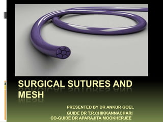
  • 1. SURGICAL SUTURES AND MESH PRESENTED BY DR ANKUR GOEL GUIDE DR T.R.CHIKKANNACHARI CO-GUIDE DR APARAJITA MOOKHERJEE
  • 3. History of Sutures  Animal sinew such as tendons and ligaments were the first recorded sutures.  Egyptian literature mentions the use of linen strips coated with a mixture of honey and flour  Sushruta described sutures and needles for the first time
  • 4. History of Sutures  Early wound closure devices were made of natural materials such as flax, silk, linen strips, and cotton. These natural materials were lubricated in oil and wine prior to application so as to reduce tissue drag and create a cleaner wound closure procedure  Another early form of a wound closure technique involved the use of the mandibles of soldier ants
  • 5. History of Sutures  Galen, a Roman surgeon, first commented on using catgut as a suture material  Lister first used sterilized suture material by using carbolic acid and then presenting the suture material in a glass tube  Halsted is still remembered for his rules of surgery advocating gentle handling of tissues, meticulous haemostasis, and interrupted silk sutures
  • 6. History of Sutures  By 1900 the catgut industry was firmly established in Germany due to the use of sheep intestines in their sausage industry  George Merson developed atraumatic sutures  1940s- uniform preparation and sterilization of sutures  1950s- sutures attached to pre attached needles  1960s- synthetic absorbable sutures introduced
  • 7. Definition of Suture  The word suture describes any material used to approximate tissue edges together or ligate blood vessels.  A suture attached to a needle is called as a ‘Stick tie’  A single strand of suture handed to the surgeon is called a ‘Free tie’  A free tie placed on the tip of a forceps or an instrument is called a ‘Tie on a passer’
  • 8. Atraumatic sutures  When a suture is attached to an eyeless needle, it is called an atraumatic suture.  Developed first by George Merson of Edinburgh. Hence these sutures are also called as ‘Mersutures’ in his memory.
  • 9. Suture size  Defined by US pharmacopoiea  The size reflects suture diameter  As the suture number increases, suture diameter decreases  Suture size is selected on the basis of tissue being sutured  The smallest size required should always be chosen to reduce injury to the tissue.
  • 10. Knot strength  Measured as force in pounds a suture will withstand before it breaks when knotted  The tensile strength of the suture material should not exceed the tensile strength of the tissue.
  • 11. Characteristic of Ideal suture material  Sterile and cost efficient  Non electrolytic, not allergenic, non carcinogenic  Inert  Easy to handle  Capable of securely holding the tissue layers throughout the healing process  Minimally reactive to tissue  Absorbed completely with minimal tissue reaction once the wound is healed  Should have uniform diameter  Should have adequate tensile strength until the purpose is over Unfortunately an ideal suture material does not exist.
  • 12. Suture strands Monofilament Multifilament  Goes through the tissue  Several strands braided with less drag or together resistance  Greater tensile strength,  Less chance of suture pliability, flexibility line infection- hence  Are coated to reduce the suture of choice for drag vascular surgeries  May act as a cove for organisms, hence never  Greater chance of getting used in infected wounds crushed
  • 13. Suture strands Monofilament Multifilament  Due to its smoothness,  Knots are more secure knots more likely to slip  Fraying on cutting edges  No fraying of the ends on cutting  Less tissue reaction  Eg Vicryl, Silk  Eg. Prolene, PDS II
  • 14. Cross section of sutures
  • 15. Sutures Absorbable sutures Non absorbable sutures  Are digested by enzymes  Are used to suture at and phagocytosed eg. Catgut sites where tensile  Or get hydrolysed and strength needs to be removed by phagocytosis maintained for at least a eg. Vicryl year  Are used to hold the wound edges in approximation  Made of non temporarily until the wound biodegradable materials is healed sufficiently to withstand normal stress that are ultimately encapsulated or walled off by body’s fibroblasts
  • 16. Sutures Absorbable sutures Non absorbable sutures  Are used to suture  Are used at exposed internal structures in the sites in the body from body where suture where they can be removal is not removed whenever possible/feasible needed eg. Skin  Eg. Catgut, Vicryl  Eg. Nylon, Silk, polypropylene
  • 17. Classification of suture materials  Monofilament  Polyfilament  Natural Absorbable  Absorbable  Catgut  Natural  Synthetic Absorbable  Synthetic  Monocryl  Polyglactin (Vicryl)  Caprosyn  Dexon II  Bondek  Biosyn  Maxon  Non absorbable  PDS II  Natural  Cotton  Non absorbable  Silk  Synthetic  Synthetic  Polypropylene(prolene)  Nurolon  Nylon (ethilon)  Mersilene  Stainless steel  Tevdek
  • 19. Catgut  Natural absorbable surgical suture  It is a brand name- manufactured by Ethicon division f Johnson and Johnson; other similar sutures include Trugut, Progut, etc.  It is 99% collagen  Absorbed by enzymatic digestion by proteolytic enzymes  Easy to handle and knots well
  • 20. Catgut  Derived from submucosa of sheep’s intestine or serosa of cattle’s intestine. The layers of intestine are scraped off to leave only submucosa. It is then treated by a fat solvent to wash of any fat. It is then dried and cut into various size. This is plain catgut.
  • 21. Catgut  If this is further treated with 20% chromic acid, it is called chromic catgut. Chromic acid increases the tissue life of the suture and thereby the tensile strength of the suture is maintained for a longer time.  Sterilized with gamma radiation and packed with isopropyl alcohol.
  • 22. Plain Catgut  Loses 50% tensile strength in 3 days and 100% strength by 15 days  Gets completely absorbed in tissue by 60 days Uses: 1. To tie small subcutaneous vessels 2. To approximate subcutaneous tissue during closure of incision 3. In circumcision to suture the cut margins of the prepuce 4. To repair the wound of lip or the oral cavity.
  • 23. Chromic Catgut  Loses 50% tensile strength in 7 days and 100% strength by 28 days  Gets completely absorbed in tissue in 90-100 days  Uses 1. To suture muscles, bowel anastomosis, peritoneum 2. During appendicectomy, to tie the mesoappendix and the base of appendix 3. The inner layer during two layer anastomosis of the small gut or anastomosis during gastrojejunostomy
  • 24. Chromic Catgut 4. During cholecystectomy, to stop bleeding from gall bladder bed. For most of the above said uses however, polygalactin sutures are replacing catgut.
  • 25. Polyglecaprone 25 (Monocryl)  Monofilament  Copolymer of 75% glycolide and 25% caprolactone  Undyed or dyed violet  Double the strength of chromic catgut  Good handling properties and smooth surface- easy passage through tissue
  • 26. Monocryl  Tensile strength  Uses maintained for 21 days 1. Can be used instead of and absorbed in 90-120 catgut days by hydrolysis 2. Intestinal anastomosis  Is sterilized by ethylene oxide 3. For closure of peritoneum 4. Subcutaneous tissue apposition 5. Urological procedures- pyeloplasty, ureter repair
  • 27. Polydioxanone Suture (PDS-II)  Polymer of paradioxanone  Is dyed violet  Is soft pliable and smooth and allows easy passage through tissues.  Tensile strength maintained for about 8 weeks and complete absorption in 180-210 days  Well suited for paediatric, CVS, orthopaedic, ophthalmic, plastic and gastro-intestinal surgeries
  • 28. Polypropylene( Prolene)  Synthetic monofilament non absorbable suture  Is inert with extremely low tissue reactivity  Low coefficient of friction, so it passes through the tissue plane easily.
  • 29. Polypropylene( Prolene)  The suture can extend upto 30% before breaking and hence is useful in situations where post operatively some elasticity is required on the part of the suture to accommodate post operative swelling and thereby helps prevent tissue strangulation.  High tensile strength for an indefinite period of time
  • 30. Polypropylene( Prolene)  Uses  In repair of posterior inguinal wall in hernia operations  Closure of skin incisions  Repair tendon injuries  Single layer closure of abdomen in case of intra abdominal infections/ obese patients  To secure prolene meshes
  • 31. Polyamide (Ethilon)  Synthetic, non  High tensile strength absorbable,  Loses 25 % tensile monofilamentous strength after 1 year  Very low coefficient of  Knot security is poor friction; hence passes through the tissues easily  Minimal tissue reaction
  • 32. Ethilon  Uses  Closure of skin incision  For hernioraphhy  Vascular surgery
  • 33. Stainless Steel Suture  Is composed of 316L  Non needled or stainless steel affixed to needles conforming to with permanent American Society for attachment Testing and Materials techniques or by  Available in precut ROTOGRIP lengths  Get encapsulated by  Very high tensile fibroblast inside the strength body
  • 34. Stainless Steel Suture  Indicated for  Contraindicated in abdominal wound people with known closure, intestinal metal allergies anastomosis, hernia  May interfere with repair, sternal closure certain and some orthopedic radiodiagnostics procedures like tendon repair
  • 35. Stainless Steel Suture  Requires expert technique for suturing  Sterilized by autoclaving
  • 36. Polyglycolic acid suture (dexon)  Delayed absorbable polyfilament  Green in colour  Maintains tensile strength for about 30 days and gets absorbed in 80-90 days  Dexon is a brand name for the suture synthesized by US surgical. Other brand names are Petcryl and Maxon.
  • 38. Polygalactin Sutures (Vicryl)  Is a copolymer of glycolide (90%) and lactide (10%)  Is digested by hydrolysis and not enzymatic reaction, hence there is less tissue reaction  Tensile strength lasts for 28-30 days and gets completely absorbed by 80-90 days.  Vicyl is a brand name for the polyglactin suture made by Ethicon. Other analogues are Truglyde, Centricryl and Safil.
  • 39. Dexon and Vicryl Uses 1. Indicated in all the situations where catgut is used 2. Better suited than catgut to suture infected wounds Disadvantages 1. Fraying of ends 2. Roughness
  • 40. Vicryl Rapide Suture  Polyglactin 910 irradiated with gamma rays  This leads to a suture material with a lower molecular weight and hence faster absorption
  • 41. Vicryl Rapide Suture  Maintains tensile strength for 10-12 days and absorbed by 42 days  Is undyed  Use 1. Subcuticular suturing 2. Mucosal sutures 3. In circumcision 4. Episiotomy repair
  • 42. Silk  Natural non absorbable braided suture  Dyed black  Derived from cocoon of silk worm larva. Is covered with a an albuminous layer which is removed during processing by a process called degummation.
  • 43. Silk  Handling property is very  Causes tissue reaction good and knots securely leading to PMN  Maintains tensile infiltration and fibrous strength for a long time capsule formation (2 years) around the silk  Is supplied as a reel or  Although classified as over atraumatic needles non absorbable, it loses (as Mersilk) its tensile strength in 1 year and cannot be  Sterilized by gamma detected after 2 years, radiation/ autoclaving hence may be re-  Loses tensile strength classified as delayed when wet absorbable suture
  • 44. Silk  Uses  In cholecystectomy to ligate cystic duct and cystic artery  Ligating mesenteric vessels during bowel resection  To ligate pedicles in nephrectomy and splenectomy  Ligation of Vagus nerve trunks during trunkal vagotomy  May be used for skin closure  To repair posterior wall of inguinal canal during hernioraphy  To secure drains/ tubes
  • 45. Cotton  Weakest non absorbable suture  Gains tensile strength when wet  Indicated for retracting vessels in CVS or pediatric surgeries  For tying off umbilicus in newborns
  • 46. SOFSILK  Multifilament, non absorbable, natural  Composed of natural, proteinaceous silk fibres called fibroin obtained from silk worm (Bombyx mori)  Black colour  Lubricated with wax or silicon  Used in CVS, ophthalmic or neuro-surgery  CI- Kidney, Urinary bladder surgeries as it is a nucleus for stone formation
  • 47. Misc multifilament non absorbable sutures  Ethibond excel suture- ethylene terephthalate  Mersilene- ethylene terephthtalate  Surgilon- Nylon 6 coated with silicon  Nurolon- Nylon 6  Ti-Cron- Braided polyester suture  Used in CVS, ophthalmic and neuro- surgeries
  • 48. Tevdek/ Polydek  Made of PTFE  Makes the suture inert and reduces tissue reactivity  Originally designed for heart valves implantation  Dyed green and white
  • 50. Surgidac  Braided, polyester, non absorbable made of ethelyne terephthalate  Used for general tissue approximation, cardiovascular, ophthtalmic and neuro surgeries  Undyed or dyed green
  • 52. Things to consider…  … while chosing a needle type for suturing  Type of needle- Type of tissue being closed  Curvature of the needle- based on working space in operative field
  • 53. Needle material  Initially stainless steel needles were being used  Now we use Surgalloy- a high nickel stainless steel  Gives it better resistance to bending and breakage
  • 54. Anatomy of the needle  Chord length- straight line distance from point of curve to swage  Needle length- the entire length of the needle  Radius- distance between the centre of the circle to the body of the needle if the curved needle were to make a full circle  Diameter- thickness of the needle
  • 55. Anatomy of the needle  Swage: This is the suture attachment end creates a single, continuous unit of suture and needle. This may be designed to allow easy release of the needle and suture material (pop-off)  The surgical needle may be coated with silicone to allow easier tissue passage.
  • 57. Types of suture needles
  • 58. Types of suture needles  Round bodied  minimises the tissue trauma because the needle pierces the tissue without cutting it  used for suturing peritoneum, abdominal viscera, myocardium, dura  Cutting needle  Have two opposing cutting edges  Designed to cut through tough difficult to penetrate structures  Used in plastic surgery, especially of the face
  • 59. Types of suture needles  Reverse cutting needle  Designed with cutting edge on the outer convex side  For skin, oral mucosa, tendon sheaths  Blunt needles  To dissect to friable tissue rather than cutting through it  For suturing liver and kidney
  • 60. Types of suture needles  DermaX needle  By Syneture  Contains four cutting edges with a double point tip  Moves smoothly with precision  Better control through subcuticular/ cuticular tissues like forehead, eyelid, nose, chin breast
  • 61. Shapes of sututre needles
  • 62. Packaging of sutures  Two layer cover to allow sterile field  Outer cover is made of laminate packing and is heat sealed  Inner cover is different for absorbable and non absorbable sutures  Because absorbable sutures need to be protected from atmospheric moisture, they are heat sealed in aluminium foils.
  • 68. Subcuticular suturing  Particularly useful in patients where keloid formation may be suspected.
  • 69. Running closure (Baseball stitch)  Where wound edges approximate easily and where a straight incision is being approximated  Not done where collection is suspected
  • 70. Running lock closure  Similar to Running closure except that the stitch is locked before giving the next stitch
  • 71. Purse string suture  Placed in a circular motion around a lumen and then tightened to invert the opening  Given around the stump of appendix once appendicectomy has been done  In bowel end to end anastomosis
  • 72. Traction sutures  Used to retract tissues that is not easily held back with a conventional retractor  Eg. In myocardium, sclera of the eye, tongue
  • 73. Retention suture/ Stay suture  Placed at a distance  Used as preventive from primary suture to avoid primary to relieve tension on wound disruption the primary line  Removed after the  Placed on skin, tension in the subcutaneous tissue affected area is or fascia decreased  Used for those patients where slow healing is expected
  • 74. Secondary line suture  Used to support the primary line suture, eliminate dead space or prevent fluid accumulation in an abdominal wound
  • 75. Figure of eight stick tie  Indicated for larger vessels or tissue pedicels  To prevent knot slippage  To stop haemorrhage
  • 76. Timing of Suture Removal Wound location Timing of removal (days) Face Three to five Scalp Seven to 10 Arms Seven to 10 Trunk 10 to 14 Legs 10 to 14 Hands or feet 10 to 14 Palms or soles 14 to 21
  • 81. Principles of knot tying  Ensure that the knot is firm enough to eliminate slippage  Make sure that the knot is as small as possible and the free ends are as short as feasible  Avoid damaging suture material while handling  Avoid excessive tension  Do not tie the knots too tightly- prevent strangulation of tissue
  • 84. Surgical Staples  Basic principles  Preservation of adequate tissue vascularization  Creation of adequate lumen  Prevention of leaks and fistulas  Evading of tissue tension  Haemostasis
  • 85. Surgical Staples  Advantages  Less tissue reaction  Accelerated wound healing  Efficiency  Less anesthesia and intra operative time
  • 86. Surgical Staples  Types of staplers  Linear stapler- used for closure of a linear wound  Circular or intraluminal staplers- used in GI surgeries for end-to-end, end-to-side, side- to-end or side-to-side anastomosis. They offer a circular, double staggered row of staples.  Ligating and dividing stapler- issues a double row of two staples and ligates the tissue. The tissue can then be divided between the staple lines.
  • 87. Surgical Staples  Purse string suture clamps- for placement of purse string sutures.  Skin staplers- to approximate skin edges.  Endoscopic staplers
  • 90. Fibrin Glue  Synthesized from bovine blood • Uses • Used to control bleeding and approximating tissues that are difficult to approximate by suturing, eg. Liver, spleen and lung. • Microsurgical anastomosis of blood vessels • Used in Cardiopulmonary bypass surgery • Repair ocular implants • Close superficial lacerations and fistula • Repair dural tears
  • 91. Surgical Glue  Made of cyanoacrylate  For adhesion of superficial lacerations
  • 92. III COSEAL Surgical sealant  Completely surgical vascular sealant  Used in vascular surgeries as an adjunctive haemostat  Seals immediately  It is not a substitute for sutures, staples but an adjunct  No contraindication  Should not be injected into vessels
  • 93. IV Vessel Sealing technology (LigaSure)
  • 94. Vessel Sealing technology (LigaSure)  For ligating vessels and tissue bundles  Works by fusing the collagen and elastin fibres in vessels to seal the blood flow  It uses ‘Instant Response Technology’- It is a feedback controlled response system that diagnoses the tissue type in the jaws of the instrument and delivers the appropriate amount of energy to effectively seal the vessel or tissue bundle.  Thermal spread upto 1mm  No sticking or charring of tissue.
  • 97. Adhesive skin closure tapes/strips
  • 98. Adhesive skin closure tapes/strips  Made of nylon or polypropelene  Used to reinforce subcuticular skin closure or to approximate wound edges of small incisions or lacerations  Minimal tissue reactivity  Low rate of infection  No ischemia or necrosis  Is gentler to the skin than needle  May have an additional microbiocidal
  • 99. Adhesive skin closure tapes/strips  Disadvantage- cannot be used in presence of moisture/ infection/ oily surface/ hairy surface/ skin under tension  Eg Dermabond/ Indermil
  • 101. Surgical Mesh  Surgical mesh is a woven fabric used for chest wall reconstruction, strengthening tissues, provide support for internal organs, and to treat surgical or traumatic wounds.  The most common types of surgical mesh are hernia mesh, stress urinary incontinence slings and mesh for treating prolapse.
  • 102. Surgical Mesh  Titanium mesh has been used in some back surgeries  As with any surgical implant, some complications can occur, including infection, inflammation, tissue damage, and septic shock.
  • 103. Surgical Mesh  Hernioplasty with living Fascia MacArthur’s operation  A strip of fascia 1.25 cm wide was cut from external oblique aponeurosis  The strip was left attached to at the pubic end and cut from where the aponeurosis becomes muscular  The free end was attached to a needle with a wide eye ( Gallie’s needle)  The living mesh was darned into the posterior wall of the inguinal canal
  • 104. Surgical Mesh  Gallie’s Graft If external oblique aponeurosis is thin/friable, tensor fascia lata cut from the outer side of the thigh and grafted over the posterior wall of the inguinal canal  Tantalum Gauze- made of wire darn (filigree)
  • 105. Surgical Mesh Hernioplasty with a skin graft (Mair’s procedure)- A full or a partial thickness graft was harvested and sutured to the posterior wall of inguinal canal. Side effects: formation of cysts/ dermoid cyst.
  • 106. Synthetic meshes- Non absorbable  Polypropylene mesh  Monofilament  Inert; can be used in presence of infection  Good elasticity and high tensile strength  Non absorbable  Used for  Hernia repair  Abdominal wall construction  Sterilized by gamma radiations  Fixed with prolene sutures
  • 107. Synthetic meshes- Non absorbable  Polyglactin 910(vicryl) mesh  absorbable mesh that offers temporary support during wound healing  PTFE or Gore-Tex  Soft, flexible non absorbable  Impervious to fluids  Should not be used in presence of infection
  • 108. Synthetic meshes- Non absorbable  Expanded PTFE (ePTFE)  Greatest expansion of all the meshes  Useful in abdominal and vascular surgeries  Stainless steel mesh  Rigid and difficult to work with  Causes discomfort to patient
  • 109. Synthetic meshes- non absorbable  Polyester mesh (Mersilene)  Least inert synthetic mesh  Never be used in presence of infection  Fibres may harbor bacteria  Polyethylene mesh  multifilament,; made of polyester fibre  Used for abdominal wall repair and hernia repair
  • 110. Synthetic meshes- absorbable  Polyglycolic acid; Polyglactin 910  Used for wound closure and organ support
  • 111. Mesh for inguinal hernia
  • 112. Stents  A stent is a small mesh  Stents usually are made tube that's used to treat of metal mesh, but narrowed or weakened sometimes they're made arteries in the body. of fabric. Fabric stents,  Stents help prevent the also called stent grafts, arteries from becoming are used in larger narrowed or blocked arteries. again in the months or  Some stents are coated years after angioplasty with medicines that are slowly and continuously released into the artery. These stents are called drug-eluting stents.
  • 113. Stents according to location  The most widely known stent use is in the coronary arteries with a bare- metal stent, a drug- eluting stent or occasionally a covered stent.Coronary stents are placed during angioplasty.
  • 114.
  • 115. Stents according to location  Ureteral stents are used to ensure the patency of a ureter, which may be compromised, for example, by a kidney stone. This method is sometimes used as a temporary measure, to prevent damage to a blocked kidney, until a procedure to remove the stone can be performed. Indwelling times of 12 months or longer are indicated to hold ureters open, which are compressed by tumors in the neighbourhood of the ureter or by tumors of the ureter itself.
  • 116. Stents according to location  In many cases these tumors are inoperable and the stents are used to ensure drainage of urine through the ureter. If drainage is compromised for longer periods, the kidney can be damaged. The main complications with ureteral stents are dislocation, infection and blockage by encrustation. Recently stents with coatings (e.g. heparin) were approved to reduce infection, encrustation and therefore stent exchanges.
  • 117. Stents according to location  A urethral or Prostatic stent might be needed if a man is unable to urinate. Often this situation occurs when an enlarged prostate pushes against the urethra, blocking the flow of urine. The placement of a stent can open the obstruction.
  • 118. Stents according to location  Stents are used in a variety of vessels aside from the coronary arteries. Stents may be used as a component of peripheral artery angioplasty.
  • 119. Stents according to location  Esophageal stent
  • 120. Stents according to location  Biliary stent
  • 121. Stents according to location  A stent graft is a tubular device, which is composed of special fabric supported by a rigid structure, usually metal. The rigid structure is called a stent. An average stent on its own has no covering, and therefore is usually just a metal mesh. Although there are many types of stent, these stents are used mainly for vascular intervention.The device is used primarily in endovascular surgery  Stent grafts are most commonly used in the repair of an abdominal aortic aneurysm
  • 122. Bibliography  Bailey and Love’s Short practice of Surgery edition 13, edition 25  Suture and Surgical Haemostasis by Rebecca Pieknik  Farquharson’s textbook of operative general surgery edition 9  Bedside clinics in Surgery Makhan lal Saha  www.wikisurgery.com