Weitere ähnliche Inhalte Ähnlich wie Mechanisms of Scleroderma Disease Review Ähnlich wie Mechanisms of Scleroderma Disease Review (14) Kürzlich hochgeladen (20) 1. The n e w e ng l a n d j o u r na l of m e dic i n e
review article
Mechanisms of Disease
Scleroderma
Armando Gabrielli, M.D., Enrico V. Avvedimento, M.D., and Thomas Krieg, M.D.
S
cleroderma (systemic sclerosis) is a complex disease in which ex- From the Department of Medical Science
tensive fibrosis, vascular alterations, and autoantibodies against various cellular and Surgery, Section of Clinical Medicine,
Università Politecnica delle Marche, and
antigens are among the principal features (Fig. 1 and 2).1 There are two major Ospe ali Riuniti — both in Ancona (A.G.);
d
subgroups in the commonly accepted classification of scleroderma: limited cutane- and the Department of Molecular and
ous scleroderma and diffuse cutaneous scleroderma.2 In limited cutaneous sclero- Cellular Biology and Pathology, Institute of
Endocrinology and Experimental Oncol-
derma, fibrosis is mainly restricted to the hands, arms, and face. Raynaud’s phenom- ogy, Consiglio Nazionale delle Ricerche,
enon is present for several years before fibrosis appears, pulmonary hypertension University of Naples Federico II, Naples
is frequent, and anticentromere antibodies occur in 50 to 90% of patients. Diffuse (E.V.A.) — all in Italy; and the Depart-
ment of Dermatology, University of Co-
cutaneous scleroderma is a rapidly progressing disorder that affects a large area of logne, Cologne, Germany (T.K.). Address
the skin and compromises one or more internal organs. reprint requests to Dr. Gabrielli at the De-
We believe that the acronym CREST (calcinosis, Raynaud’s phenomenon, esopha- partment of Medical Science and Surgery,
Section of Clinical Medicine, Polo Didat-
geal motility dysfunction, sclerodactyly, and telangiectasia) is obsolete, since it can- tico, Via Tronto, 10, Ancona, Italy, or at
not be assigned to only one subgroup of patients with the disease and does not a.gabrielli@univpm.it.
sufficiently indicate the burden of internal-organ involvement. In rare cases, patients
N Engl J Med 2009;360:1989-2003.
with scleroderma have no obvious skin involvement. Patients with scleroderma plus Copyright © 2009 Massachusetts Medical Society.
evidence of systemic lupus erythematosus, rheumatoid arthritis, polymyositis, or
Sjögren’s syndrome are considered to have an overlap syndrome. This classifica-
tion can be useful, but none of the proposed classifications sufficiently reflect the
heterogeneity of the clinical manifestations of scleroderma.
Scleroderma can lead to severe dysfunction and failure of almost any internal
organ. Here, too, there is considerable heterogeneity (Table 1). Involvement of
visceral organs is a major factor in determining the prognosis. The kidneys, esopha
gus, heart, and lungs are the most frequent targets. Renal involvement can be
controlled by angiotensin-converting–enzyme inhibitors. Severely debilitating esoph-
ageal dysfunction is the most common visceral complication, and lung involve-
ment is the leading cause of death.
The mechanisms underlying visceral involvement in scleroderma are unclear,
despite progress in the treatment of these complications. Relevant data on mecha-
nisms are limited, since most of the available information is derived from cross-
sectional studies and from patients in various stages of the disease, often after
treatment; moreover, there are no satisfactory animal models of scleroderma. Never-
theless, a critical evaluation of the available experimental and clinical data will help
reduce ambiguity and may provide the basis for future studies of scleroderma.
Epidemiol o gy a nd Gene t ic Suscep t ibil i t y
The results of studies of the prevalence and incidence of scleroderma are conflict-
ing because of methodologic variations in case ascertainment and geographic dif-
ferences in these measurements. The available data indicate a prevalence ranging
from 50 to 300 cases per 1 million persons and an incidence ranging from 2.3 to 22.8
cases per 1 million persons per year.6 Women are at much higher risk for scleroderma
than men, with a ratio ranging from 3:1 to 14:1. A slightly increased susceptibility
n engl j med 360;19 nejm.org may 7, 2009 1989
The New England Journal of Medicine
Downloaded from nejm.org on May 28, 2011. For personal use only. No other uses without permission.
Copyright © 2009 Massachusetts Medical Society. All rights reserved.
2. The n e w e ng l a n d j o u r na l of m e dic i n e
A B C D E
*
*
Figure 1. Clinical Signs and Histologic Features in Patients with Scleroderma.
ICM
AUTHOR: Gabrielli
Panel A shows hyperkeratosis of the nail folds of a patient in an edematous phase of 1st
RETAKE
limited cutaneous scleroderma.
2nd
Panel B shows fingertip ulceration in a F FIGURE: 1limited cutaneous scleroderma. Panel C shows a lymphohistio
REG patient with of 4
3rd
cytic infiltrate around blood vessels in a skin specimen (hematoxylin and eosin). In Panel D, a skin-biopsy specimen
CASE Revised
Line 4-C
from a patient with early diffuse disease shows intense deposition of collagenous matrix throughout the dermis,
EMail SIZE
ARTIST: ts
xtending into the subcutaneous fat layer (hematoxylin and eosin). H/T E shows intimal and medial thickening in
e Enon
H/T Panel 33p9
Combo
one interlobar artery (arrow) and two arcuate arteries (asterisks) in the kidney of a patient with scleroderma. The
glomerular tuft is partially collapsed, and the tubular epithelium NOTE:
AUTHOR, PLEASE is atrophic. Fibrosis with mononuclear-cell infiltra-
Figure has been redrawn and type has been reset.
tion is present in the interstitium (hematoxylin and eosin). carefully.
Please check
JOB: 36019 ISSUE: 05-07-09
to scleroderma among blacks has been reported.7,8 E a r ly a nd L ate L e sions
Familial clustering of the disease, the high fre-
quency of other autoimmune disorders in families Important features of the tissue lesions in vari-
of patients with scleroderma, and differences in ous stages of scleroderma are early microvascular
phenotypes among race and ethnic groups8,9 all damage, mononuclear-cell infiltrates, and slowly
suggest that genetic factors contribute to sclero- developing fibrosis (Fig. 1). In later stages of scle-
derma. Scleroderma-associated polymorphisms of roderma, the main findings are very densely packed
genes encoding cytokines, cytokine receptors, collagen in the dermis, loss of cells, and atrophy.
chemokines, and extracellular proteins have been
reported.10 Many of these variants have been linked Early Vascular and Inflammatory Alterations
to cohorts of patients, but few have been indepen- Vascular injury is an early event in scleroderma.
dently confirmed. By contrast, there is strong evi- It precedes fibrosis and involves small vessels,
dence of linkage of certain HLA class II molecules particularly the arterioles.15,16 The vascular dam-
to clinical phenotypes and particular autoanti- age, which occurs in virtually all organs,17,18 con-
bodies.11 The data provide support for the notion sists of large gaps between endothelial cells, loss
that sclero erma is not one clearly defined disease
d of integrity of the endothelial lining, and vacuo
but a syndrome encompassing various phenotypes. lization of endothelial-cell cytoplasm. In addition,
Environmental challenges (e.g., viruses, drugs, there are several basal lamina-like layers, perivas-
vinyl chloride, and silica) may induce clinical cular infiltrates of mononuclear immune cells
phenotypes that are similar or identical to sclero- (with rare lymphocytes) in the vessel wall, obliter
derma.12 Moreover, several reports indicate that ative microvascular lesions, and rarefaction of cap
during pregnancy, fetal or maternal lymphocytes illaries.15,16,19,20 The remarkable paucity of small
can cross the placenta and initiate a graft-versus- blood vessels is a characteristic finding in later
host reaction that culminates in scleroderma. stages of scleroderma.
There are clinical, serologic, and histopathologi- Notwithstanding the progressive loss of blood
cal similarities between scleroderma and chron- vessels and high plasma levels of vascular endo
ic graft-versus-host disease (GVHD), and alloge- thelial growth factor21,22 caused by the adaptive
neic cells have been detected in peripheral-blood response to hypoxia, there is a defect in vascu
and skin-biopsy specimens obtained from patients logenesis.20,23,24 The molecular mechanism (or
with scleroderma.13,14 However, rigorous evidence mechanisms) underlying this paradox is unknown:
that these cells participate in the pathogenesis of both angiogenic21,22 and angiostatic20,25,26 factors
scleroderma is lacking. have been detected in early scleroderma. Notably,
1990 n engl j med 360;19 nejm.org may 7, 2009
The New England Journal of Medicine
Downloaded from nejm.org on May 28, 2011. For personal use only. No other uses without permission.
Copyright © 2009 Massachusetts Medical Society. All rights reserved.
3. Mechanisms of Disease
A
a b c
B
Classic Autoantibodies Clinical Features New Autoantibodies Role
Anti–topoisomerase I Diffuse cutaneous scleroderma Anti–endothelial cell Induce apoptosis of endothelial cells
Anticentromere proteins Limited cutaneous scleroderma, pul- Anti–FBN 1 Activate normal human fibroblasts
monary hypertension
Anti–RNA polymerase I/II Diffuse cutaneous scleroderma, renal Anti–MMP 1 and 3 Prevent degradation of ECM proteins
involvement
Antipolymyositis, Polymyositis, calcinosis Anti-PDGFR Stimulate normal human fibroblasts
sclerosis through Ha-Ras-ERK1/2-ROS
Antifibrillarin (U3RNP) Diffuse cutaneous scleroderma, Anti–Nag-2 Induce endothelial-cell apoptosis
internal-organ involvement
Anti-Th/To Limited cutaneous scleroderma, pul-
monary fibrosis
Figure 2. Autoantibodies in Scleroderma. AUTHOR: Gabrielli RETAKE 1st
ICM
Panel A shows antinuclear-antibodyREG F FIGURE: 2 of The speckled nuclear staining 2nd
staining patterns. 4 pattern (left) can be detected in
3rd
30% of patients with diffuse scleroderma and suggests the presence of anti–topoisomerase I antibodies. The
CASE Revised
homogeneous nucleolar staining pattern (center) is detected in 25 to4-C of patients with the myositis–scleroderma
Line 50%
EMail SIZE
ARTIST: ts
overlap syndrome. Unlike this homogeneous staining pattern, a pattern characterized by clumping of the nucleoli (not
H/T H/T
Enon
Combo 33p9
shown) is highly specific for diffuse scleroderma (in 5% of patients). Nucleolar antigens are RNA polymerases, fibrillar-
in, Th/To, or PM-Scl. The anticentromere-antibody staining pattern (right) can be detected in 70 to 80% of patients
AUTHOR, PLEASE NOTE:
with limited cutaneous scleroderma and is associatedredrawn high risk of been reset. hypertension. The antigens are ki-
Figure has been with a and type has pulmonary
Please check carefully.
netochore proteins of the centromere regions of chromosomes. Panel B lists the classic and newly discovered autoanti-
bodies in scleroderma. ECM denotes extracellular-matrix protein; ERK1/2 extracellular-signal–regulated kinases 1 and
2; FBN-1 fibrillin-1; MMP 1 and 3JOB: 360metalloproteinases 1 and 3; Nag-2 UE: 05-07-09 anti-inflammatory drug–acti-
matrix 19 ISS
nonsteroidal
vated gene; PDGFR platelet-derived growth factor receptor; and ROS reactive oxygen species.
inflammatory cytokines such as tumor necrosis the early stages, whereas type I collagen accumu-
factor α can stimulate or inhibit angiogenesis de- lates in later stages.28,29
pending on the duration of the stimulus.27
Cel l T y pe s in L e sions
Fibrosis
Fibrosis gradually replaces the vascular inflam- Endothelial Cells
matory phase of scleroderma and ultimately dis- Endothelial cells are affected early in scleroder-
rupts the architecture of the affected tissue. It is ma.30 In early lesions there is endothelial-cell
the cause of the main symptoms of the disease. apoptosis, or changes of the endothelial pheno-
Fibrosis in the skin begins in the lower dermis type in the absence of endothelial-cell prolifera-
and upper subcutaneous layer and occurs together tion or precursor differentiation.20,31,32 The mo-
with loss of microvasculature, reduction of ap- bilization of endothelial precursors from bone
pendages, and loss of reticular structure and the marrow is related to disease severity, but recruit-
rete ridges. The composition of accumulated ma- ment of such cells to peripheral vasculature has
trix varies with the stage of the disease. A mix- not been shown.33 The interaction of progenitor
ture of different collagen types, proteoglycans, endothelial cells with platelets and platelet-derived
and elastic fibers including fibrillin is typical of growth factor (PDGF) is essential for the matura-
n engl j med 360;19 nejm.org may 7, 2009 1991
The New England Journal of Medicine
Downloaded from nejm.org on May 28, 2011. For personal use only. No other uses without permission.
Copyright © 2009 Massachusetts Medical Society. All rights reserved.
4. 1992
Table 1. Clinical Findings in Patients with Scleroderma in Four Countries.*
Finding Diffuse Cutaneous Scleroderma Limited Cutaneous Scleroderma
United States France Germany Italy United States France Germany Italy
The
(N = 119) (N = 30) (N = 484) (N = 177) (N = 128) (N = 97) (N = 674) (N = 565)
percentage of patients percentage of patients
Calcinosis 23 16 NR 20 42 36 NR 22
Raynaud’s phenomenon 97 100 94.2 94 99 99 96.3 96
Articular involvement 98 70 56.6† 22 78 65 44.9† 16
Esophageal dysmotility 67 79 69.3 69 67 63 59.2 55
Lung fibrosis 30 57 56.1 71‡ 37 30 20.8 53‡
Isolated pulmonary arterial 2 12 27.7 NR 31 9 60.0 NR
hypertension
Heart involvement 11§ 10§ 23.0¶ 32‖ 19§ 14§ 12.0¶ 23‖
n e w e ng l a n d j o u r na l
The New England Journal of Medicine
of
Reduced LVEF 20** 15** NR NR 6** 12** NR NR
Renal crisis 17 7 15.9 12 2 0 9.1 6
n engl j med 360;19 nejm.org may 7, 2009
* Data are from Meyer et al.,3 Hunzelmann et al.,4 and Ferri et al.5 The study in Italy included a third subgroup of patients who were said to have intermediate cutaneous scleroderma;
data on these patients are not shown in the table. LVEF denotes left ventricular ejection fraction, and NR not reported.
Copyright © 2009 Massachusetts Medical Society. All rights reserved.
m e dic i n e
† This value includes patients with both muscle and articular involvement.
‡ This value includes patients who also had isolated pulmonary hypertension.
§ In the country listed, heart involvement was defined by the presence of arrhythmia requiring treatment.
¶ This value includes patients with one of the following: palpitations, a conduction disturbance, or diastolic dysfunction.
‖ This value includes patients with one of the following: pericarditis, congestive heart failure, severe arrhythmia, or a conduction disturbance.
** The LVEF was less than 50% on echocardiography or there was diastolic dysfunction.
Downloaded from nejm.org on May 28, 2011. For personal use only. No other uses without permission.
5. Mechanisms of Disease
tion and recruitment of endothelial precursors.34,35 an excess of reactive oxygen species. The origin
The perivascular space is a preferred site of early of activated fibroblasts in the skin and internal
lesions in scleroderma. Progressive wall thicken- organs of patients with scleroderma is still debat-
ing and perivascular infiltrates are features of the ed. Fibroblasts may undergo local activation or
vascular lesions in this compartment, indicating originate from resident pericytes, mesenchymal
the involvement of vascular smooth-muscle cells stem cells, or progenitor cells (e.g., fibrocytes) re-
and pericytes. cruited from the circulation.46
Pericytes and Smooth-Muscle Cells Mononuclear Cells
Small vessels contain vascular smooth-muscle The cellular infiltrates in the early lesions of scle-
cells and pericytes. Pericytes have the potential roderma consist mostly of T cells, macrophages,
to differentiate into vascular smooth-muscle cells, B cells, and mast cells.47-49 T cells in skin lesions
fibroblasts, and myofibroblasts (specialized con- are predominantly CD4+ cells,49 display markers
tractile cells expressing α–smooth-muscle actin of activation,50 exhibit oligoclonal expansion,51
and the ED-A splice variant of fibronectin)36-38 and are predominantly type 2 helper T (Th2)
and to influence endothelial-cell proliferation.39 cells.52 These characteristics parallel the increased
Increased thickness of the vascular wall, serum levels of cytokines derived from Th2 cells
caused by the proliferation of vascular smooth- in scleroderma.53,54 CD20-positive B cells are also
muscle cells, indicates that these cells are re- found in skin lesions.48 They may contribute to
sponding to scleroderma-induced injury. Pericytes the pathogenesis of fibrosis through the secre-
in the lesion overexpress several cytokine recep- tion of interleukin-6 and TGF-β55,56 and the pro-
tors, including PDGF receptor (PDGFR),40 but this duction of autoantibodies.
occurs only in early lesions and in patients with
Raynaud’s phenomenon and antinuclear antibod- Solubl e Medi at or s
ies. These cells proliferate and contribute to in-
creased wall thickness.41 Collectively, the cellular
changes in early lesions are loss of endothelial Cytokines and Growth Factors
cells, proliferating pericytes and vascular smooth- Genomewide transcription profiles of skin-biopsy
muscle cells, and immune cells in the perivascu- specimens obtained from patients with sclero-
lar space. Endothelial cells are the only mesen- derma have provided direct evidence of the involve-
chymal cell type that undergo apoptosis in early ment of cytokines in the activation of fibroblasts.
scleroderma, whereas vascular smooth-muscle Within the limitations of such an approach (e.g.,
cells and pericytes proliferate vigorously. variations according to the site of the biopsy,
mixed cell populations, and post-transcriptional
Fibroblasts regulation), the data indicate systemic changes of
Fibroblasts appear to orchestrate the production, gene transcription in endothelial cells, fibroblasts,
deposition, and remodeling of collagens and other and B and T lymphocytes in scleroderma. These
extracellular-matrix components. Fibroblasts in studies have shown transcriptional changes in
scleroderma are heterogeneous in terms of col- clinically affected and unaffected skin.48
lagen synthesis.42 Overproduction of collagen is
due to enhanced transcription or increased sta- TGF-β
bility of collagen-specific messenger RNA.43 Up- TGF-β is a potent profibrotic cytokine.57 DNA mi-
regulated transcription of collagen genes in scle- croarray analysis indicates that a group of TGF-
roderma cells is autonomous and maintained in β–dependent genes are overexpressed in biopsy
vitro over several passages.44 Fibroblasts in sclero- specimens from skin lesions in patients with scle-
derma can convert to myofibroblasts,38 and they roderma.48 TGF-β is also the strongest inducer of
overexpress several cytokines (e.g., transforming myofibroblasts, and it modulates the expression
growth factor β [TGF-β] and monocyte chemoat- of various cytokine receptors, including recep-
tractant protein 1) and TGF-β receptors.45 These tors for TGF-β and PDGF.45,58 In scleroderma
findings underscore the role of autocrine loops fibroblasts, TGF-β further up-regulates connec-
in sustaining the fibrotic reaction. In addition, tive-tissue growth factor (CTGF), a cysteine-rich
fibroblasts in patients with scleroderma contain modular protein belonging to the CCN family of
n engl j med 360;19 nejm.org may 7, 2009 1993
The New England Journal of Medicine
Downloaded from nejm.org on May 28, 2011. For personal use only. No other uses without permission.
Copyright © 2009 Massachusetts Medical Society. All rights reserved.
6. The n e w e ng l a n d j o u r na l of m e dic i n e
matricellular growth factors (CYR61, CTGF, and Extracellular-Matrix Components and Their
NOV [nephroblastoma overexpressed])59 that has Receptors
biologic activities similar to those of TGF-β. En- The hallmark of scleroderma is excessive deposi-
hanced TGF-β and CTGF expression has been tion of extracellular-matrix components, caused
detected in scleroderma lesions, and enhanced by overproduction of collagen and other glyco-
TGF-β signaling in fibroblasts causes skin fi- proteins (e.g., fibronectin and fibrillin).42,43 The
brosis in a mouse model that appears to reca- macromolecular arrangement of collagens in scle-
pitulate the clinical and histologic features of roderma is altered by cross-links that are normal-
scleroderma.60 ly seen in bone but not skin collagen matrix; these
Smad-dependent or Smad-independent signal- cross-links are formed by lysyl hydroxylase 2, the
ing downstream of TGF-β has been extensively level of which is increased in scleroderma.71
characterized in scleroderma cells (Fig. 3).61 In- Extracellular-matrix molecules modulate cellu
hibition of protein kinase C delta, geranyl trans- lar responses by regulating the activity of cytok-
ferase 1, or stress-activated protein kinase p38 ines and growth factors. For example, TGF-β–
eliminates the expression of collagen I and III in fibrillin interaction is required for fibroblast
scleroderma cells.62,63 TGF-β, produced as inac- activation in scleroderma. The extracellular ma-
tive precursor, can be activated by thrombospon- trix also provides points of adhesion, which are
din and by αv 3 integrin, underscoring the inter-
β bound by integrins, transmembrane receptors
action among cytokines, extracellular matrix, and connecting the extracellular-matrix environment
integrins. The expression of all these molecules to the cytoskeleton, thereby mediating outside-in
is induced in scleroderma.64,65 and inside-out signaling.72 Integrin α1β1 elicits
signals to down-regulate collagen synthesis by
PDGF fibroblasts; α1β1-knockout mice have enhanced
PDGF, which is linked to wound healing and fi- collagen synthesis in wounds.73 Fibroblasts in
brosis, may have a role in scleroderma. The pres- patients with scleroderma have reduced surface
ence of stimulatory antibodies to PDGFR in serum levels of α1β1 integrin, resulting in the failure of
from patients with scleroderma, the strong stimu- integrin to down-regulate collagen synthesis.74
lation by PDGF of the pericyte-to-fibroblast tran Impairment of integrin signaling may amplify
sition,38 the presence of high levels of PDGF and fibrosis in scleroderma. There is accumulating
its beta receptor in skin lesions from patients with evidence that crosstalk between different integrins
scleroderma,66,67 and the beneficial effects of se- and extracellular-matrix molecules determines the
lective inhibitors of PDGF signaling on dermal activity of many cytokines and growth factors that
fibrosis68 all indicate the importance of PDGF in interact directly with responding target cells.64,65
scleroderma. PDGF inhibitors may thus have a Overall, the altered extracellular matrix in sclero-
therapeutic benefit in fibrosis. derma probably provides an environment that
amplifies receptor-mediated cell activation.
Other Cytokines and Biologically Active Substances
Endothelin-1 acts in concert with TGF-β to con- Autoantibodies
vert fibroblasts into myofibroblasts.69 The bene- Scleroderma is associated with several autoanti-
ficial effect of endothelin-1–receptor inhibitors on bodies, some of which are important diagnostic
pulmonary hypertension in patients with sclero- markers. Tests for autoantibodies against topoi-
derma indicates that endothelin-1 is an important somerase I (Scl-70), centromere-associated proteins,
signaling molecule in this disease. Inhibition of and nucleolar antigens can be useful in facilitat-
endothelin signaling may alleviate the overstim- ing the diagnosis and formulating a prognosis. Al
ulation of TGF-β in scleroderma.70 Many other though the autoantibodies correlate with disease
cytokines have been implicated in the angiogene- severity and the risk of specific organ complica-
sis, angiostasis, fibrosis, and localized inflam- tions, their pathogenetic relevance is unclear. Re-
mation in scleroderma. To date, there is no com- cently, autoantibodies against nonnuclear antigens
pelling evidence linking the levels and activity of have been described (Fig. 2), including antibodies
these cytokines to one or more specific patho- against cell-surface antigens. Antibodies against
genic events in this condition (Table 2). PDGFR appear to be agonistic, since they stimu-
1994 n engl j med 360;19 nejm.org may 7, 2009
The New England Journal of Medicine
Downloaded from nejm.org on May 28, 2011. For personal use only. No other uses without permission.
Copyright © 2009 Massachusetts Medical Society. All rights reserved.
7. Mechanisms of Disease
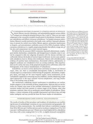
Figure 3. Activation of Fibroblasts in Scleroderma.
External factors such as interleukins, chemokines, thrombin, endothelin-1, growth factors, reactive oxygen species
(ROS), and activating antibodies trigger signaling cascades in fibroblasts. For example, the phosphorylation of Smad2
triggers a signaling cascade from Smad3 to Smad1, which interacts with Smad4 and regulates gene transcription in
the nucleus. Activation of transforming growth factor β (TGF-β) receptors (TGF-βR) also results in the activation of
pathways not involving Smad proteins,61 modulating transcription factors. These pathways intersect with pathways
induced by activation of platelet-derived growth factor receptors (PDGFR), leading to a complex intracellular signal-
ing network. Production of extracellular-matrix protein, cytoskeleton, cytokines, and cytokine receptors is thereby
stimulated; these participate in regulatory loops to sustained fibroblast activation. CTGF denotes connective-tissue
growth factor, ERK1/2 extracellular-signal–regulated kinases 1 and 2, α-SMA α–smooth-muscle actin, and SRE serum-
responsive element.
late a specific signaling cascade.75 However, the signaling inhibitors has been reported in resistant
specificity of these stimulatory auto ntibodies
a cases of sclerodermatous GVHD.76
remains to be established. The same type of auto
antibodies with PDGF agonistic activity has been R e ac t i v e Ox ygen Specie s
detected in crude immunoglobulin derived from
the serum of patients with sclerodermatous GVHD, High levels of reactive oxygen species and oxida-
and a significant beneficial effect of PDGFR- tive stress have been directly or indirectly impli-
n engl j med 360;19 nejm.org may 7, 2009 1995
The New England Journal of Medicine
Downloaded from nejm.org on May 28, 2011. For personal use only. No other uses without permission.
Copyright © 2009 Massachusetts Medical Society. All rights reserved.
8. Table 2. Cytokines, Growth Factors, and Biologically Active Substances Involved in the Pathogenesis of Scleroderma.*
1996
Variable Main Cell Source Pathogenic Relevance Effect in Scleroderma
Interleukin-1 Macrophages, monocytes Has a role in production of interleukin-6 and Constitutively expressed in skin fibroblasts
PDGF-α by fibroblasts
Interleukin-4 Th2 lymphocytes Stimulates fibroblast proliferation, chemotaxis, Increased levels in serum; increased protein and
and collagen synthesis; stimulates production gene expression in skin and in cultured fibro-
of TGF-β, CTGF, and TIMP-1; up-regulates ex- blasts; increased number of interleukin-4–
pression of adhesion molecules by endothelial producing T lymphocytes
cells
Interleukin-6 Fibroblasts, macrophages, endothelial cells, Stimulates collagen and TIMP-1 synthesis; pro- Increased levels in tissue and serum; enhanced
B cells, T cells motes a Th2-polarized immune response production in vitro by PBMC and cultured fi-
broblasts
Interleukin-8 Alveolar macrophages, lung fibroblasts, skin fibro- Serves as a potent chemoattractant and activator Elevated levels in serum, skin specimens, and
blasts of neutrophils; promotes fibroblast chemotaxis bronchoalveolar-lavage fluids
Interleukin-10 Activated B cells, monocytes Promotes a predominant Th2 immune response Increased levels in serum
that induces collagen synthesis
The
Interleukin-13 Th2 lymphocytes Induces fibrosis through a TGF-β–dependent and Increased levels in serum
TGF-β–independent mechanism
Interleukin-17 Th1 and Th2 lymphocytes Induces proliferation of fibroblasts; stimulates fi- Increased levels in serum; overexpressed in skin
broblast production of collagen, interleukin-6,
and PDGF by stimulating macrophage produc-
tion of TNF-α and interleukin-1; induces en-
dothelial-cell production of interleukin-1 and
increased expression of interleukin-6, ICAM-1,
and VCAM-1
TGF-β Macrophages, fibroblasts, T cells, B cells, platelets, Induces proliferation of fibroblasts and production Elevated levels of TβRI in vivo; increased levels of
endothelial cells of CTGF and endothelin-1; stimulates synthesis TGF-β in skin in some studies; elevated ex-
of collagens, fibronectin, proteoglycans; inhib- pression and phosphorylation levels of
n e w e ng l a n d j o u r na l
The New England Journal of Medicine
its extracellular-matrix degradation by reduced Smad2 or Smad3 effectors of TGF-β–
of
synthesis of MMP and induction of TIMP-1; signaling pathway
stimulates expression of TGF-β and PDGF re-
ceptors
n engl j med 360;19 nejm.org may 7, 2009
CTGF (CCN2) Fibroblasts, endothelial cells, smooth-muscle cells Induced by TGF-β, interleukin-4, and VEGF; induc- Elevated levels in serum; increased gene expres-
es proliferation and chemotaxis of fibroblasts sion in skin and in fibroblasts in vitro
Copyright © 2009 Massachusetts Medical Society. All rights reserved.
m e dic i n e
and stimulates production of extracellular
matrix
TNF-α Macrophages, T cells, B cells, endothelial cells, fi- Stimulates a profibrotic or antifibrotic response, Contradictory outcomes in patients with sclero-
broblasts, vascular smooth-muscle cells depending on experimental conditions derma treated with TNF-α antagonists
Downloaded from nejm.org on May 28, 2011. For personal use only. No other uses without permission.
MCP-1/CCL2 Macrophages, fibroblasts, endothelial cells Stimulates collagen production in part through Elevated levels in serum; increased spontaneous
TGF-β; regulates migration of monocytes and production by PBMC; increased expression in
Th2 cells lesional skin
MCP-3 Mononuclear cells, skin fibroblasts Promotes leukocyte movement; activates proa2(I) Increased expression in skin-biopsy specimens
collagen promoter–reporter gene constructs from patients with early scleroderma and in fi-
broblasts cultured from skin-biopsy speci-
mens
9. Mechanisms of Disease
cated in scleroderma.77-79 The origin and the
growth factor, TβRI transforming growth factor β (TGF-β) receptor type I, Th1 type 1 helper T cells, Th2 type 2 helper T cells, TIMP-1 tissue inhibitor of MMP 1, TNF-α tumor necrosis
monocyte chemoattractant protein 1, MCP-3 monocyte chemoattractant protein 3, MMP matrix metalloproteinases, PBMC peripheral-blood mononuclear cells, PDGF platelet-derived
Increased levels in serum; increased gene expres-
lavage biologic fluids; increased expression in
sion in cultured fibroblasts; increased expres-
increased levels in bronchoalveolar-lavage bi-
Increased gene and protein expression in lung fi-
* CCL2 denotes chemokine ligand 2, CTGF connective-tissue growth factor (also known as CCN2), ICAM-1 intercellular adhesion molecule 1, IGF-II insulin-like growth factor II, MCP-1
broblasts; increased immunostaining in scle-
perturbation of cellular reactive oxygen species
sion in skin-biopsy specimens from patients
Elevated expression of PDGF and PDGF in skin;
Increased levels in serum and bronchoalveolar-
appear to be specific for scleroderma. In almost
all inflammatory diseases, the increase in levels
with limited cutaneous scleroderma
of cellular reactive oxygen species is a direct con-
sequence of the activation of mononuclear blood
roderma-related lung disease
cells.80 In scleroderma, the high levels of reactive
oxygen species in mesenchymal cells are relative-
ly independent of the inflammatory status; they
persist in vitro in the absence of growth factors
ologic fluids
and cytokines, render cells sensitive to stress, and
induce DNA damage.81 The source of reactive
tissues
oxygen species is the membrane NADPH oxidase
system, which is stimulated in all cell types with-
in or surrounding the vessel wall in response to
injury.82-84 Furthermore, free radicals have direct
blasts; induces synthesis of collagen, fibronec-
ates fibroblasts into myofibroblasts; increases
proliferation and chemotaxis of macrophages
Serves as mitogen and chemoattractant for fibro-
and vascular smooth-muscle cells; differenti-
extracellular-matrix production by fibroblasts
Activates vascular smooth-muscle cells; induces
profibrogenic effects on fibroblasts,77-85 and they
Stimulates production of type I collagen and fi-
tin, proteoglycans; stimulates secretion of
bronectin in scleroderma lung fibroblasts
contribute to the release of mediators implicated
in fibrosis.86,87
Increases production of type I collagen
TGF-β type I, MCP-1, interleukin-6
the Im mune S ys tem, Ox idat i v e
S t r e ss, a nd Fibrosis
The hierarchy and relevance of the cells and solu-
ble mediators described above in the pathogene-
factor α, VCAM-1 vascular-cell adhesion molecule 1, and VEGF vascular endothelial growth factor.
sis of scleroderma are not clear. We present a
in vitro
plausible series of events that lead to scleroderma,
based on links among the immune system, oxida-
tive stress, and fibrosis.
We do not know the primary triggering event
in scleroderma. It is probably an autoimmune
Platelets, macrophages, endothelial cells, fibro-
Endothelial cells, fibroblasts, vascular smooth-
process against mesenchymal cells.88 Whatever
the primary trigger, at the cellular level, a slight
increase in reactive oxygen species generates mild
oxidative stress early in the disease, coinciding
with endothelial-cell abnormalities and initial
perivascular inflammation.15,16,89 These abnor-
malities, which are likely to be mild, are respon-
sible for subtle vascular dysfunction that is not
clinically manifested (Fig. 4A). Low and persis-
muscle cells
Skin fibroblasts
tent levels of superoxide, converted to hydrogen
Fetal cells
blasts
peroxide, can traverse lipid membranes. High
levels of hydrogen peroxide in a single cell are
sufficient to activate neighboring normal cells
and to generate an inflammatory focus releasing
a large array of mediators (Fig. 4). Low levels of
reactive oxygen species are responsible for the
down-regulation of proteasome activity in pri-
Angiotensin II
Endothelin-1
mary cells, mimicking the slow decay of protea-
some activity seen in senescent cells.92 Several
PDGF
IGF-II
proteins are stabilized by impaired proteasome
function,81,93 and the increase in levels of Ras
n engl j med 360;19 nejm.org may 7, 2009 1997
The New England Journal of Medicine
Downloaded from nejm.org on May 28, 2011. For personal use only. No other uses without permission.
Copyright © 2009 Massachusetts Medical Society. All rights reserved.
10. The n e w e ng l a n d j o u r na l of m e dic i n e
1998 n engl j med 360;19 nejm.org may 7, 2009
The New England Journal of Medicine
Downloaded from nejm.org on May 28, 2011. For personal use only. No other uses without permission.
Copyright © 2009 Massachusetts Medical Society. All rights reserved.
11. Mechanisms of Disease
[ERK]) 1 and ERK 2 (ERK1/2) (protein kinases
Figure 4 (facing page). Lesions in Different Stages
of Scleroderma. that are important in cell proliferation) in the
As shown in Panel A, microvascular injury is one of the phosphorylated, active state. The NADPH oxidase
early events in the pathogenesis of scleroderma and is subunits p67 and p47 undergo phosphorylation
characterized by endothelial-cell damage, the prolifera- by ERK1/2 and stimulate the production of re-
tion of basal-lamina layers, occasional entrapment of pe- active oxygen species.95 These events generate
ripheral-blood mononuclear cells in the vessel wall, and
an autoamplification circuit linking Ras with
initial perivascular mononuclear-cell infiltrates. Endothe-
lial cells show signs of increased programmed cell death. ERK1/2 and reactive oxygen species,81 which in
One or more reactive oxygen species (ROS)–generating turn amplifies and maintains the cytokines and
triggering agents could be responsible for this stage. growth factors and their cognate receptors in an
ROS may be generated inside the vascular lumen by pe- autocrine loop (Fig. 4B).94 These events have been
ripheral-blood cells47,78 or within the vessel wall by mac-
detected in primary scleroderma fibroblasts, which
rophages, endothelial cells, vascular smooth-muscle
cells, or adventitial fibroblasts in response to one or generate reactive oxygen species–Ras–ERK1/2
more noxious agents. Although low levels of ROS are when cultured in low serum and after several
necessary for normal vascular function, excessive pro- passages in vitro. Inhibition of any component
duction is responsible for functional and structural dam- of this loop abolished reactive oxygen species,
age. As shown in Panel B, uncontrolled production of
DNA damage, and collagen synthesis.81 Under
ROS activates local mesenchymal cells, inducing
chemotaxis, proliferation, extracellular-matrix produc- normal conditions, overstimulation of receptors
tion, and the release of cytokines and growth factors that is prevented by receptor down-regulation and
amplify the inflammatory focus.90 An autocrine circuitry desensitization. In scleroderma, the initial signal
(Ha-Ras–extracellular-signal–regulated kinases 1 and 2 is long-lasting, persistent, and not subjected to
[ERK1/2]/ROS) maintains ROS at levels that are high be-
down-regulation, because it is less intense than
cause of the reduced turnover of cytokine receptors.
Structural and functional abnormalities of vessel walls under normal conditions and continuous.
and intravascular changes occur, leading to overt clinical In vivo, the reactive oxygen species–Ras–ERK1/2
symptoms. As shown in Panel C, the next stage is domi- circuitry can be induced and maintained in vas-
nated by fibrosis, derangement of visceral-organ archi- cular smooth-muscle cells and fibroblasts by the
tecture, rarefaction of blood vessels, and consequently,
diffusion of hydrogen peroxide from fibroblasts,77
hypoxia,91 which contributes to the maintenance of fibro-
sis. As shown in Panel D, once the single or multiple migration of monocytes through endothelial-cell
mechanisms responsible for mesenchymal-cell activa- gaps,47,78 and exposure of membrane-bound anti-
tion subside or recede or mesenchymal cells themselves bodies in lymphocytes to specific cellular anti-
undergo senescence or apoptosis,81 the disease burns gens (Fig. 4A). In this context, endothelial cells
out. The clinical picture is dominated by internal-organ
may succumb to the stress induced by reactive
derangement. Triggering, amplifying, and maintenance
factors are not necessarily confined to a single stage. En- oxygen species that are produced by lymphocyte–
vironmental, local, and genetic factors can influence the mesenchymal-cell interactions, while in the same
disease progression. In the inset, coupling of the NA- area, pericytes, fibroblasts, and smooth-muscle
DPH oxidase to the glutathione (GSH) cycle is shown. cells proliferate in a Ras-dependent manner, lead-
Glucose metabolism, in particular G6PD, generates NA-
ing to vessel-wall thickening.96 This crucial event
DPH/H+, which is rapidly oxidized by NADPH oxidase
enzymes to NADP+ H+ -e-. H+ enters the GSH cycle: oxi- exacerbates hypoxia under conditions of stress
dized GSH (GSSG) is reduced by GSH reductase (GRH) (e.g., cold) and depletes ATP. In normal condi-
to GSH, which is oxidized back to GSSG by GSH peroxi- tions, in the presence of ATP, the NADPH-oxi-
dase. This enzyme uses as a preferred substrate H2O2 dase system is coupled to glutathione (GSH) syn-
(2GSH + H2O2 → GS–SG + 2H2O), produced by SOD and
thesis. Even partial loss of ATP uncouples the
superoxide generated by the NADPH oxidase cycle. GSH
is synthesized from amino acids by the enzyme γ-gluta system and reduces cellular GSH (Fig. 4B and
myl-cysteine synthetase, a rate-limiting reaction, which is 4C).97 Under these conditions, reactive oxygen
tightly dependent on ATP. ATP depletion reduces GSH species cannot be buffered, and they cause fur-
synthesis, increases peroxides, and unleashes the NA- ther damage to endothelial cells and persistent
DPH oxidase cycle, which generates a large excess of
activation of vascular smooth-muscle cells, peri-
ROS, unbuffered by GSH.
cytes, and fibroblasts. The process is further am-
plified by the nonspecific stabilization of several
protein accounts for the sensitivity of cells to cytokine receptors by reactive oxygen species.92
growth factors.81,93 Reactive oxygen species also This step probably corresponds to the first
inhibit tyrosine phosphatases94 and maintain symptom of scleroderma. Recurrent Raynaud’s
MEK (MAP–extracellular-signal–regulated kinase phenomenon could be the direct consequence of
n engl j med 360;19 nejm.org may 7, 2009 1999
The New England Journal of Medicine
Downloaded from nejm.org on May 28, 2011. For personal use only. No other uses without permission.
Copyright © 2009 Massachusetts Medical Society. All rights reserved.