An Atlas of Musculoskeletal Oncology: Volume 3
•Als PPT, PDF herunterladen•
8 gefällt mir•1,171 views


Empfohlen
Weitere ähnliche Inhalte
Was ist angesagt?
Ähnlich wie An Atlas of Musculoskeletal Oncology: Volume 3
Ähnlich wie An Atlas of Musculoskeletal Oncology: Volume 3 (18)
Kürzlich hochgeladen
Kürzlich hochgeladen (20)
An Atlas of Musculoskeletal Oncology: Volume 3
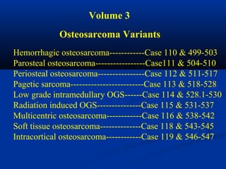
- 1. Volume 3 Osteosarcoma Variants Hemorrhagic osteosarcoma------------Case 110 & 499-503 Parosteal osteosarcoma-----------------Case111 & 504-510 Periosteal osteosarcoma----------------Case 112 & 511-517 Pagetic sarcoma-------------------------Case 113 & 518-528 Low grade intramedullary OGS------Case 114 & 528.1-530 Radiation induced OGS---------------Case 115 & 531-537 Multicentric osteosarcoma------------Case 116 & 538-542 Soft tissue osteosarcoma--------------Case 118 & 543-545 Intracortical osteosarcoma------------Case 119 & 546-547
- 3. Hemorrhagic Osteogenic Sarcoma
- 4. Hemorrhagic (Telangiectatic) Osteosarcoma The hemorrhagic (OGS), an extremely lytic and hemorrhagic variant of the osteosarcoma, presents in the same age group and location as a classic osteosarcoma but has a radiographic appearance almost identical to that of an aggressive aneurysmal bone cyst, making for a very difficult differential consideration for the radiologist. At the time of biopsy the tumor is very hemorrhagic and has the gross appearance of an aneurysmal bone cyst. Even microscopically, many areas of the hemorrhagic OGS will have the appearance of an aneurysmal bone cyst with only an occasional mitotic figure. For this reason, it is very important for the surgeon who performs the biopsy to obtain an adequate specimen with good sampling by means of an open biopsy as apposed to a simple needle biopsy. The microscopic features of the hemorrhagic OGS is a large number of benign- appearing giant cells and thus the terminology “giant cell rich”
- 5. osteosarcoma that is used by many pathologists. There is very little evidence of osteoblastic acitivity in the hemorrhagic OGS and, because it is so lytic in character, it frequently presents with a pathologic fracture early in the course of the disease and with that come potential problems for the treating orthopedic surgeon who must deal with the major contamination that occurs during the fracture. Because of the possible complications, one might consider an early limb salvage procedure before the fracture occurs. It was once felt that the prognosis for the hemorrhagic OGS was worse than that of the classic OGS because of its lytic dest- uctive nature. However, since the advent of systemic chemotherapy, the prognosis for survival is no different than for a classic OGS.
- 6. CLASSIC Case #110 23 year male hemorrhagic OGS proximal humerus Aneurysmal lesion
- 7. hemorrhagic tumor Coronal T-1 MRI
- 8. Coronal T-1 MRI tumor thru path fracture
- 9. Resected tumor cut in path lab
- 10. Photomic showing giant cells and malignant cells
- 11. osteoid blood Photomic showing hemorrhagic response
- 12. Neer allograft Post op x-ray with alloprosthetic reconstruction
- 13. 18 year followup x-rays
- 14. Case #499 15 year male hemorrhagic OGS distal femur
- 15. Lateral view
- 16. Bone scan
- 18. hemorrhagic tumor Coronal T-2 MRI
- 19. tumor Axial T- 2 MRI
- 20. blood Photomic
- 21. osseo- integration 3 yrs post op Compress total knee reconstruction CPS
- 22. Case #500 19 year male hemorrhage OGS proximal femur Looks like ABC
- 23. Lateral view
- 24. Initial biopsy reveals aneurysmal bone cyst
- 25. 6 weeks later shows lysis of outer shell Repeat biopsy reveals hemorrhagic OGS
- 26. femoral head tumor Hip disarticulation specimen
- 28. Case #501 6 year female path fracture thru unicameral bone cyst
- 29. cystic lesion Lateral view
- 30. 7 weeks after cyst steroid injection
- 31. 1 month later and progressive lytic destruction
- 32. Biopsy here shows hemorrhagic OGS
- 33. Case #501.1 Telangiectatic OGS 19 year old male with acute onset of pain 2 wks ago in right hip
- 34. PO 1 mo 2 mo 3 mo
- 35. Cor T-1 T-2
- 36. Axial T-1 T-2 Gad
- 37. Sag T-2 Gad
- 38. Case #502 4 year male cystic looks like lesion unicameral bone cyst
- 39. Progressive lysis after steroid injection
- 40. 2 months later with progressive lysis and looking malignant
- 41. Biopsy reveals hemorrhagic OGS
- 42. Clinical appearance before shoulder disarticulation
- 43. Case #503 17 year female hemorrhagic OGS C-3
- 44. AP view
- 45. CT scan
- 46. Photomic
- 47. 6 years later with spontaneous fusion and no tumor
- 49. Parosteal Osteosarcoma The parosteal (OGS) is a low grade variant arising from the surface of a long bone that presents as an exophytic mass with dense fibro- osseous tissue. It carries an excellent five year survival prognosis of 85% and accounts for about 4% of all osteosarcomas. This tumor has very little, if any, medullary involvement which clearly separates it from the classic OGS. It is seen more commonly in females than males and is found in a slightly older age group than the classic OGS. By far the most common location for this tumor is in the posterior aspect of the distal femur where it is frequently presents with minimal symptoms of pain but with a palpable tumor mass that might have been present many years before medical advise was sought. Histologically, this tumor has a very low mitotic index and in many cases can be confused with a normal healing fracture callous with occasional areas of cartilage being seen. Because this tumor is extremely low grade, it is not
- 50. responsive to adjuvant therapy such as chemotherapy or radiation therapy. The treatment consists of a wide surgical resection that must have safe margins, otherwise the recurrence rate will be quite high. Recurrence can occur 10 to 15 years after the surgery. In many cases the lesion can be resected without sacrificing the adjacent joint, but in larger lesions the best approach is a total joint replacement similar to that used for the classic OGS.
- 51. CLASSIC Case #111 32 year male parosteal OGS distal femur
- 52. AP view
- 53. Bone scan
- 54. Sagittal T-1 MRI
- 56. Axial T-1 MRI
- 58. Photomic
- 59. Higher power
- 60. Case #504 18 year male parosteal OGS distal femur
- 61. tumor AP view
- 62. Bone scan
- 63. tumor Axial T-2 MRI
- 65. tumor Macro section
- 66. Photomic
- 67. osseointegration Compress total knee reconstruction 2 years later
- 68. 10 years later with recurrence as a high grade dedifferentiated parosteal OGS tumor
- 69. Another view tumor
- 71. Close up of osseointegration of Compress implant
- 72. Case #505 32 year male parosteal OGS tumor proximal humerus
- 74. tumor CT scan
- 75. Amputation specimen cut in path lab
- 76. Photomic
- 77. Case #506 25 year male parosteal OGS distal femur
- 78. Distal femoral resection specimen tumor
- 79. tumor Cut specimen in path lab fatty marrow
- 80. Case #507 13 year male parosteal OGS mid femur AP view
- 81. Lateral view
- 82. CT scan
- 84. Autoclaved bone replaced with IM nail fixation
- 85. Post op x-ray 2 years later autoclaved bone
- 86. Case #508 17 year male with parosteal OGS mid tibia
- 87. Lateral x-ray
- 88. tumor CT scan
- 89. Bone scan
- 90. biopsy site Segmental resection mid tibial lesion
- 91. tumor Surgical specimen cut in path lab
- 92. Allograft reconstruction over IM nail
- 93. X-ray 1 year later
- 94. Case #509 41 year female parosteal OGS humerus
- 95. CT scan
- 96. tumor Resected cut specimen in path lab
- 97. Case #509.1 Parosteal OGS pseudotumor M.O. 10/06 3/07 17 year male with football injury 9/06
- 98. Sag T-1 Sag Gad
- 100. Case #510 32 year female with high grade parosteal OGS femur
- 101. Macro section tumor
- 102. Photomic
- 104. Periosteal Osteosarcoma The periosteal osteosarcoma is another surface type OGS that tends to be low grade to intermediate with potential for pulmonary metastasis in about 25% of cases. It accounts for 2% of all OGS’s and, compared to the parosteal OGS, has a much higher percentage of cartilagenous tissue in the tumor to the point where it can look like a periosteal chondroma but with a much higher mitotic index. One must find a few areas of osteoid formation to classify this as a periosteal OGS. It is seen typically in the second decade of life and is slightly more common in females than males. It arises from long bones, typically the tibia or femur, and has a higher incidence in diaphyseal bone than does OGS. Like the parosteal OGS, this lesion is treated by aggressive wide local resection that often can spare the adjacent joint. In most cases chemotherapy is not utilized unless the clinical picture is more aggressive than usual.
- 105. CLASSIC Case #112 15 year female with periosteal OGS tibia
- 106. CT scan
- 108. Axial T-2 MRI
- 109. Photomic
- 110. Post op x-ray following wide resection and allograft reconstruction
- 111. Case #511 30 year male with periosteal OGS prox tibia
- 112. CT scan
- 113. Sagittal T-2 MRI
- 114. edema tumor Axial T-1 MRI
- 115. Wide resection proximal tibia tumor bulge
- 116. tumor Cut specimen in path lab
- 117. Photomic
- 118. Proximal tibia resected ready for reconstruction
- 119. Post op x-ray with alloprosthetic TKA reconstruction allograft
- 120. Case # 512 9 year female periosteal OGS tibia
- 121. AP x-ray
- 122. Lateral view
- 123. Cut specimen in path lab following AK amputation
- 124. Photomic
- 125. Higher power
- 126. Case #513 14 year male periosteal OGS
- 127. Sagittal T-2 MRI
- 128. Axial T-1 MRI
- 129. Axial T-2 MRI
- 130. Case #514 26 year female periosteal OGS distal femur
- 131. Lateral view
- 132. stress shielding X-ray 10 years following wide resection and cemented prosthetic reconstruction
- 133. Case #515 12 year female periosteal OGS tibia
- 134. Bone scan
- 135. Axial T-1 MRI
- 136. Photomic
- 137. Case #516 15 year male periosteal OGS distal tibia
- 138. CT scan
- 139. Bone scan
- 140. Photomic
- 141. Case #517 39 year female periosteal OGS pseudotumor In fact is a Nora’s lesion or bizarre parosteal osteochondromatous proliferation (BPOP)
- 142. Bone scan
- 143. CT scan
- 144. edema Axial Gad contrast MRI
- 145. Sagittal PD
- 146. Sagittal T-2 MRI edema
- 147. Axial gad contrast MRI
- 148. Pagetic Sarcoma
- 149. Pagetic Sarcoma There are multiple diseases of the skeletal system that can result in a secondary form of OGS most likely brought about by a second mutation at a later age in a patient with chronic benign disease. These diseases include Paget’s disease, osteoblastoma, fibrous dysplasia, benign giant cell tumor of bone, bone infarcts, and chronic osteomyelitis. The most common of this group is Paget’s disease, a non-specific inflammatory osteomyelitis of bone seen in older patients that may be induced by a virus infection. Approx- imately 1% of patients with Paget’s disease can go on to Pagetic OGS which accounts for 3% of all OGS. The most common location for this secondary form of OGS is in the humerus, followed next by the pelvis and femur. The patients typically have a long history of dull, aching pain from their inflammatory Paget’s disease but then suddenly develop an acute new pain in the area of the older pain with x-ray evidence of recent lysis and destruction
- 150. of old Pagetic reactive bone. The prognosis for survival in this secondary form of OGS is extremely poor with only about 8% surviving, mainly because the older age group in which the disease occurs make it impractical to implement the aggressive protocols used in younger age groups.
- 151. CLASSIC Case #113 tumor 80 year female with Pagetic sarcoma pelvis
- 152. Bone scan
- 153. tumor Axial T-2 MRI
- 154. osteoid Photomic
- 155. Post op internal hemipelvectomy
- 156. Case#518 tumor 83 year female Pagetic sarcoma pelvis
- 157. tumor CT scan
- 158. tumor Another CT cut
- 159. Photomic
- 160. Case #519 85 year female with Paget’s disease pelvis
- 161. Same disease in lumbar spine
- 163. Same disease in tibia Advancing osteolytic wedge
- 164. old Paget’s Same patient with Pagetic sarcoma humerus new tumor
- 165. Macro section from amputation specimen tumor
- 166. Photomic
- 167. Post op x-ray following forequarter amputation
- 168. Case # 520 73 year female Pagetic sarcoma skull ready for resection
- 169. Lateral view of skull
- 170. Occipital view
- 171. Tangential view
- 172. tumor Resected specimen cut in path lab
- 173. Photomic
- 174. Case #521 old Paget’s with prior fracture 82 year male Pagetic sarcoma distal humerus
- 175. tumor Close up of new tumor
- 176. Photomic
- 177. Case #522 80 year female Pagetic sarcoma distal humerus
- 178. Case #523 femur humerus 84 male with multi focal Pagetic sarcoma
- 179. Case #524 83 year male Pagetic sarcoma femur
- 180. Case #525 60 year male Pagetic sarcoma femur
- 181. Lateral view
- 182. Photomic
- 183. Photomic
- 184. Case #526 78 female Pagetic sarcoma proximal tibia
- 185. Lateral view tumor
- 186. Case #527 92 year male Pagetic sarcoma tibia
- 187. Case #528 78 year female Pagetic sarcoma lumbar spine
- 188. Low Grade Intramedullary Osteogenic Sarcoma
- 189. Low Grade Intramedullary OGS Low grade intramedullary OGS is another rare low grade fibro- osseous variant of OGS that is unique because it is totally confined within the cortical anatomy of a long bone, most typically around the knee joint. It is found in an older age group than the classic OGS and is typically seen between the ages of 15 and 55 years; it affects males and females equally. The radiologic picture is that of a diffuse sclerotic change within the metaphysis of the long bone with no periosteal response or lytic destruction of the cortical anatomy. The smoky appearance of metaphyseal bone suggests the diagnosis of chronic osteomyelitis or perhaps fibrous dysplasia. Microscopically, the tumor has a histological appearance similar to parosteal OGS and because of this carries the same excellent prognosis for survival as we see in parosteal sarcoma. Likewise, treatment is similar without the use of chemotherapy or radiation. These lesions must be treated with complete wide resection that frequently involves a TKA, similar as in the classic OGS.
- 190. CLASSIC Case #114 63 year female intramedullary OGS distal femur
- 191. Lateral view
- 192. Bone scan
- 193. tumor CT scan
- 194. tumor Macro section from resected specimen
- 195. Photomic
- 196. Photomic
- 197. Case #528.1 51 year female tumor low grade intramedullary OGS distal femur
- 198. Bone scan
- 199. Coronal T-1 MRI tumor
- 200. tumor Axial T-1 MRI
- 201. tumor Resected distal femur cut in path lab
- 202. Photomic
- 203. Case #529 32 year female tumor low grade intramedullary OGS distal femur
- 204. Lateral view
- 205. CT scan
- 206. Photomic
- 207. Case #530 56 year male low grade tumor intramedullary OGS distal tibia
- 208. Lateral view tumor
- 209. Bone scan
- 210. Photomic
- 211. Radiation-induced Osteogenic Sarcoma
- 212. Radiation-induced Osteosarcoma One of the most malignant forms of OGS is the secondary type induced by radiation therapy, usually over 3000 rads, for some type of either benign or malignant disease process in the past. One of the most common types of radiation-induced OGS is seen in patients with breast cancer who receive local radiation following radical mastectomy and than develop OGS in the shoulder girdle area. Other malignant diseases that can result in OGS after radiation therapy include Ewing’s sarcoma and lymphomas. Benign diseases that can result in OGS from radiation therapy include GCT,ABC, and fibrous dysplasia. The average delay for the occurrence of secondary OGS is 15 years, with a range from 3 to 55 years. The prognosis for this variant is extremely poor, similar to Pagetic OGS. It has a very high rate of metastasis to the lung for which chemotherapy is not very effective.
- 213. CLASSIC Case #115 33 year female radiation-induced sarcoma scapula
- 214. tumor Widely resected specimen cut in path lab
- 215. scapula tumor Close up
- 216. Photomic
- 217. Higher power
- 218. Post op x-ray following scapular wing resection
- 219. Case #531 tumor 35 year female with radiation sarcoma prox femur prior radiation treatment for Hodgkin’s 20 yrs ago
- 220. Frog leg lateral tumor
- 221. Shortly after with pathologic fracture
- 223. Higher power
- 224. Case #532 72 year male radiation sarcoma pelvis Prior radiation therapy for prostate cancer 3 years before
- 225. Another view at a different date with hip dislocation
- 226. Photomic
- 227. Case #532.1 Radiation induced OGS 79 yr male with prior prostate CA radiation therapy and now presents with radiation OGS
- 228. Coronal Anterior CT Posterior CT
- 229. L Sagittal CT scan R Sagittal CT scan
- 230. Low axial CT cut thru L hip showing large tumor Upper CT cut thru SI area showing tumor R post ilium
- 231. Metastatic disease seen on chest x-ray
- 232. Case #533 56 year female with radiation sarcoma scapula Prior history of radiation for breast cancer
- 233. Oblique view
- 234. Bone scan
- 235. Photomic
- 236. Case #534 tumor 63 year female with radiation sarcoma scapula with prior radiation treatment for breast CA 12 yrs ago
- 237. Case #535 44 year female with radiation sarcoma proximal humerus 2nd to prior radiation for tumor breast cancer
- 238. Photomic with radiation OGS
- 239. Case #536 76 year male radiation sarcoma femur Prior history of radiation therapy for soft tissue tumor 10 years ago
- 240. Bone scan
- 242. Case #537 Elderly M.D. with long history working under X-ray fluoroscope Now skin cancer and radiation sarcoma index finger
- 243. X-ray of index finger sarcoma tumor
- 246. Multicentric Osteosarcoma The multicentric variant of OGS is an extremely rare variant occurring in approximately 1% of all OGS. It has two distinct categories: (1) Synchronous multicentric OGS occurring in child- hood and adolescence. This is the more severe variant, considered to be extremely high grade with a very poor prognosis associated with it. This form presents with multiple sclerotic lesions seen in a fairly symmetrical fashion in long bones, mostly in the lower extremities and because of the heavy tumor burden associated with multiple lesions throughout the skeleton, the alkaline phos- phatase is frequently elevated. (2) Metachronous multicentric OGS occurring mainly in adults is less aggressive than the synchronous form seen in children, presenting usually with a solitary lesion. Then, later on, more lesions develop that are considered multi- focal in nature. The possibility of metastasis can not be ruled out. These forms of OGS are quite resistant to chemotherapy and surgical treatment is frustrating because of the multi focal disease.
- 247. CLASSIC Case #116 8 year female multicentric OGS
- 248. Close up distal femur
- 249. Lateral view
- 250. Bone scan
- 251. Close up bone scan
- 252. Upper body bone scan
- 253. Coronal T-1 MRI
- 254. Another coronal cut T-1 MRI
- 255. Sagittal T-1 MRI
- 256. Photomic
- 257. Another photomic
- 258. Case #538 18 year female multicentric OGS pelvis and femur
- 259. tumor tumor Gad contrast coronal MRI
- 260. Another Gad contrast cut
- 261. pelvic tumor Axial T-2 MRI
- 262. Internal Hemipelvectomy Recon plate placed across pelvic ring surgical defect
- 263. Placement of air screws just prior to cementation
- 264. cement Placement of cement around screws and plate
- 265. femoral implant total hip Constrained total hip in and securing muscles to custom proximal femoral replacement implant
- 266. acetabulum tumor bulge ilium Outer face of resected specimen
- 267. tumor bulge ilium Inner face
- 268. Closure
- 269. recon plate and screws Post op x-ray
- 270. Case #539 16 year female multicentric OGS tumor Proximal tibial lesion
- 271. Lateral view with skip lesion in distal tibia
- 272. Bone scan showing two lesions in tibia
- 273. Bone scan showing iliac lesion
- 274. Bone scan showing sternal lesion
- 275. Photomic from tibial biopsy
- 276. Proximal tibial resection and tumor total knee bulge reconstruction
- 277. Proximal tibial prosthesis in position ready for relocation and closure
- 278. Reconstruction completed and ready for closure
- 279. Case #540 Multicentric OGS femur and sacrum 20 year male
- 280. Lateral view
- 281. Coronal T-1 MRI showing tumor at both ends of femur
- 282. Sagittal T-1 MRI distal femur tumor
- 283. tumor Axial T-2 MRI distal femur
- 284. Another axial T-2 MRI
- 285. Bone scan
- 286. Coronal T-1 MRI
- 287. Axial T-1 MRI
- 288. tumor Coronal gad contrast MRI showing sacral lesion
- 289. Photomic from femoral biopsy
- 290. Case #541 10 year female with multicentric OGS femur and tibia
- 291. tumor Lateral view
- 292. skip lesion AP view femur tumor
- 293. tumor Coronal T-2 MRI distal femur
- 294. tumor Sagittal T-2 MRI distal femur tibial lesions
- 295. tumor Coronal T-1 MRI knee joint tumor
- 296. tumor Coronal T-1 MRI showing multicentric involvement
- 297. Case #542 tumor 15 year male with multicentric OGS tibia and femur
- 298. tumor Coronal T-1 MRI tumor
- 299. Coronal T-2 MRI tumor
- 300. Sagittal T-1 MRI tumor
- 301. tumor Axial T-2 MRI view of distal femur
- 303. Soft Tissue Osteosarcoma OGS can be seen in soft tissue outside the skeletal system. It accounts for 4% of all OGS and is typically in large muscle groups around the pelvis and thigh area. It occurs most often in patients over 40 years of age and hits males and females equally. Soft tissue OGS, with its mature appearing bone in the central area of the lesion and aggressive, poorly mineralized tissue at the periphery, must be differentiated from myositis ossificans, which has a typical zonal pattern with peripheral maturation of bone formation. As with any soft tissue sarcoma, the treatment consists of wide local resection. Because of the poor prognosis, worse than that of bone osteosarcoma, systemic chemotherapy is utilized extensively as one would use for a typical medullary OGS.
- 304. CLASSIC Case #118 tumor 67 year male soft tissue OGS calf
- 305. AP view
- 306. Sagittal T-1 MRI tumor
- 307. Axial T-1 MRI
- 308. Cut surgical specimen in path lab
- 309. Photomic
- 310. Case #543 76 year female soft tissue OGS calf
- 311. Lateral view
- 312. CT scan
- 313. Bone scan
- 314. Axial T-1 MRI
- 315. Sagittal T-1 MRI tumor
- 316. Photomic
- 317. Case #544 60 year female with soft tissue OGS leg
- 318. Oblique view
- 319. Bone scan
- 320. Case #545 63 year male soft tissue OGS hand tumor
- 321. Lateral view
- 322. tumor Axial T-1 MRI
- 323. tumor Axial T-2 MRI
- 326. Intracortical Osteogenic Sarcoma
- 327. Intracortical Osteosarcoma The intracortical OGS is perhaps the rarest variant of OGS with only 14 cases described in the world literature since 1960. It occurs between the ages of 10 and 47 years, equally between males and females, and is seen most typically in the femur or tibia as a metadiaphyseal lesion with a radiographic appearance very similar to that of osteoid oasteoma. The prognosis is usually quite good with a total of three deaths in the world literature. It is usually treated by wide resection without chemotherapy. A few cases are higher grade and carry a poor prognosis similar to the classic OGS.
- 328. CLASSIC Case #119 42 year female with intracortical OGS femur
- 329. Bone scan
- 330. Axial PD MRI
- 331. Sagittal T-2 MRI
- 333. X-ray 18 months after curettement with recurrence
- 334. Bone scan at time of recurrence
- 335. Axial Gad contrast MRI same time
- 336. tumor Sagittal PD MRI same time
- 337. tumor Unicortical segmental wide resection
- 338. Photomic of resected specimen
- 339. Post op x-ray following allograft unicortical resection and allograft recon
- 340. PD T-2 tumor Sagittal PD & T-2 MRI 18 months later with met to C-spine
- 341. Case #546 43 year female intracortical OGS distal femur
- 342. Lateral view
- 343. Sagittal T-1 MRI
- 345. Biopsy photomic
- 346. Photomic
- 347. Case #547 47 year female intracortical OGS humerus
- 348. Lateral view
- 349. CT scan
- 351. Post op x-ray after wide resection and allograft recon
Hinweis der Redaktion
- AP radiograph of the proximal humerus of a 23-year-old male. There is extensive aneurysmal lytic destruction of the proximal epiphyseal-metaphyseal portion of the humerus with a pathologic fracture and extensive permeative lysis of the upper diaphysis of the humerus suggesting aggressive destructive disease, similar to what is seen with an aneurysmal bone cyst. There is very little evidence of the tumor osteoid formation that is seen in a classic osteosarcoma.
- Coronal T1-weighted MRI reveals a very destructive neoplasm arising from the proximal humeral metaphyseal-epiphyseal area with an aneurysmal blow-out of the lateral cortex with large hemorrhagic cyst formation creating a Codman's reactive triangle at the lower pole of the tumor as it bulges under the deltoid muscle. The fleshy portion of the tumor in the medullary canal is low signal on this T1-weighted image with sharp contrast to the high signal fatty marrow below.
- Coronal T1-weighted MRI shows the pathologic fracture through the tumor area just below the neck of the humerus.
- Gross appearance of the tumor following a wide resection demonstrating the hemorrhagic aneurysmal appearance of the lesion filled with clotted blood. It looks almost identical to the appearance of an aneurysmal bone cyst.
- Low power photomicrograph reveals extensive hemorrhage into an aggressive, highly malignant osteoblastic tumor that is conspicuous because of the large number of benign-appearing macrophages or giant cells seen throughout the tissue with a small amount of osteoid formation. The large number of hyperchromatic pleomorphic osteoblasts clearly separates this pathology from that of an aneurysmal bone cyst.
- Low power photomicrograph shows large hemorrhagic areas in a highly anaplastic osteoid-forming sarcoma with malignant-appearing osteoblasts demonstrating hyperchromicity and pleomorphism.
- AP radiograph shows the postoperative appearance following a wide resection of the proximal third of the humerus. It has been reconstructed with a cemented Neer prosthesis. This patient was treated successfully with systemic chemotherapy and is alive and well 18 years later with excellent function of his upper extremity.
- Here we see the radiographic results 18 years later with no evidence of subluxation at the shoulder joint. There is considerable evidence of stress shielding in both the allograph and the lower humerus but no signs of loosening. He has normal hand function and no pain problems.
- A 15-year-old boy presented with symptoms of pain in his distal femur for three months. This AP radiograph of the distal femur shows diffuse permeative lysis of the entire distal femoral metaphyseal area with a laminated periostitis surrounding the entire distal shaft of the femur, suggesting the diagnosis of either Ewing's sarcoma, aneurysmal bone cyst, or hemorrhagic osteogenic sarcoma. A biopsy proved this lesion to be a hemorrhagic osteosarcoma.
- This lateral radiograph again shows permeative lytic changes in the distal metaphysis and cortical breakthrough anteriorly, suggesting a malignant diagnosis.
- Technetium bone scan performed as part of the staging process shows a strong pickup of the isotope in the entire distal femoral metaphysis compatible with either a benign or malignant disease.
- Sagittal T2-weighted MRI shows a large, high intensity signal abnormality filling the entire distal femoral metaphysis up to the growth plate with multiple cystic loculations that would suggest hemorrhage into the medullary canal, as would be seen in aneurysmal bone cyst or in hemorrhagic osteosarcoma.
- Coronal T2-weighted MRI again demonstrates the high signal abnormality in the central portion of the distal metaphysis of the femur with a loculated pattern, suggesting a hemorrhagic lesion, but with high signal abnormality seen as well in the periosteum surrounding the distal metaphysis.
- Axial T2-weighted MRI shows high signal in the hemorrhagic lesion located in the distal femoral metaphysis and high signal abnormality in the surrounding periosteal tissue, suggesting either an aneurysmal bone cyst or hemorrhagic osteogenic sarcoma.
- At the time of the biopsy, the surgeon reported a large amount of dark red blood in a mossy lining surrounding the hemorrhagic lesion. This low power photomicrograph of a biopsy specimen taken from the distal femoral metaphysis reveals osteoid formation by an aggressive, malignant-appearing osteoblastic stem cell and giant cell activity suggests the diagnosis of a hemorrhagic or telangiectatic osteogenic sarcoma that frequently have the appearance of a benign aneurysmal bone cyst.
- After two cycles of adjuvant chemotherapy this patient was taken to the operating room and a wide resection of the distal femur was performed. Reconstruction was carried out with a rotating hinge prosthesis attached to the femur with a ComPreSs (compression) device so as to avoid stress shielding in the future. This x-ray taken three years following that procedure shows excellent osseointegration of the implant to the distal femur and no evidence of recurrent tumor. There was also no evidence of pulmonary metastases. This patient did have an early failure of the compression fixation device that required a simple revision within the first six months after the primary procedure.
- A 19-year-old male had symptoms of pain and swelling in his proximal thigh for three months. AP radiograph of the proximal femur shows an aneurysmal bone-forming lesion bulging from the medial aspect of the upper femur with evidence of extensive permeative lysis of the subadjacent bone and medial cortex. There is a fairly sharp and mature outer lining to the aneurysmal lesion that would suggest the diagnosis of a benign aneurysmal bone cyst. Early on, a biopsy suggested that diagnosis.
- This lateral radiograph shows in better detail the ossifying aneurysmal lesion with a fairly mature shell of bone at the outer edge, suggesting the diagnosis of a benign aneurysmal bone cyst.
- This low power photomicrograph taken at the time of the original biopsy shows early bone formation with a large amount of hemorrhage and giant cell response within the osteoid tissue. There is no evidence of malignant osteoblastic cells suggesting the diagnosis of a benign aneurysmal bone cyst.
- Six weeks later the patient was complaining of increased pain and swelling of his thigh. This AP radiograph taken at that time shows a breakdown of the benign pattern of the ossifying lesion, especially at the lower pole of the lesion, with progressive lytic destruction of the cortex that would suggest a more malignant diagnosis such as hemorrhagic osteogenic sarcoma. Indeed, a biopsy proved that diagnosis. The patient was then placed on adjuvant chemotherapy.
- Surgical specimen resulting from a hip disarticulation shows a very large, hemorrhagic, friable reddish-brown tumor surrounding the entire upper end of the femur that is compatible with the diagnosis of a hemorrhagic osteogenic sarcoma.
- Photomicrograph taken from the amputation specimen shows benign-appearing giant cells surrounded by aggressive, malignant-appearing osteoblastic-forming young osteoid tissue that is strongly suggestive of a diagnosis of hemorrhagic osteogenic sarcoma. This patient eventually developed pulmonary metastases and died two years later.
- A 6-year-old girl presented with a pathological fracture through what was felt to be a benign solitary bone cyst. This radiograph of the proximal humerus shows a fairly geographic lytic process involving the proximal end of the humerus in a metaphyseal location with evidence of chronic thinning of the outer cortex and a smoky appearance to the medullary contents compatible with the radiographic diagnosis of a solitary bone cyst.
- Lateral radiograph of the proximal humerus with a benign, cystic-appearing lesion dilating the thin cortex of the proximal metaphysis and a smoky appearance to the medullary contents. There is periosteal reaction to a pathological fracture through what appeared to be a benign bone cyst. Assuming it was a benign process, this lesion was treated with a cortisone injection into the lesion.
- This radiograph obtained six weeks later still has the clinical appearance of a benign bone cyst in a healing phase, but the patient still complained of pain in the area.
- The girl continued to complain of pain and swelling in the area. On this radiograph obtained a month later, there is evidence of extensive lytic destruction of the upper humerus which is certainly not the appearance of a benign bone cyst and should strongly suggest the diagnosis of a hemorrhagic osteosarcoma or possible infection in the area after injection.
- The patient was taken to the operating room and a open biopsy revealed a malignant-appearing hemorrhagic tumor. This photomicrograph shows benign-appearing giant cells surrounded by very aggressive, pleomorphic, hyperchromatic-appearing osteoblastic cells that are attempting to form osteoid and thus the diagnosis of a giant cell rich hemorrhagic osteosarcoma. This patient was treated with adjuvant chemotherapy and a forequarter amputation. She died one year later from pulmonary metastases.
- This 19 year old male patient presented with a two week history of spontaneous pain in the right hip. On this initial x-ray one can see a pathologic fracture thru the base of the femoral neck which was felt to be the result of a pre-existing benign solitary bone cyst.
- The patient was taken to the OR and a combined DHS and single cannulated hip screw were placed thru the fracture site as one can see in the PO x-ray. No biopsy was taken of the so-called cystic area. Follow-up x-rays were obtained at one, two and three months which shows progressive lytic destruction around the fracture site which suggested the possibility of infection or perhaps the presence of an aggressive tumor at the fracture site.
- Because of the alarming changes seen in the 3 month PO x-ray this MRI study was obtained which on the coronal images suggested a large fluid collection about the fracture site which might be blood from a hematoma or aneurysmal bone cyst or even an abscess.
- The same findings are seen on these axial images that clearly show a large fluid space with a fluid-fluid level suggesting blood from a hematoma or ABC.
- The sagittal images show the same at which point it was decided to biopsy the area and were very surprised to find histologic evidence for the diagnosis of a hemorrhagic OGS for which the patient was place on a system chemotherapy program following which the entire proximal third of the femur was resected widely and reconstructed with a Compress system bipolar prosthesis.
- A 4-year-old boy presented with a pathological fracture through what was felt to be a benign solitary bone cyst. This AP radiograph of the shoulder shows chronic enlargement of the proximal metaphysis of the humerus with thinning of the surrounding cortical structure and a fairly benign-appearing geographic process that suggests the diagnosis of a benign bone cyst for which the patient was treated with cortisone injections.
- On this radiograph obtained one month later, the process still looks like a benign bone cyst with a thin cortical structure in the dilated proximal humerus and a smoky appearance of the medullary canal.
- Radiograph obtained two months later reveals evidence of extensive lytic destruction of the upper humeral metaphysis and fairly heavy osteoblastic activitiy within the medullary canal that would now suggest the diagnosis of a hemorrhagic osteogenic sarcoma.
- This low power photomicrograph from a biopsy specimen shows osteoid tissue being formed by hyperchromatic and pleomorphic-appearing osteoblasts, as well as extensive hemorrhage into the tissue, very compatible with the diagnosis of hemorrhagic osteogenic sarcoma.
- This patient was placed on adjuvant chemotherapy during which time the tumor continued to grow aggressively, causing severe pain. This photograph shows the large, aneurysmal-appearing sarcoma located just beneath the biopsy site. A forequarter amputation was performed, however the patient died two years later from pulmonary metastases.
- A 17-year-old female presented with severe pain in the neck associated with paraparesis. This lateral radiograph of the cervical spine shows extensive lytic destructive changes within the third cervical vertebra with permeative lysis that would suggest the diagnosis of either an aneurysmal bone cyst, giant cell tumor, Ewing's sarcoma or hemorrhagic osteosarcoma. In this case, a biopsy proved the diagnosis to be osteosarcoma.
- AP radiograph also shows the extensive lytic permeation of the vertebral body of C3.
- CT scan through the third vertebra of the cervical spine shows lytic destructive changes that seem to be originating from the pedicle on one side. Bulging into the vertebral canal resulted in paraparesis.
- Low power photomicrograph from one of the biopsy specimens shows numerous benign-appearing giant cells in a field of aggressive osteoblastic osteoid formation that should make the diagnosis of osteogenic sarcoma -- a giant cell rich type.
- The patient was treated with multiple surgical debriedements to decompress the cervical spinal cord. After one of these procedures, she developed a significant infection in the area. The parents refused adjuvant chemotherapy or radiation therapy. Following the last procedure with the extensive infection, the lesion went on to involute spontaneously. This is the radiographic appearance six years later with a spontaneous fusion of the entire area around the C3 vertebrae. The patient experienced no recurrence and no pulmonary metastases, and without the use of adjuvant chemotherapy. She was alive and well 10 years after the initial onset of disease.
- A 32-year-old male presented with a lump that had been present for several years, blocked normal flexion at the knee and caused minimal symptoms of pain. This lateral radiograph shows a dense fibro-osseous tumor mass arising from the posterior surface of the distal femoral metaphysis with very little evidence of medullary involvement. The lesion has the appearance of a sessile-based osteochondroma, or perhaps a juxtacortical chondroma.
- This AP radiograph does not shows the lesion well because it lies directly beneath the patellar shadow. There is no evidence of a Codman's reactive triangle in the medial and lateral cortex as would be seen with a classic osteosarcoma.
- The bone isotope scan shows hot focal pickup of the technetium-99 isotope in the area of the surface osteoid tumor in the back of the distal femur. Note that there is minimal pickup in the medullary canal indicating that this is indeed a surface tumor.
- Sagittal T1-weighted MRI shows the low-signal characteristics of a dense fibro-osseous exophytic tumor mass arising from the posterior surface of the distal femoral epiphyseal-metaphyseal area with absolutely no reaction in the normal fatty marrow beneath the posterior cortex.
- Sagittal STIR MRI shows the high signal characteristics of the parosteal sarcoma on the posterior aspect of the distal femur with several areas of signal void where dense ossification is occurring. Note that the low signal features of the bone marrow show no evidence of reaction to the surface tumor.
- Axial T1-weighted MRI shows the low signal features of the parosteal sarcoma growing posteriorly and laterally beneath the hamstring tendons with no signal abnormality in the medullary canal. The tumor lies directly beneath the popliteal artery seen in the midline posteriorly.
- Axial STIR MRI reveals the high signal features of the parosteal sarcoma in the popliteal space protruding laterally without involvement of the medullary canal.
- On this low power photomicrograph there is extensive osteoblastic activity with young tumor osteoid being formed in an organized fashion by osteoblastic cells that are hyperchromatic but with a low mitotic index, There is absolutely no evidence of giant cell activity or hemorrhage in the tissue and it could conceivably be confused with a healing fracture callous.
- High power photomicrograph shows tumor osteoid being formed by hyperchromatic osteoblastic cells with a low mitotic index.
- This 18 year male noted a slightly tender mass in his popliteal space for one year that resulted in this initial x-ray showing an exophytic bone mass arising from the posterior cortex of the distal femur which is the classic appearance and location of a parosteal OGS. One might also consider a sessile based osteochondroma. A biopsy proved the diagnosis of parosteal OGS.
- AP x-ray at the same time.
- A bone scan revealed a strong pickup suggesting a malignant diagnosis.
- This axial T-2 MRI shows a surface lesion with no high signal fat extending into the base of the lesion ruling out the diagnosis of osteochondroma and suggesting parosteal OGS.
- This sagittal T-2 MRI shows a low signal lesion typical of a dense fibro-osseous lesion such as parosteal OGS.
- A distal femoral resection was carried out and here we see a macro section of the surgical specimen with no evidence of medullary involvement.
- Here we see a photomicrogragh of the biopsy specimen showing malignant osteoblasts forming tumor osteoid but with a low mitotic index and thus no chemotheraoy was advised.
- Following the wide distal femoral resection the limb was reconstructed with a Compress rotating hinge TKA. Here we see the x-ray appearance two years later with excellent osseointegration of bone to the spindle of the implant.
- Ten years later the tumor recurred as seen in this x-ray. A biopsy revealed a dedifferentiated parosteal OGS which required a marginal resection of the tumor along with chemotherapy.
- Here we see another view of the tumor recurrence.
- The biopsy specimen shows a high grade OGS compared to the low grade appearance of the tumor 10 years ago.
- Close-up of the favorable osseointegration between the prosthetic spindle and the distal femur not seen with conventional cemented long stem devices. The wound became infected following the last procedure which cleared with surgical debriedment and antibiotics and three years later the patient is doing well without recurrence or infection.
- This 32 year male presented with a two year history of a slow growing bony mass in his proximal humerus which on biopsy revealed an aggressive parosteal OGS.
- This axillary view shows circumferential involvement.
- This CT scan proves this to be a surface lesion but with extensive invasion of the surrounding soft tissues making the consideration of a safe limb salvage procedure difficult.
- Because of the high risk of local recurrence a shoulder disarticulation was performed and here we see the cut surgical specimen. No chemotherapy was used and the patient was alive five years later without evidence of disease.
- The biopsy specimen seen here shows a low grade bone forming tumor.
- A 25-year-old male gave a history of a slightly tender but firm tumor mass arising from the posterolateral aspect of his distal femur that has been present for two years. AP radiograph shows an ossifying mass immediately adjacent to the lateral cortex of the distal femur with no evidence of permeation into the subadjacent bony structure, characteristic of a low-grade parosteal osteogenic sarcoma.
- The patient was taken to the operating room and the distal 6 inches of the femur was resected in preparation for the placement of a rotating hinge knee prosthesis with a conventional cemented stem. The specimen seen from the back side shows a bulging, ossifying tumor mass lying directly on the surface of the posterolateral femur with the femoral condyles below.
- The surgical specimen was cut with a band saw in the pathology lab. The tumor lies directly on the posterolateral cortex of the femur but does not invade the subadjacent medullary canal, characteristic of parosteal osteogenic sarcoma.
- A 13-year-old boy presented with a tumor mass that had been growing slowly over the past two years. This radiograph of the mid-portion of the femur shows a surface-type lesion arising from the posteromedial cortex with a sclerotic reactive bone at the base but no medullary involvement. There is neoplastic bone formation above the sclerotic base that strongly suggests the diagnosis of a parosteal osteosarcoma.
- Closer view of the lesion which is fairly circumferential in nature. It arises from the cortex surface without permeation into the subjadjacent bone.
- CT scan at the mid-portion of the lesion shows neoplastic bone growing from the surface of the femoral diaphysis.
- The patient was taken to the operating room and a segmental resection of the middle third of the femur was carried out. The surgical specimen shows a good margin of normal muscle lying on the surface of the tumor.
- The surgical specimen was stripped of all its soft tissue and the remaining intact femoral diaphysis was autoclaved for 3 minutes at 130 degrees centigrade, killing the tumor completely and leaving the bony remains for an allograft reconstruction. This photograph shows the autoclaved autograft placed back into the patient and fixed with an intramedullary nail.
- This radiograph obtained two years later shows that the autoclaved bone has healed nicely at both ends, similar to the result seen with a conventional allograft. No recurrence of the tumor is noted.
- Photograph of the lower leg of a 17-year-old male shows a large tumor mass arising beneath the skin at the mid-portion of the tibia. The mass had been growing for a year and a half with minimal symptoms of pain.
- This radiograph shows a heavily ossified parosteal lesion arising from the surface of the tibia anteriorly with the characteristic features of a low-grade parosteal osteogenic sarcoma.
- CT scan through the mid-portion of the lesion shows the heavily ossified tumor on the surface of the tibia without permeation into the subadjacent bony structures.
- Technetium bone scan shows heavy pickup in the area of the parosteal osteosarcoma.
- The patient was taken to the operating room and a segmental resection of the upper portion of the tibia diaphysis was carried out, leaving the knee joint intact. The biopsy site is intact with the subadjacent tumor and a wide resection was performed around the entire lesion.
- The surgical specimen after being cut with a band saw in the pathology lab shows the characteristic gross features of the parosteal sarcoma lying on the anterior cortical surface of the tibia. There is no evidence of medullary involvement.
- Operative photograph showing the reconstruction being carried out by means of an allograft that has been used to replace the segmental defect.
- One year postoperative radiographic appearance of the allograft in position with an IM nail for fixation, This patient did not have a local recurrence or develop pulmonary metastases. No chemotherapy was necessary.
- A 41-year-old female presented with a surface-type, ossifying lesion of the upper humeral diaphysis that had been growing for six months. This AP radiograph of the humerus shows fairly mature neoplastic bone arising circumferentially around the upper diaphysis of the humerus with a small area of lysis in the center of the tumor suggesting a more aggressive pattern to the disease, A biopsy of this area revealed a high-grade but surface type osteogenic sarcoma, necessitating the use of adjuvant chemotherapy along with surgical resection.
- CT scan through the central portion of the lesion shows the heavily ossified tumor mass lying circumferentially around the upper humeral diaphysis.
- The gross specimen shows the surface characteristics of this lesion that microscopically revealed high-grade osteogenic sarcoma. The humeral defect was reconstructed with a segmental allograft along and the patient received adjuvant chemotherapy. The patient did not develop local recurrence or pulmonary metastases.
- This 17 year old football player sustained an injury to his arm in Sept. of 06. A month later we see the radiographic appearance in the left side image which shows new subperiosteal bone formation that could suggest the diagnosis of a parosteal osteogenic sarcoma. However, 5 months later we see the appearance to the right with mature bone formation in the fascial compartment of the biceps muscle that strongly supports the diagnosis of traumatic myosytis ossificans and not parosteal OGS.
- On this sagittal T-1 and gad contrast MRI we see a low signal lesion demonstrating the characteristics of mature bone formation with minimal perfusion unlike that of a malignant bone forming tumor.
- The same low signal activity is seen in these axial images at different levels.
- A 32-year-old female gave a history of a firm tumor mass in her distal thigh for eight months. These AP and lateral radiographic views of the distal femur show a heavily ossified circumferential surface-type tumor mass arising from the distal metadiaphyseal portion of the femur that on biopsy proved to be a high-grade surface-type osteogenic sarcoma.
- TION :This was prior to the advent of chemotherapy and the patient was treated with an amputation at a high thigh level. A gross macrosection of the surgical specimen shows the surface characteristics of this tumor without any evidence subadjacent medullary involvement.
- Low power photomicrograph taken through the area of the tumor shows neoplastic osteoid formation being created by a high-grade osteoblastic stem cell.
- A 15-year-old girl had mild symptoms of pain and a lump in her tibia for nearly a year. These AP and lateral radiographs show a surface tumor arising from the mid-diaphysis of the tibia. There are minimal lytic changes seen within the anterior cortex of the tibia with a low-grade chondroid-appearing mass arising from the surface of the diaphysis with flocculated calcification similar to what might be seen in a periosteal chondroma or a Nora's lesion.
- CT scan shows in better detail the surface characteristics of this periosteal sarcoma. There is a fair amount of dense reactive bone at the base but with a chondroid-appearing cap measuring about 2 cm in diameter, similar to what is seen in a periosteal chondroma or perhaps a sessile-based osteochondroma. There is no evidence of medullary involvement.
- This CT scan of the resected specimen shows in better detail the chondroid appearance of the surface lesion with mild reactive bone at the base but no evidence of permeation into the adjacent cortex or medullary canal. This looks very much like a periosteal chondroma.
- Sagittal T2-weighted MRI shows the high signal features of a high water content chondroid lesion arising subperiosteally on the surface of the tibia with no significant invasion into the subadjacent diaphyseal bone and minimal reactive edema in the surrounding soft tissue. The reactive bone seen at the base is very low signal.
- Low power photomicrograph of the biopsy specimen shows a low-grade cartilaginous tumor similar to a chondrosarcoma with immature cartilage matrix being produced by fairly aggressive chondroblasts with hyperchromatic nuclei but no mitotic activity. The diagnostic feature is the bone formation seen in the lower right corner that is tumor osteoid with calcification, a feature which distinguishes it from the typical chondrosarcoma and thus classifies it as an osteosarcoma variant.
- This lateral radiograph obtained following surgical treatment shows an allograft reconstruction supported by a side plate after wide resection of two-thirds of the diaphysis of the mid-portion of the tibia. No further adjuvant therapy was considered and the patient went on to heal nicely without local recurrence and without pulmonary metastases.
- A 30-year-old man gave a history of pain about the knee joint for about 3 months. This radiograph revealed a calcifying lesion arising off the posterior cortex of the proximal tibia that on biopsy turned out to be a periosteal osteogenic sarcoma.
- CT scan through the center of the lesion shows a calcifying surface mass arising from the posterior cortex of the tibia with slight erosion into the cortical structure and a calcific pattern that might suggest the diagnosis of a cartilaginous tumor, such as periosteal chondroma or a periosteal osteosarcoma as was the case here.
- Sagittal T2-weighted MRI shows a high signal lesion arising from the posterior cortex of the proximal tibia with a low signal component in the central area that represents the signal voids created by calcification within the cartilaginous tumor.
- This axial T1-weighted image shows the low signal characteristics of this cartilaginous tumor arising from the posterior cortex of the tibia. It is sharply marginated with a slight sclerotic base.
- The resected proximal 4 inches of the tibia, including an area off the posterior aspect of the tibia where the tumor is bulging into the popliteal space.
- The specimen has been cut in the pathology lab so that the calcifying cartilaginous tumor can be seen arising from the posterior cortex of the proximal tibia, characteristic of a periosteal osteogenic sarcoma.
- This low power photomicrograph reveals a low-grade cartilaginous tumor but with areas in the central portion that show osteoid being produced by the tumor as well. Osteoid production is a necessary finding in order to make the diagnosis of a periosteal osteogenic sarcoma in a mostly a chondrogenic tumor.
- Surgical appearance following resection of the proximal 4 inches of the tibia with the remaining normal structures still intact.
- Lateral radiograph taken shortly after surgery shows the result of the surgical reconstruction consisting of a total knee implant with an allograft placed over the proximal portion of the tibial component and a short side plate to control rotation of the allograft.
- Photograph of the upper portion of the leg of a 9-year-old girl who presented with a tender lump over the anterior portion of her tibia.
- AP radiograph shows vague evidence of periosteal activity around the proximal part of the tibia, suggesting periosteal irritation.
- Lateral radiograph of the proximal tibia shows a surface tumor arising from the anterior cortex of the tibia. There are patchy areas of calcification compatible with a calcifying chondroid tumor such as a periosteal osteogenic sarcoma.
- Because of the diffuse nature of this sarcoma, an above-knee amputation was performed. The pathological specimen shows a subperiosteal tumor surrounding nearly the entire upper half of the tibial diaphysis that has the appearance of cartilaginous tissue. Notice that there is no evidence of involvement of the subadjacent cortex or medullary canal.
- Low power photomicrograph taken from the surgical specimen shows a low-grade cartilaginous tumor that might suggest a chondrosarcoma. However, osteoid was found in a few areas tumor and thus the diagnosis of a periosteal osteogenic sarcoma.
- High power photomicrograph shows in better detail the pleomorphic and hyperchromatic appearance of the chondroblastic nuclear pattern which is compatible with an aggressive, malignant cartilaginous tumor.
- AP radiograph of the proximal tibia of a 14-year-old boy shows vague and poorly defined lytic activity in the proximal tibial cortex. It is not well visualized on this AP film.
- Sagittal T2-weighted MRI shows in much better detail the high signal characteristics of a periosteal osteosarcoma arising from the anterior proximal tibial cortex and bulging under the periosteum to produce a palpable mass anteriorly, just below the knee joint. Histologically the lesion was confirmed to be a periosteal osteogenic sarcoma.
- Axial T1-weighted MRI shows the low signal characteristics of this chondroid tumor arising beneath the periosteum of the anterior medial aspect of the proximal tibia with no evidence of any medullary invasion.
- Axial T2-weighted MRI shows the high signal features of the chondrogenic tumor lying on the surface of the proximal tibial cortex. The lesion was treated initially by an attempt at a wide resection. Shortly thereafter, there was an aggressive local recurrence that mandated an above-knee amputation supplemented with adjuvant chemotherapy. The patient was alive and well five years later without recurrence or pulmonary metastases.
- AP radiograph of the distal femur of a 26-year-female shows a calcifying surface lesion arising from the medial and distal cortex of the femur that on biopsy turned out to be a periosteal osteogenic sarcoma.
- Lateral view shows in better detail the calcifying pattern of this chondrogenic tumor lying on the posterior surface of the distal femur, similar to the appearance of a parosteal OGS except for a larger percentage of cartilage tissue and far less blastic activity. Calcification here is seen within the chondroid portion of the tumor.
- The patient was treated by a distal femoral resection and reconstruction with a customized tumor prosthesis with a total knee implant. This radiograph was taken 10 years following that procedure demonstrating continued stability to the cemented stem but showing evidence of stress shielding.
- A 12-year-old female presented with a tender, firm nodularity to the anterior portion of the right proximal tibia that on this radiograph shows lytic changes within the cortical structures of the upper diaphysis. There is evidence of periosteal lifting on the medial side fo the tibia.
- Technetium bone scan shows increased activity in the proximal diaphysis of the tibia.
- Axial T1-weighted MRI shows a signal abnormality of a surface lesion arising from the subcutaneous portion of the proximal tibia with several speckled areas of signal void representing calcification within the tumor.
- Low power photomicrograph shows what appears to be a low-grade chondrosarcoma but with some areas of osteoid formation and calcification seen typically in a periosteal osteogenic sarcoma. This lesion was treated by an aggressive local resection, following which there was no local recurrence or pulmonary metastases.
- A 15-year-old boy presented with a tender mass over his distal anterior tibia just above the ankle joint that on this radiograph shows a surface lesion with aneurysmal characteristics that might suggest the diagnosis of an aneurysmal bone cyst. A biopsy proved this tumor to be a periosteal osteogenic sarcoma.
- N :CT scan through the epicenter of the lesion shows the aneurysmal-appearing lesion arising from the anterior distal portion of the tibia. There is matrix calcification not found in aneurysmal bone cysts but typically seen in periosteal osteogenic sarcoma.
- Bone scan performed prior to surgery shows extensive uptake in the aneurysmal lesion in the distal tibia.
- High power photomicrograph shows chondroid tissue being formed by aggressive-appearing osteoblastic cells, as demonstrated by the hyperchromatic and pleomorphic nuclear patterns. The presence of osteoid formation in a few areas determined the diagnosis of periosteal osteogenic sarcoma. This patient was treated with a below-knee amputation.
- A 39-year-old female presented with a firm, tender nodule beneath the skin attached to her mid-shaft tibia. This AP radiograph of the tibia shows a calcifying lesion attached to the cortical structures of the tibia that might suggest the diagnosis of a periosteal osteosarcoma. In this case, a biopsy proved the process to be a bizarre parosteal osteochondromatous proliferation, an idiopathic reactive lesion that may be the result of incidental trauma. It is not a neoplastic condition but has a high local recurrence rate of about 30% following local resection. These lesions never become malignant.
- Technetium bone scan shows increased focal uptake in the area of the calcific lesion.
- CT scan cut through the center of lesion shows a surface lesion arising from the medial cortex of the tibia. The slight cortical erosion and fluffy dystrophic calcification within the lesion can also be seen in periosteal osteogenic sarcoma which was the concern here.
- Axial gadolinium contrast MRI shows the high signal features of this chondroid tumor on the surface of the upper tibia. The low signal voids secondary to calcification might be seen with a stress fracture or periosteal osteogenic sarcoma.
- Sagittal proton density MRI shows the intermediate signal features of the surface lesion arising from the cortex of the upper tibial diaphysis with no evidence of any cortical invasion or medullary involvement.
- Sagittal T2-weighted MRI shows the high signal features of the surface lesion, compatible with a chondroid tumor. There are inflammatory changes seen in the adjacent soft tissues beneath the skin
- This gadolinium contrast study shows minimal uptake by the tumor tissue which is devoid of vascular supply but does have a slightly increased signal, compatible with a chondroid tumor. This lesion was treated by an excisional biopsy to prove the diagnosis of a Nora's lesion, following which there was no local recurrence.
- An 80-year-old female who had Paget's disease for many years and then developed acute pain in the right pelvic area. This AP radiograph reveals old Paget's disease involving the entire right hemipelvis and degenerative osteoarthritis of the hip joint typical of Paget's disease. However, the acute lytic change seen in the supraacetabular area is a new finding that strongly suggests a conversion of the Paget's disease into a hemorrhagic osteosarcoma.
- In this bone isotope scan, there is extensive pickup of the isotope in the right hemipelvic area, typical of Paget's disease. It is in no way diagnostic of a Pagetic sarcoma.
- Axial T2-weighted MRI shows a very large hemorrhagic, cystic tumor ballooning out of the right hemipelvis. This might suggest an aneurysmal bone cyst in a younger patient or a hemorrhagic metastatic carcinoma in an older person, or a hemorrhagic osteosarcoma in a patient with Paget's disease as seen here.
- On this low power photomicrograph there is microscopic evidence of tumor osteoid formation with fairly heavy calcification and osteoblastic activity that takes on the appearance of an osteosarcoma because of the pleomorphic hyperchromatic nature of the large nuclei in the osteoblasts.
- AP radiograph of the patient several months following a palliative total joint reconstruction utilizing large amounts of methyl methacrylate and Steinman pins to stabilize the defective innominate bone and a conventional cemented femoral component. This procedure made the patient more comfortable for the remaining six months of her life. She died of extensive disseminated disease and pulmonary metastases. She was not considered for chemotherapy because of her poor health and age.
- An 83-year-old female underwent total hip replacement for arthritis and Paget's disease which she had had for 25 years. She then developed new symptoms of pain in the hip. This AP radiograph of the hip shows a new, permeative, lytic lesion involving the entire iliac crest and body of the ilium down to the hip joint on top of old, benign Pagetic inflammatory bone in the lower half of the pelvis. This picture suggests the diagnosis of a Pagetic sarcoma arising within old Paget's disease of the pelvis.
- CT scan through the epicenter of the tumor shows a large bone-forming mass arising both inside and outside the iliac portion of the pelvis. It has all the earmarks of an osteogenic sarcoma arising out of Paget's disease.
- Another CT cut through the pelvis shows a large aneurysmal-appearing, ossifying mass that has the appearance of an osteosarcoma. The changes seen in the sacroiliac area are compatible with old Paget's disease.
- Low power photomicrograph from a biopsy of the ilium reveals old inflammatory osteomyelitis with a characteristic high turnover mosaic pattern that strongly suggests the diagnosis of Paget's disease. In the soft tissue immediately above this pagetic bone, there is evidence of a high-grade sarcoma that is rich in giant cells and confirms the diagnosis of an osteosarcoma arising out of Paget's disease. The prognosis for this condition is very poor. Because of the advanced age of the patient, chemotherapy could not be used and she died of disseminated disease after an attempt was made at a local resection of the painful mass in her right hemipelvis.
- AP radiograph of the pelvis of an 85-year-old female with known Paget's disease for many years shows the characteristic chronic inflammatory changes but no recent lytic areas to suggest sarcoma.
- Lateral radiograph of the lumbar spine shows chronic deformation of L4 and L5 with a heavy course trabecular pattern and slight aneurysmal change in these two vertebrae, compatible with the diagnosis of Paget's disease.
- On this AP radiograph of the skull there are lytic changes as well as a patchy blastic response in the center of the lytic area that is compatible with the diagnosis of Paget's disease.
- Lateral radiograph of the tibia shows early pagetoid changes within the proximal two-thirds of the tibia with slight dilatation of the cortex anteriorly and an advancing wedge of osteolysis progressing distally. This is referred to as the advancing blade of grass sign, very characteristic for early Paget's disease.
- Radiograph of the humerus shows more recent lytic destructive change within the mid-portion of the diaphysis, resulting in new symptoms of pain in the area, that is strongly suggestive of sarcomatous degeneration within the pre-existing Paget's disease seen in the entire upper third of the humerus.
- A biopsy revealed the diagnosis of pagetic osteogenic sarcoma. The patient was taken to the operating room and a forequarter amputation was performed. This macrosection of the humerus shows a sarcomatous-appearing lesion arising from the mid-portion of the humeral diaphysis, eating its way through the cortex and presenting into the surrounding soft tissue.
- Low power photomicrograph from the surgical specimen is dominated by neoplastic osteoid formation created by aggressive-appearing osteoblastic cells that clearly suggests the diagnosis of an osteosarcoma arising from pre-existing Paget's disease.
- Postoperative radiograph of the chest taken after the forequarter amputation demonstrating no evidence of metastatic disease. This patient was alive and well several years following the amputation.
- This photograph shows a patient being prepped on the operating table for resection of a pagetic osteogenic sarcoma of the occipital area. The patient, a 73-year-old female with Paget's disease for 25 years, had recently developed symptoms of pain in the skull associated with a mass that grew over the past three months.
- Lateral radiograph shows chronic pagetic changes seen throughout the entire calvarium with patchy osteoblastic changes and diffuse osteolysis of the calvarium. However, in the occipital area there is a bulging mass posteriorly that on biopsy proved to be an osteosarcoma arising from Paget's disease.
- On this special radiograph of the skull emphasizing the occipital area, an osteoblastic lesion that respresents osteosaroma can be seen arising from the calvarium on the left side.
- Tangential view across the apex of the pagetic sarcoma that is forming malignant osteoid tissue on the surface of the calvarium.
- The patient was taken to the operating room and a craniotomy was performed to resect the osteosarcoma from the occipital area. The resected specimen demonstrates pagetic changes in the calvarium with evidence of a pagetic osteogenic sarcoma mass lying on the surface of the widened calvarium bone.
- This low power photomicrograph of the surgical specimen shows the classic changes of pagetic infllammatory disease with a high turnover reversal mosaic pattern. Immediately adjacent to this blastic-appearing bone there is evidence of pagetic sarcoma lying immediately on the surface of thickened calvarial bone.
- An 82-year-old male with Paget's disease for many years developed pain in his elbow area over the last three months. This AP radiograph of the humerus and elbow shows a lytic lesion located in the supracondylar area of the humerus, just proximal to the elbow. Plates are in place from a previous pathological fracture.
- Close-up radiograph of the elbow shows the recent lytic changes within the distal humerus, extending down to the elbow joint with cortical breakthrough seen circumferentially on top of old pagetic changes. This picture is characteristic for pagetic sarcoma.
- The patient was treated by amputation of the extremity at the shoulder level. A photomicrograph of the lesion gives diagnostic evidence of pagetic sarcoma arising on top of old Paget's disease. There is an abundance of malignant osteoblasts and giant cell activity.
- AP radiograph of the humerus in an 80-year-old female with a known history of Paget's disease shows the chronic hypertrophic changes of the entire humerus secondary to Paget's disease of long standing. In addition, a new change is seen distally just above the elbow where a painful soft tissue mass occurred over the past several months. A biopsy proved it to be a Pagetic sarcoma for which the patient underwent an amputation at the shoulder level.
- Lateral radiograph of the femur and AP radiograph of the humerus in an 84-year-old male with known Paget's disease show a soft tissue sarcoma arising out of the old Paget's disease of the humerus that on biopsy was proven to be an osteosarcoma. The lytic lesion in the distal femoral metaphysis was also proven to be osteogenic sarcoma arising out of Paget's disease. Because of the age of the patient palliation was the only concern. He underwent radiation to the sites of the two painful lesions and died six months later of disseminated disease.
- AP radiograph of the femur of an 83-year-old male with known Paget's disease shows a large tumor mass arising from the mid-portion of the femur. There is a recent pathological fracture on top of the classic changes of chronic Paget's disease of the upper and lower portion of the femur. Neoplastic bone formation is present within the tumor mass and a biopsy proved this to be an osteosarcoma arising out of Paget's disease. The patient was treated by means of a hip disarticulation.
- AP radiograph of the distal femur of a 60-year-old male with known Paget's disease shows a recent pathological fracture through a lytic destructive change on top of old pagetic disease of the entire distal femur. The pathological fracture through the lesion shows tumor osteoid forming in the central portion of the femur that on biopsy proved to be an osteogenic sarcoma arising from Paget's disease.
- Lateral radiograph again shows the pathological fracture through the motheaten, lytic destructive lesion that is bursting into the soft tissue posteriorly with neoplastic bone formation, characteristic of osteogenic sarcoma arising from Paget's disease.
- Low power photomicrograph of the surgical biopsy specimen shows the characteristic reversal mosaic pattern of Paget's disease surrounded in the soft tissue by a sarcomatous spindle cell pattern, diagnostic of a Pagetic sarcoma. This patient was treated with a high level amputation through the femur and died eight months later of disseminated disease.
- Low power photomicrograph taken from the same surgical biopsy specimen shows a combination of malignant spindle cells and a few areas of osteoid formation being produced by obviously malignant osteoblasts, typical of osteosarcoma in Paget's disease.
- A 78-year-old female with known Paget's disease presented with new sympoms of pain in the knee of four months duration. AP radiograph of the knee joint shows chronic inflammatory changes in the proximal tibia suggestive of Paget's disease. There is a new lytic process just beneath the medial tibial plateau that on biopsy proved to be a Pagetic sarcoma arising from the tibia.
- Lateral radiograph of the proximal tibia reveals the course bone pattern of chronic Paget's disease with a lytic change in the central area resulting from a sarcomatous degeneration. This patient was treated by an above-knee amputation but died of pulmonary metastases two years later.
- Radiograph of the amputation specimen of the tibia in a 92-year-old male who had chronic Paget's disease for 30 years and then developed an acute sarcomatous change in the mid-portion of the tibia with the geographic lytic change on top of old Pagetic osteomyelitis. The process was proven on biopsy to be an osteogenic sarcoma arising from Paget's disease.
- AP radiograph of the lumbosacral spine in a 78-year-old female with known Paget's disease shows a recent onset, painful osteoblastic mass arising out of the upper lumbar spine and growing out into the paraspinous area. On biopsy, this mass proved to be osteogenic sarcoma. Treatment consisted of local debriedement of the tumor followed by postoperative radiation therapy for palliation. The patient died 18 months later of disseminated disease.
- An AP radiograph of the distal femur in a 63-year-old female with low-grade symptoms of pain for six months. There is evidence of diffuse sclerotic changes throughout the entire distal femoral metaphysis. This picture suggests chronic low-grade inflammatory disease or, perhaps, fibrous dysplasia.
- Lateral radiograph of the same patient with a more lytic appearance, chronic thinning of the anterior cortex and bony sclerotic response at the periphery of the lytic lesion, There is evidence of matrix calcification that would suggest the diagnosis of fibrous dysplasia.
- On the bone isotope scan, there is a very strong pickup of the isotope in the distal femoral metaphysis suggesting active disease such as chronic low-grade osteomyelitis or fibrous dysplasia.
- CT scan of both distal femori shows evidence of chronic dilatation of the distal femoral metaphysis with sclerotic response seen at the periphery of a lytic lesion with matrix calcification that could suggest fibrous dysplasia or even chondrosarcoma. There is no evidence of any extracortical activity that one would expect to see in osteosarcoma.
- This patient had a wide local resection and a prosthetic reconstruction with a rotating hinge total knee device. This macrosection of the distal femoral metaphysis shows diffuse infiltrative tumor activity filling the entire medullary canal with no evidence of normal fatty marrow. There is some evidence of cortical erosion and permeation that would suggest a low-grade neoplasm such as a chondrosarcoma or osteosarcoma. This picture is also compatible with fibrous dysplasia.
- Low power photomicrograph of the specimen shows definite evidence of infiltrative osteoid-forming neoplasm flowing through the trabecular pattern of the metaphyseal bone suggesting malignancy. There is young osteoid formation that is poorly mineralized with osteoblasts that suggest low-grade sarcoma.
- Low power photomicrograph showing tumor osteoid formation being produced by malignant osteoblasts with a low mitotic index quite characteristic for a low-grade intramedullary osteosarcoma.
- A 51-year-old female presented with symptoms of pain of a dull aching nature in her lower thigh. This lateral radiograph of the distal femur shows a diffuse sclerosis of the entire distal metadiaphyseal portion ot the femur with a slight suggestion of periosteal activity that initially suggested the diagnosis of low-grade infection. A biopsy proved the lesion to be a low-grade intramedullary osteogenic sarcoma.
- Skeletal technetium bone scan shows heavy pickup of the isotope in the area of the distal femur where the tumor is located.
- Coronal T1-weighted MRI shows the low signal features of the signal abnormality in the distal femoral metaphysis that stops abruptly at the old growth scar and shows minimal if any evidence of involvement in the periosteal sleeve around the distal femur. This is the typical appearance for low-grade intramedullary OGS.
- Axial T1-weighted MRI shows the low signal abnormality filling the entire distal femoral metaphysis with minimal if any breakthrough into the soft tissue posteriorly.
- The patient was taken to the operating room and a wide resection of the distal 5 inches of the femur was carried out. The surgical specimen has been cut with the band saw in the pathology lab. Note the diffuse tumor involvement of the distal femoral metaphysis with no evidence of any soft tissue involvement outside the cortex, typical of low-grade intramedullary OGS.
- Low power photomicrograph shows tumor osteoid formation being produced by low-grade osteoblastic cells with a very low mitotic index. This patient was treated with a limb salvage resection of the distal femur and reconstruction with a rotating hinge total knee implant. Because of the low-grade nature of the tumor, chemotherapy was not used . The patient was alive and well five years later with no recurrence.
- A 32-year-old female gave a history of dull aching pain in her knee for the past year, AP radiograph of the distal femur shows a diffuse sclerotic process within the medullary canal in the distal femur with no evidence of cortical erosion or breakout into the surrounding soft tissue.
- Lateral radiograph shows a lytic process involving a large portion of the distal femoral diaphyseal-metaphyseal area with thinning of the cortex on one side only and a sclerotic base on the opposite side. This lesion was biopsied and proved to be a low-grade intramedullary osteogenic sarcoma.
- ION :CT scan through two-thirds of the femoral metaphysis with osteoid formation in the lytic component, characteristic of osteosarcoma.
- Low power photomicrograph taken from the biopsy specimen. The tumor, which is osteoid-producing with an osteoblastic cell, looks malignant but has a very low mitotic index. The patient was treated by a wide resection of the distal femur and reconstruction with a cemented stem rotating hinge prosthesis. No chemotherapy was indicated because of the low-grade nature of the disease.
- A 56-year-old male presented with dull aching pain in his ankle area for a year-and-a-half. AP radiograph of the distal tibia and ankle shows diffuse sclerotic changes within the entire distal tibial metaphysis and epiphysis but without evidence of cortical erosion or breakthrough into the surrounding soft tissue. This picture is strongly suggestive of the diagnosis of low-grade intramedullary OGS.
- Oblique radiograph of the distal tibia again shows motheaten erosion within the distal tibial epiphysis and metaphysis and diffuse sclerotic response filling the entire distal third of the tibia.
- Technetium bone scan shows strong pickup of the isotope in the distal portion of the tibia.
- Low power photomicrograph from the biopsy specimen shows tumor osteoid formation being produced by low-grade osteoblastic neoplastic cells with a low mitotic index. The patient was treated by a below-knee amputation with no adjuvant chemotherapy. He was alive and well 5 years later.
- A 33-year-old female had a prior history of a benign hemangioma removed from the skin overlying her scapula when she was 3 years old. She was treated with low dose radiation postoperatively to avoid keloid formation. Thirty years later she developed a painful mass in the area for which this radiogragh was taken. This tangential radiograph of the scapula reveals permeative lytic destruction of the scapular wing with aneurysmal dilatation of the cortex posteriorly and reactive new bone formation at the periphery of the lesion, similar to that seen in aneurysmal bone cyst.
- This is the gross appearance of the tumor after wide resection of the entire scapular wing, including the tumor and adjacent muscle.
- Closer view of the gross specimen reveals the aggressive neoplastic tumor arising from the scapular wing and invading the surrounding muscle tissue. It has a tannish appearance, characteristic of high-grade sarcomas.
- Low power photomicrograph shows extensive high-grade sarcoma infiltrating the muscle belly surrounding the scapula.
- High power photomicrograph shows high-grade sarcoma with a nuclear pattern similar to that seen in Ewing's sarcoma, with very little evidence of osteoid formation. The pathologists gave the diagnosis of a small cell osteogenic sarcoma induced by radiation therapy 30 years before.
- On this radiograph taken after surgery, there is evidence of a subtotal resection of the scapular wing with the glenoid body still intact. Shortly after this the patient developed pulmonary metastases and died from her disease which was resistant to chemotherapy.
- A 35-year-old female gave a history of Hodgkin's disease 20 years before that was treated with chemotherapy and low radiation therapy to both inguinal areas. Six months prior to this x-ray examination she developed pain in the left hip. This AP radiograph of the pelvis and left hip shows diffuse lytic changes in the head, neck and intertrochanteric area of the left proximal femur with small areas of osteoid formation in the medullary canal that on biopsy proved to be osteogenic sarcoma arising from out the irradiated field from her old Hodgkin's disease.
- Lateral radiograph showing the motheaten appearance of the upper end of the femur, characteristic of a sarcoma.
- Radiograph taken at a slightly later date reveals a pathological fracture through the irradiated bone.
- The biopsy material was used to prepare this scanning lens photomicrograph that shows evidence of a high-grade sarcoma arising in the medullary bone of the proximal femur. It is very anaplastic, typical of irradiation osteogenic sarcoma.
- A soft tissue area of the biopsy specimen shows early osteoid formation and aggressive anaplastic-appearing osteoblastic cells. This patient was treated by a hip disarticulation plus adjuvant chemotherapy. A poor prognosis would be expected with this type of irradiation sarcoma and the patient was dead within a year-and-a-half following her surgery.
- A 72-year-old male gave a history of prostate cancer that was treated with local radiation therapy three years before. The man had also undergone a total hip replacement for osteoarthritis. He had developed recent symptoms of pain in the hip that were progressive over the past three months. This AP radiograph shows neoplastic activity in the para-acetabular area and posterior column of the hip extending down into the ischium with lytic changes as well as blastic activity within the ischium. Biopsy proved the biopsy of osteosarcoma arising in the area of previous radiation therapy.
- Radiograph taken after a spontaneous dislocation of the total hip implant. This again shows evidence of tumor activity in the ischial area with soft tissue involvement as well.
- Low power photomicrograph of the biopsy specimen shows osteogenic sarcoma with tumor osteoid being produced by malignant osteoblastic cells. Because of the poor prognosis for survival, no attempt was made to remove the tumor. The patient died a year later from metastatic disease.
- This 79 year old male presented with a history of radiation therapy for prostate cancer 12 years ago and now has a painful mass in the inguinal area seen as a blastic lesion on this x-ray and on biopsy turned out to be a radiation osteosarcoma.
- On these anterior and posterior CT scans we can see the major portion of the tumor anteriorly and on the posterior cut we can see a smaller and separate focus in the right posterior ilium which makes this a case of multifocal osteosarcoma making it impossible to consider a surgical resection.
- On these sagittal CT scans we see the large tumor about the left hip area and smaller multifocal areas in both sacroiliac areas in the posterior ilium.
- Here we see an upper and lower level CT cut which reveals the major tumor about the anterior pelvis and left hip area at the top and a small separate lesion in the right posterior iliac area seen in the lower image.
- As is typical of radiation induced and multifocal osteosarcoma, the prognosis is very poor with early pulmonary involvement as we see in this chest x-ray. Because of this and the older age of the patient the treatment should consist only of palliation without chemotherapy and perhaps local radiation therapy for control of local pelvic pain.
- A 56-year-old female who gave a history of prior radiation therapy to the right shoulder area for breast cancer had recently developed symptoms of pain in the shoulder. This AP radiograph shows motheaten destruction within the body of the scapula with a sclerotic response and possible pathological fracture through the area. A biopsy revealed osteogenic sarcoma arising from previous radiation therapy.
- ON :Oblique radiograph showing the lytic destructive changes within the body of the scapula.
- Technetium bone scan shows increased activity in the body of the scapula where the tumor was located.
- Low power photomicrograph of the biopsy specimen shows tumor osteoid produced by a malignant-appearing osteoblastic cell. This patient was treated by a forequarter amputation but died from pulmonary metastases a year later.
- A 63-year-old female gave a history of breast cancer twelve years before for which she was treated with surgery and radiation therapy to the area. This AP radiograph of the shoulder shows a calcific lesion in the scapular area on the right side that initially was felt to be calcific bursitis or tendonitis and treated with multiple injections of cortisone. At this point there was a suspicion of sarcoma and a biopsy revealed osteogenic sarcoma arising from an area of previous radiation therapy, Her treatment consisted of a forequarter amputation. She died of her disease a year-and-a half later.
- A 44-year-old female who gave a history of breast cancer treated with local radiation therapy developed a painful mass in her upper arm three months before this x-ray was taken. This AP radiograph reveals extensive ossification around the proximal humerus in a lobulated pattern that is strongly suggestive of osteosarcoma. A biopsy confirmed the diagnosis and a forequarter amputation was performed.
- Low power photomicrograph taken from the surgical specimen shows tumor osteoid being produced by malignant osteoblasts.
- A 76-year-old male who gave a history of a soft tissue sarcoma in his thigh that was treated by surgical resection and radiation therapy ten years ago presented with symptoms of pain in the thigh area for the past three months. This AP radiograph shows a bone-forming tumor arising from the mid-diaphysis of the femur. It has a fluffy chaotic pattern strongly suggestive of the diagnosis of osteosarcoma which was proven on biopsy.
- This bone scan shows heavy pickup of isotope in the mid-portion of the femur where the tumor was located.
- Low power photomicrograph from the biopsy specimen shows neoplastic osteoid being produced by malignant-appearing osteoblasts. This patient was treated with a hip disarticulation and adjuvant chemotherapy.
- An 82-year-old retired physician gave a history of using a fluoroscope for manipulation of patients in the 1930's and 40's without protecting his hands during this radiation treatment of his patients. Approximately one year prior to this photograph he developed pain in his hand, This photograph shows severe irradiation changes in the fingers with distortion of the nails and small tumor masses arising beneath the skin that on biopsy proved to be high-grade sarcomas arising from his irradiation dermatitis.
- Radiograph of the hand showing extensive destructive changes within the phalanges of the index finger and soft tissue swelling about the area induced by irradiation sarcoma.
- Low power photomicrograph from the biopsy specimen shows a very high-grade sarcoma with small areas of osteoid being formed with very bizarre pleomorphic nuclear patterns, characteristic of irradiation-induced osteogenic sarcoma. The patient was treated by an amputation of the hand. He developed pulmonary metastases one year later and died.
- An 8-year-old girl presented with symptoms of pain in the lower extremity for 6 months and an elevated alkaline phosphatase. This AP radiograph of both femori reveals multiple sclerotic lesions stippled throughout the diaphyses of both femoral shafts with a greater involvement on the right side where there is a dense sclerotic lesion in the distal femoral metaphysis, compared to the smaller lesion on the opposite side. There is minimal evidence of cortical or periosteal involvement.
- This AP radiograph of the distal right femur shows in better detail the sclerotic nature of the synchronous involvement of the entire distal half of the femur with its greatest magnification in the distal metaphyseal area that could be misconstrued as a chondrosarcoma or perhaps fibrous dysplasia.
- Lateral radiograph of the right femur showing the sclerotic appearance of the entire distal half of the femur with a smaller skip lesion in mid shaft.
- The bone isotope scan demonstrates the pickup of the synchronous lesions seen mostly in the right femur but also in both hips and multiple areas in both sides of the pelvis.
- This isotope scan shows both femori with multiple lesions, fairly symmetrical on both sides.
- On this isotope scan of the upper half of the body there is evidence of involvement in both proximal humeri and multiple focal involvement of the maxillofacial and cranial structures.
- On this T1-weighted MRI, one can see the low signal features of multiple synchronous sclerotic lesions seen in both femori, with the greatest involvement on the right side in the distal femoral metaphysis.
- A T1-weighted MRI showing the femori as well as the pelvic bone with multiple low signal lesions seen on both sides in the femur and pelvic area.
- A sagittal T1-weighted MRI of the right distal femur shows multiple lesions with the major involvement being the distal femoral metaphysis.
- Low power photomicrograph shows osteoid formation surrounded by aggressive malignant-looking osteoblastic cells with severe hyperchromacity and pleomorphism that strongly suggests the diagnosis of a high-grade osteosarcoma.
- High power photomicrograph shows in better detail the malignant osteoid formation being produced by aggressive osteoblastic cells. This patient was treated with aggressive systemic chemotherapy which controlled the symptoms in the skeletal system. Because of the poor prognosis, no surgery was performed and the patient succumbed to the disease within a year after its clinical onset.
- An 18-year-old female gave a history of severe pain in the right hip area for two months. This AP radiograph shows a large permeative lytic destructive lesion involving the entire supra-acetabular on the right side. There is a soft tissue mass bulging into the pelvis in the sciatic notch area that on biopsy proved to be a high-grade osteogenic sarcoma. Another consideration might be Ewing's sarcoma. There is the suggestion of a 2x3 cm sclerotic lesion located in the intertrochanteric area of the proximal femur on the same side that turned out to be a second focus of osteosarcoma making this diagnosis multicentric osteosarcoma.
- Coronal gadolinium contrast MRI shows a mixed high and low signal abnormality in the supra-acetabular area as well as in the area of the proximal femur. The low signal areas represent osteoid formation with the high signal areas being the more anaplastic portion of the tumor.
- Coronal T2-weighted MRI shows the two separate foci of the tumor, one being in the supra-acetabular area above the femoral head and the other being in the intertrochanteric area, just lateral to the lesser trochanter. Both foci are fairly high signal intensity except for the signal voids where bone is being formed within the tumor.
- Axial T2-weighted MRI shows the high signal abnormality displacing the entire supra-acetabular portion of the hip on one side, The mass bulges both outside and inside the pelvic wall, measuring about 3x4 inches in girth.
- The patient was placed on two cycles of preoperative chemotherapy following which an internal hemipelvectomy was performed. This surgical photograph was taken after the entire periacetabular area was removed from the pelvis. It shows the early reconstruction with a reconstruction plate being placed between the remaining upper part of the pelvis at the sciatic notch level and extending down to its attachment to the superior pubic ramus.
- A large number of heavy 6.5 mm screws are in position in the recon plate, ready for reconstruction with methyl methacrylate.
- The cement is in position over the large 6.5 mm screws. This construction has restored the anatomy of the pelvic ring.
- Here we see the placement of a custom proximal femoral replacement implant with multiple holes for securing the gluteal and vastus lateralis muscles. The THA is a constrained type.
- The entire hemipelvis with the iliac crest is seen to the right and the cut through the ischium and pubic ramus to the left. Note the bulge in the bone just above the acetabulum where the tumor is contained by normal soft tissue.
- Photograph of the internal portion of the resected specimen, looking at the inner wall of the pelvis with the iliac crest to the left and a large bulge in the center of the specimen. This represents in the intrapelvic bulging portion of the tumor that was pressing up against the bladder wall.
- A curvilinear incision was used to expose the area for the hemipelvectomy. The upper iliac area is on the left side of the picture and the right side is the extension down over the proximal femoral area.
- This postoperative radiograph shows the reconstruction with four large 6.5 mm screws going up into the posterior iliac crest area near the sacroiliac joint. Another screw is placed into the anterior pubic ramus and a recon plate connects the anterior pubic ramus to the screws placed into the proximal remaining ilium. The remaining area has been reconstructed with bone cement and a routine cemented total hip implant is shown with the proximal femoral replacement component seen at the bottom. This patient did very well following the reconstruction and was able to walk with a slight limp and no pain. However, she went on to develop other multicentric lesions in the lumbar spine and the opposite pelvis, as well as pulmonary metastatases, and died one year later.
- A 16-year-old female gave a history of pain about the knee for six months. This AP radiograph of the upper tibia shows diffuse blastic replacement of the upper third of the tibia, right to the joint surface. There is slight periosteal activity circumferentially around the upper metaphysis, strongly suggesting the diagnosis of osteogenic sarcoma. A biopsy proved that to be the case.
- Lateral radiograph of the tibia shows the blastic changes in the proximal third of the tibia as well as a small focus of osteoblastic activity in the medullary canal at the distal end of the diaphysis, suggesting a multicentric involvement or a so-called skip metastasis.
- Technetium bone scan of the lower extremity shows the increased activity in the proximal tibial metaphysis as well as the small skip lesion located further down the diaphysis.
- Technetium bone scan of the trunk shows multicentric lesions involving the hip joint, the iliac crest and the lumbodorsal spine area.
- Technetium bone scan of the thorax demonstrating multiple lesions of the dorsal spine and a lesion of the proximal humerus and sternum.
- Low power photomicrograph from the biopsy of the tibia shows malignant osteoid being formed by aggressive -appearing osteoblasts. The diagnosis of multicentric osteogenic sarcoma was made. The patient was placed on chemotherapy and, at a later date, a resection of the proximal tibia and total knee arthroplasty was performed.
- Low power photomicrograph from the biopsy of the tibia shows malignant osteoid being formed by aggressive -appearing osteoblasts. The diagnosis of multicentric osteogenic sarcoma was made. The patient was placed on chemotherapy and, at a later date, a resection of the proximal tibia and total knee arthroplasty was performed.
- Photograph showing the proximal tibial replacement cemented into the distal end of the tibia. The femoral component has been cemented on the distal end of the femur, ready for articulation.
- The completed total knee reconstruction ready for wound closure. Following this procedure, the patient did very well in regard to her knee replacement. However, after the completion of the chemotherapy, she died of disseminated disease one year later.
- A 20-year-old male gave a history of pain in his thigh for six months. This AP radiograph of the femur shows a bone-forming tumor arising from the distal diaphyseal portion of the femur with extension into the soft tissue medially and patchy ossification extending a distance of 2 cm away from the surface of the femur. This picture strongly suggests the diagnosis of osteogenic sarcoma which was proven on biopsy.
- Lateral radiograph shows diffuse sclerotic changes throughout the mid-portion of the femoral diaphysis created by an osteogenic sarcoma.
- Coronal T1-weighted MRI of both femori shows the low signal abnormality in the distal metadiaphyseal portion of the femur with extensive involvement of soft tissue on both the medial and lateral sides, characteristic for osteosarcoma.
- Sagittal T1-weighted MRI of the same femur shows the low signal abnormality in the medullary canal of the distal femoral diaphysis with a large soft tissue extension posteriorly into the upper portion of the popliteal space.
- Axial T2-weighted MRI shows the high signal features of the osteosarcoma arising from the medullary canal and permeating through the cortex into the surrounding soft tissue, especially on the posteromedial aspect of the distal femoral diaphysis. There are areas of signal void where heavy calcification is occurring within the tumor.
- Axial T2-weighted image at a higher level again shows the high signal features of the soft tissue component of the osteosarcoma that bulges 3-4 cm away from the cortex of the femur, especially in the posteromedial direction.
- Technetium bone scan, part of the staging process, shows increased uptake in both the distal and proximal ends of the femur with normal bone in-between. There is also heavy pickup in the sacrum on the same side, showing at least 3 separate focal areas of involvement and confirming the diagnosis of multicentric osteosarcoma.
- Coronal T1-weighted MRI shows the low signal abnormality in the left sacral wing caused by tumor involvement there, as well as low signal activity in the upper end of the femur on the same side.
- Axial T1-weighted image at the hip level shows the low signal abnormality of the tumor involving the upper portion of the femur at the trochanteric level.
- Coronal gadolinium contrast MRI shows a very bright signal replacing the entire left half of the sacrum right up to the S-I joint, secondary to infiltration of osteosarcoma in that area.
- Low power photomicrograph of the surgical biopsy specimen shows a high-grade osteosarcoma forming tumor osteoid with multiple mitotic figures suggesting high-grade disease. This patient was treated with systemic chemotherapy for a period of 12 months. Because of the extensive multicentric nature of the disease it was elected not to perform a surgical procedure. It would be virtually impossible to remove all the disease process. The patient died of his disease two years later.
- A 10-year-old girl presented with symptoms of pain in her right knee. This AP radiograph of both femori shows diffuse osteoblastic activity in the distal femoral metaphysis on the right side with minimal evidence of periosteal reaction or soft tissue involvement outside the femur.
- Lateral radiograph of both femori shows osteoblastic changes in distal femoral metaphysis on the right side only with definite evidence of soft tissue involvement anteriorly, just above the growth plate, where there is a 1 cm elevation of the periosteum under the quadriceps tendon. The lesion was biopsied and proved to be osteogenic sarcoma.
- Further X-rays of the entire diaphysis revealed skip lesions at multiple levels throughout the diaphysis of the right femur, suggesting the diagnosis of multicentric osteogenic sarcoma.
- Coronal T2-weighted MRI shows extensive mixed high and low signal abnormality in the distal femoral metaphysis. The area of low signal is the ossifying portion of the tumor and the higher signal represents the more anaplastic, less calcified portion of the tumor, The tumor is definitely bulging out of the cortex on both sides.
- Sagittal T2-weighted MRI shows extensive signal abnormality filling the entire distal femoral metaphysis and epiphysis with cortical breakthrough, both anteriorly and posteriorly. The middle of the tumor is heavily ossified, demonstrated by low signal activity. Notice the small foci located just beneath the articular surface of the femoral condyle as well as the proximal tibial epiphysis, characteristic of multicentric disease.
- Coronal T1-weighted MRI shows the two small multicentric foci just beneath the proximal tibial growth plate.
- Coronal T1-weighted MRI shows in better detail the two small foci just beneath the growth plate in the proximal tibia and a smaller lesion just beneath the articular surface near the tibial spine. After two cycles of chemotherapy, this patient underwent a wide resection of the distal femur and a rotating hinge total knee device, However, she died of disseminated disease a year-and-a-half later.
- A 15-year-old boy presented with symptoms of pain below his knee of four months duration. This AP radiograph of the proximal tibia shows a sclerotic lesion within the center of the proximal tibial metaphysis just beneath the growth plate. There is no evidence of any periosteal activity on this view.
- Coronal T1-weighted MRI shows a low signal abnormality replacing the entire upper metaphysis of the tibia. There is also involvement of the distal metaphysis of the femur above. Biopsy proved the lesions to be multicentric osteogenic sarcoma.
- Coronal T2-weighted MRI shows a mixed high and low signal abnormality replacing the upper metaphysis of the tibia without evidence of any cortical breakthrough.
- Sagittal T1-weighted MRI shows the low signal abnormality of the proximal tibial metaphysis with extension into the epiphysis anteriorly across the growth plate and cortical breakout anteriorly in the area of the tibial tuberosity. On biopsy the lesion proved to be osteogenic sarcoma.
- Axial T2-weighted MRI image of the mid-portion of the femur shows a tumor arising from the medullary canal and breaking out into the subperiosteal tissue circumferentially. There is a significant bulge into the vastus lateralis muscle to the side. The patient was placed on adjuvant chemotherapy for two cycles, following which an attempt was made to resect the involved portions of the tibia and the femur. Replacement was by means of a total knee implant but unfortunately multicentric osteogenic sarcoma carries a poor prognosis and the patient died two years later.
- Lateral radiograph of the leg in a 67-year-old male who had a painful lump in his calf for 6 months. This radiograph shows dense ossification within a large soft tissue mass that has a zonal pattern just the opposite of myositis ossificans with heavy sclerosis at the central area compared to a more radiolucent appearance at the periphery of the lesion. Note that the subadjacent tibia is not involved.
- AP of the proximal tibia showing the dense ossification in the area of the medial head of the gastroc muscle.
- On this coronal T1-weighted MRI, there is a black signal void in the area of the gastroc muscle with dense ossification within the soft tissue, suggesting a bone-forming lesion.
- Axial T1-weighted MRI shows again the low signal characteristics of bone formation in the medial area and a higher signal abnormality at the periphery of the central signal void.
- The gross specimen demonstrates features of a sarcoma with dense bone formation centrally located and a softer nodular tissue with less mineralization at the periphery of the lesion. This is just the opposite of the zonal pattern seen in myositis ossificans.
- Low power photomicrograph of the lesion showing osteoid formation with osteoblastic activity at the periphery of the mineralizing osteoid that suggests an osteogenic sarcoma, similar to osteogenic sarcoma of bone.
- A 76-year-old female presented with a firm, non-tender mass that had grown gradually over the past year. This AP radiograph of the lower leg shows a bone-forming soft tissue lesion laying in the anterior compartment of the lower leg just anterior to the fibula. The zonal pattern suggests a soft tissue osteosarcoma because the mature, calcific portion of the lesion is centrally located as opposed to a myositis ossificans lesion where the mature ossification is seen at the periphery of the lesion. This was proven on biopsy to be an osteogenic sarcoma.
- Lateral radiograph of the ossifying soft tissue lesion located in the anterior compartment of the lower leg.
- CT scan through the epicenter of the tumor shows the characteristic zonal pattern for a malignant soft tissue osteosarcoma with the mature, calcifying portion of the lesion located centrally and the more radiolucent portion at the periphery, just the reverse of myositis ossificans. There is no attachment to any subadjacent bone and there was no prior history of injury.
- On this technetium bone scan of the lower extremities there is strong pickup on the area of the ossifying soft tissue lesion.
- Axial T1-weighted MRI through the center of the lesion shows a fairly low signal lesion located within the anterior compartment of the leg, just beneath the skin, showing dense ossification within the tumor. There is minimal if any evidence of inflammatory activity at the periphery of the tumor.
- Sagittal T1-weighted MRI shows a low signal characteristic to this heavily calcified and ossifying tumor with very little evidence of inflammatory edema at the periphery.
- Photomicrograph obtained from the resected tumor shows tumor osteoid being produced by malignant osteoblasts. This tumor was resected with a wide margin, following which the patient received local radiation therapy but no chemotherapy because of her advanced age. She had no recurrence of her tumor three years following this procedure.
- A 60-year-old female presented with a slightly tender soft tissue mass in her medial calf area that had been growing gradually over the past year. She had no prior history of trauma. This AP radiograph of the lower leg shows a fairly dense ossifying lesion occurring within the medial gastroc area with no evidence of involvement of the subadjacent tibia. On biopsy the lesion proved to be a soft tissue osteogenic sarcoma.
- Lateral radiograph shows the ossifying nature of the soft tissue mass in the calf area. It does not have the peripheral mature ossification pattern seen in myositis ossificans.
- Technetium bone scan has marked uptake of the isotope in the bone-forming tumor in the soft tissue with no evidence of activity within the adjacent tibia. This patient was treated by a wide local resection of the tumor and local radiation therapy, but no chemotherapy. She showed no recurrence of the tumor five years later.
- A 63-year-old male presented with a tender but that had been present for five months. This radiograph of the hand shows an ossifying mass arising within the web space between the first and second metacarpal. The mass has a zonal pattern that would strongly suggest the diagnosis of a malignant osteogenic sarcoma.
- Lateral radiograph of the hand shows again the ossifying mass within the first web space between the thumb and index finger.
- On this axial T1-weighted MRI there is a low signal abnormality within the tumor mass that fills the entire thenar space between the thumb and index finger metacarpal.
- Axial T2-weighted MRI shows a mixed high and low signal abnormality in the thenar space of the hand with the low signal in the central area representing the mature ossifying portion of the malignant osteosarcoma. The outer peripheral part is the higher grade, high signal portion of the lesion. There is no attachment to any of the subadjacent bones.
- Coronal T2-weighted MRI through the tumor lesion shows again the low signal characteristics of the heavily ossified portion of the tumor in the middle. The high signal, more anaplastic aggressive portion of the osteosarcoma is at the periphery. This patient was treated with a combination of adjuvant chemotherapy and an above-the-elbow amputation. Despite this treatment, he developed pulmonary metastases.
- This chest x-ray shows extensive metastatic disease in both lung fields. The patient died shortly after this study.
- AP and lateral radiographs of the femur in a 42-year-old female who had symptoms of dull, aching pain in her thigh for the previous six months. This radiograph has all the features of an intracortical osteoid osteoma with a lytic nidus measuring approximately 11 mm in diameter but with minimal sclerotic response as we see in a typical osteoid osteoma.
- On the bone isotope scan, there is a small focal pickup in the area of the lytic lesion that has all the features of a osteoid osteoma.
- Axial proton density MRI shows a nidus within the cortex of the femur posteriorly measuring approximately 11 mm in diameter with an intermediate signal, quite characteristic of an intracortical osteoid osteoma. There is no evidence of dense reactive bone over the outer periphery of the nidus which is typical for intracortical osteosarcoma.
- Sagittal T2-weighted MRI demonstrates the high signal features of the nidus of the lesion in the posterior cortex of the femur with a slight amount of reactive edema in the adjacent bone marrow that would strongly suggest the diagnosis of an osteoid osteoma.
- On this low power photomicrograph, there is an osteoid-forming tumor being produced by osteoblastic cells with a low mitotic index without an aggressive pattern. This is compatible with the diagnosis of osteoid osteoma, Note that there are no giant cells.
- Lateral radiograph of the femur taken a year and a half after curettement of the nidus of what was thought to be an osteoid osteoma. There is a painful recurrence with a lytic area twice as large as the original nidus.
- Bone isotope scan taken a year and a half after the curettement shows hot pickup that is twice the size of the original nidus.
- This gadolinium contrast T1-weighted MRI shows a signal abnormality in the posterior cortex of the femur that is still separate from the medullary canal but is bulging posteriorly into the posterior compartment with a signal void in the central area suggesting bone formation.
- On this proton density T2-weighted MRI shows the soft tissue tumor mass growing out the back side of the femur with lytic destruction of the cortex and a slight bulge into the medullary canal with an intermediate signal compatible with the diagnosis of an osteosarcoma.
- Specimen after the posterior two-thirds of the femoral diaphysis was widely resected. The tumor is growing out the back side of the femur a distance of 2-3 cm.
- Low power photomicrograph shows a neoplastic osteoid formation by osteoblastic cells that have the appearance of an low grade osteosarcoma.
- Postoperative AP radiograph of the operated femur showing cortical allograft in position following resection of 70% of the diaphysis of the femur with a side plate for stability.
- Because this tumor was considered aggressive, the patient was placed on postoperative adjuvant chemotherapy but, despite this, a year and a half later she developed a metastatic lesion to the lower cervical spine that is seen on this sagittal T2-weighted proton density MRI. There is tumor growing into the vertebral canal from behind causing paraparesis, The patient died shortly after this image was taken from desseminated disease.
- A 43-year-old female presented with symptoms of mild, dull aching pain in her knee. This AP radiograph of the distal femur shows a very small cortical lytic lesion measuring about 4x6 mm arising from the posterior cortex of the femur that has the appearance of a benign osteoid osteoma.
- Lateral radiograph shows again the small intracortical lytici lesion in the posterior aspect of the distal femur with a sharp sclerotic periphery suggesting the diagnosis of a benign osteoid osteoma.
- Sagittal T1-weighted MRI shows in better detail the intracortical lesion expanding out into the popliteal space with a sizable soft tissue mass. On biopsy, the mass proved to be a malignant osteogenic sarcoma arising from a focus in the posterior cortex of the femur.
- Sagittal T2-weighted MRI shows the high signal features of the osteosarcoma arising from a focus in the posterior cortex of the femur.
- High power photomicrograph of the biopsy specimen shows tumor osteoid being produced by malignant osteoblasts. This patient was treated by a combination of chemotherapy and a wide resection of the distal femur. The knee joint was replaced with a rotating hinge device. Five years later the patient had no local recurrence and no pulmonary metastases.
- Another area of the surgical specimen that histologically shows a more chondroid-appearing portion of the intracortical osteosarcoma.
- A 47-year-old female presented with symptoms of mild pain in the arm of four months duration, This AP radiograph shows a benign-appearing lytic lesion arising within the cortex of the humerus about five inches above the elbow that on biopsy proved to be an intracortical osteogenic sarcoma.
- Lateral radiograph of the humerus shows in better detail the intracortical nature of the lesion that has the appearance of a benign, fibrous cortical defect with a fairly sharp sclerotic base. This is not the usual picture for a malignant tumor.
- CT scan through the epicenter of the lesion shows the intracortical nature of the lesion posteriorly. There has been an attempt to form a shell of reactive bone around the periphery of the nidus that appears to be a benign osteoblastoma.
- Sagittal T1-weighted MRI image shows extension of the tumor into the soft tissue adjacent to the mid-diaphysis of the humerus.
- The patient was taken to the operating room and a wide resection of the distal portion of the humeral diaphysis was carried out, leaving the elbow joint intact. The defect was reconstructed with an allograft fixed into position with a recon plate. The patient did well following this procedure and did not develop pulmonary metastases or local recurrence.