2. Subtalar Dislocations: a Report of Two Cases
112
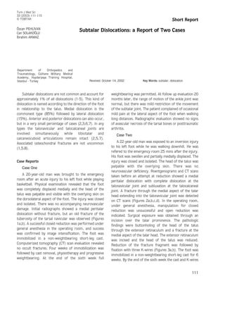
Figure 1. Radiographs showing the complete dislocation of a) talocalcaneal and b) talonavicular joints.
(a)
(b)
3. Ö. PEHL‹VAN, C. SOLAKO⁄LU, ‹. AKMAZ
113
(a) (b)
(c) (d)
Figure 2. a) and b) radiographs showing talocalcaneal and talonavicular irregularity, c) and d) coronal CT topogram and scan showing the medial
talonavicular dislocation and fracture at the medial side of the head of the talus.
4. were removed without anesthesia, followed by
physiotherapy and progressive weightbearing. At the end
of the eighth week full weightbearing was permited. The
patient was monitored for 26 months. At the last follow-
up examination he was currently pain- free in his daily
activities, but he had mild to severe pain on the lateral
side of the foot when walking long distances and at the
forced inversion of the foot. There was no limitation of
movement of the ankle joint, but the range of motion of
the subtalar joint was diminished by 25% compared with
the right side. There was no radiographic evidence of
arthritis or avascular necrosis of the tarsal bones.
Discussion
Medial peritalar dislocation most commonly occurs by
a severe inversion of the foot. The sustentaculum tali act
as a fulcrum causing first talonavicular dislocation,
followed by talocalcaneal dislocation (2,4-7). Lateral
dislocation is a result of a severe eversion force. The
anterior calcaneal process acts as a fulcrum, causing
subtalar dislocation first and talonavicular dislocation last
(2,4,6).
Associated fractures of the tarsal bones, the base of
the fifth metatarsal, and both malleoli are common with
peritalar dislocations. The frequency of tarsal bone
fractures varies from 20 to 60%, with lateral and open
types being more likely to contain a fracture (2,4-8). In
case one presented in this report, a rare condition of pure
medial peritalar dislocation, without fracture, was
treated.
Dislocation must be reduced as soon as possible in
order to avoid soft tissue and circulatory complications.
Subtalar Dislocations: a Report of Two Cases
114
(a) (b)
Figure 3. a) and b) radiographs after open reduction of peritalar dislocation and ORIF of the fracture of the head of the talus.
5. Closed reduction, under general anesthesia, is usually
successful in the medial type of dislocation (1-7). In the
case of unsuccessful closed reduction, open reduction is
mandatory. The most common obstacles to closed
reduction are buttonholing of the talar head through the
extensor retinaculum or extensor digitorum muscle,
interlocking osteochondral fractures in the talonavicular
joint and interposition of extensor digitorum brevis
muscle (2-5,7). To our knowledge there have been no
reports of the buttonholing of the talar head accompanied
by osteochondral fracture of the head of the talus, as
presented in this report in case two.
Complications in peritalar dislocations depend on the
type and severity of the dislocation. Lateral or open
dislocations have more chance of complications (1-3,7).
Early complications in peritalar dislocations are skin
necrosis, deep infection and neurovascular compromise.
The frequency of these complications varies from 0.0 to
10%. Therefore, early diagnosis and accurate reduction
are essential to avoid these early complications (2-4). Late
complications include avascular necrosis of the tarsal
bones, osteoporosis and posttraumatic arthritis.
Avascular necrosis is a very rare condition and
osteoporosis is related to long- term of immobilization
(2,4). Among the late complications the most common is
posttraumatic arthritis, which causes pain and restriction
of the subtalar joint movements (1,2,7). Dislocations
with associated intra-articular fractures would likely
result in arthritic changes causing varying degrees of
stiffness of the subtalar joint. It has been reported that
intra-articular fractures involving talocalcaneal or
talonavicular joints can cause a significant amount of
subtalar arthrosis (2,7,8). In order to minimize the
degree of stiffness of the subtalar joint and related
symptoms, uncomplicated medial subtalar dislocations
should not be immobilized longer than 4 weeks and
immobilization of dislocations with an associated fracture
should not exceed 6 weeks. Immobilization should be
followed by immediate mobilization, physiotherapy and
full weightbearing (2,4,7).
Correspondence author:
Özcan PEHL‹VAN
‹lyas Bey Caddesi, No: 49/51 D.5
34310 Yedikule, ‹stanbul - Turkey
Ö. PEHL‹VAN, C. SOLAKO⁄LU, ‹. AKMAZ
115
1. Bak K, Koch SS. Subtalar dislocation in
a handball player. Br J Sports. Med
25(1): 24-5, 1991.
2. Freund KG. Subtalar dislocations: a
review of the literature. J Foot Ankle
Surg 28(5): 429-32, 1989.
3. Heck BE, Ebraheim NA, Jackson WT.
Anatomical considerations of irreducible
medial subtalar dislocation. Foot Ankle
Int 17(2): 103-6, 1996.
4. Marcinko DE, Zenker CC. Peritalar
dislocation without fracture. J Foot
Ankle Surg 30(5): 489-93, 1991.
5. Pierre RKST, Velazco A, Fleming LL,
Whitesides T. Medial subtalar
dislocation in an athlete: a case report.
Am J Sports Med 10(4): 240-4, 1982
6. K›n›k H, Oktay O, Ar›kan M, Mergen E.
Medial subtalar dislocation. Int Orthop
23: 366-7, 1999.
7. Merianos P, Papagiannakos K, Hatzis A,
Tsafantakis E. Peritalar dislocation: a
follow-up report of 21 cases. Injury 19:
439-42, 1988.
8. Bohay DR, Manoli A. Occult fractures
following subtalar joint injuries. Foot
Ankle Int 17(3): 164-9, 1996.
References