Rickets and osteomalacia
•Als PPTX, PDF herunterladen•
2 gefällt mir•6,832 views

A 40-year-old male presented with 4 months of generalized muscle discomfort, particularly in the shoulders, without relief. Examination showed mild weakness in arms and legs but normal reflexes. Tests revealed low calcium and phosphate levels and high alkaline phosphatase, suggestive of osteomalacia. Osteomalacia is a condition characterized by reduced bone mineralization due to vitamin D deficiency, phosphate depletion, or metabolic acidosis. This patient's presentation and test results indicate osteomalacia likely due to vitamin D deficiency.

Empfohlen
Weitere ähnliche Inhalte
Was ist angesagt?
Was ist angesagt? (20)
Andere mochten auch
Andere mochten auch (20)
Ähnlich wie Rickets and osteomalacia
Ähnlich wie Rickets and osteomalacia (20)
Mehr von Afrina Qureshi
Mehr von Afrina Qureshi (20)
Rickets and osteomalacia
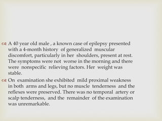
- 1. A 40 year old male , a known case of epilepsy presented with a 4-month history of generalized muscular discomfort, particularly in her shoulders, present at rest. The symptoms were not worse in the morning and there were nonspecific relieving factors. Her weight was stable. On examination she exhibited mild proximal weakness in both arms and legs, but no muscle tenderness and the reflexes were preserved. There was no temporal artery or scalp tenderness, and the remainder of the examination was unremarkable.
- 2. Subsequent investigation demonstrated haemoglobin 10.9 g/dL,ESR 16mm/h, plasma glucose (postprandial) 7.6mmol/L, glycated haemoglobin 5.6%, plasma urea andelectrolytes within normal limits, plasma calcium 7.4mg/dl(N 8.5-10.5),phosphate 2.5mg/dl(N 4.5-6.5) and plasma alkaline phosphatase 198IU/L (normal <110IU/L)
- 3. What is the most likely explanation for her symptoms and what other causes should be considered?
- 4. This patient's presentation with myalgia associated with a combination of mild hypocalcaemia, hypophosphataemia and elevation of alkaline phosphatase is strongly suggestive of Osteomalasia.
- 5. Rickets and osteomalacia are conditions characterized by pathological defects in bone matrix mineralization. Rickets refers specifically to osteomalacia, where the defect occurs in growing bone. The aetiological factors are diverse, but the end result is an increased quantity of unmineralized bone matrix (osteoid). RICKETS AND OSTEOMALACIA
- 6. RICKETS & OSTEOMALACIA Def.: reduction in bone mineralization !
- 7. The conditions may arise in three distinct situations: • Deficiency or abnormal metabolism of vitamin D • Phosphate depletion • Chronic metabolic acidosis. aetiology of osteomalacia
- 8. Vitamin D deficiency is numerically by far the mostimportant cause of osteomalacia (or rickets): it leads to acombined deficiency of calcium and phosphorus. There is intestinal malabsorption of calcium and, to a lesser extent Phosphorus. Secondary hyperparathyroidism results from the fall in plasma calcium, and the ensuing phosphaturia accelerates the phosphate deficiency.
- 9. Lack of vitamin D is often due to a combination of poor exposure to sunlight and dietary deficiency. The elderly are particularly at risk. The reason for this is multifactorial and incompletely understood, but dietary deficiency of vitamin D and calcium, together with binding of calcium by the phytate contained in chapati flour, may contribute. In addition,deeply pigmented skin generates less vitamin D3 in response to a given amount of ultraviolet exposure, an dthis may be disadvantageous in temperate climates. Vitamin D deficiency may complicate fat malabsorption from any cause. Finally, calcium deficiency enhances the rate of degradation of 25(OH)D in the liver, further exac-erbating the shortage of vitamin D and its metabolites. abnormality of the 1,25(OH)2D receptor in target tissues (vitamin D-dependent rickets type II).
- 10. Phosphate depletion is a less common cause of rickets than deficiency or altered metabolism of vitamin D. It arises as a result of a reduced maximal capacity for renal tubular phosphate reabsorption. This may either be inherited as an X-linked recessive characteristic (vitamin D-resistant rickets, X-linked hypophosphataemia) or occur aspart of Fanconi's syndrome - a more generalized hereditary or acquired renal tubular defect typified by aminoaciduria and glycosuria (p. 1057). Rarely, excessive ingestion of antacids may restrict intestinal phosphate absorption to the point where clinically important phos-phate depletion, and even osteomalacia or rickets, may appear.
- 11. Chronic metabolic acidosis, usually resulting from renal tubular disorders, may lead to rickets or osteomalacia. The bone lesions usually heal satisfactorily following treatment to correct the acidosis.
- 13. In children, failure of bone mineralization gives rise to classic bone deformities, which include widening of the metaphyses, prominence of costochondral junctions (socalled 'rickety rosary'), and varus or valgus abnormalities of the knee joints. The bones are painful and statural growth is reduced. In adults, bone pain and tenderness are the most prominent features. A characteristic proximal myopathy may develop, and is more common in cases due to vitamin D deficiency. Clinical features
- 14. Biochemical In osteomalacia due to deficiency or abnormal metabolism of vitamin D, investigations typically demonstrate a low normal plasma calcium (leading to secondary hyper- parathyroidism), a low plasma phosphate due to phospha- turia as a result of increased PTH secretion, and a raised alkaline phosphatase indicating increased numbers of osteoblasts . Vitamin D deficiency may be confirmed by measurement of 25- hydroxyvitamin D in plasma. X-linked hypophosphataemia (vitamin D-resistantrickets), on the other hand, is characterized by normocal-caemia, hypophosphataemia, and little or no alteration in plasma alkaline phosphatase level. Investigation
- 15. RICKETS, OSTEOMALACIA XRAY FINDINGS: RICKETS Thickening and widening of physes, Cupping of metaphysis, Wide metaphysis, Bowing of diaphysis, Blurred trabeculae.
- 16. RICKETS, OSTEOMALACIA XRAY FINDINGS: OSTEOMALACIA Loosers zones - incomplete stress # with healing lacking calcium, on compression side of long bones. Codfish vertebrae due to pressure of discs Trefoil pelvis, due to indentation of acetabulae stress #s
- 17. Occasionally a bone biopsy may be needed (following double tetracycline labelling) to confirm a diagnosis of osteomalacia whhen biochemical investigations are equivocal. This is particularly the case when seeking possible treatable factors in elderly patients with decreased bone density. Histological
- 18. Dietary vitamin D deficiency is treated by oral replace- ment of vitamins D2 or D3 in physiological quantities(500- 1000 units/day). This can conveniently be given as combined calcium with vitamin D tablets. Alternatively,a single i.m. does of 150 000IU of D2 in oil is effective prophylaxis and treatment for at least 6 months. The possibility of intestinal malabsorption should be borne in mind, and appropriate investigation initiated if necessary. Osteomalacia associated with anticonvulsant therapy requires higher doses of vitamin D (3000-6000 units/day)unless a change in anticonvulsant is clinically feasible. Management
- 19. Treatment of X-linked hypophosphataemia is generallyunsatisfactory. The logical treatment is with oral phosphatesupplements, but the doses needed are large and they maybe poorly tolerated because of gastrointestinal side-effects.Oral phosphate works better when combined with phar-macological doses of vitamin D, or (and probably best) incombination with 1,25(OH)2D3. When 1,25(OH)2D3 orlaOHD3 is given, great care must be taken to adjust the dose against the serum calcium, as hypercalcaemia may easily be induced. Management